. 2005 Dec;25(23):10379-90.
doi: 10.1128/MCB.25.23.10379-10390.2005.

RBP-Jkappa/SHARP recruits CtIP/CtBP corepressors to silence Notch target genes

Affiliations

RBP-Jkappa/SHARP recruits CtIP/CtBP corepressors to silence Notch target genes

Franz Oswald et al. Mol Cell Biol. 2005 Dec.

Abstract

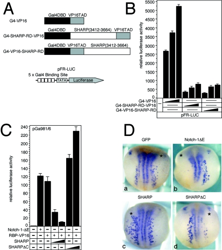
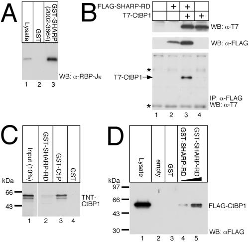
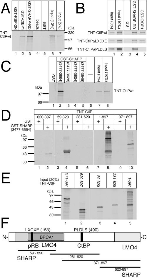
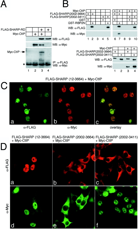
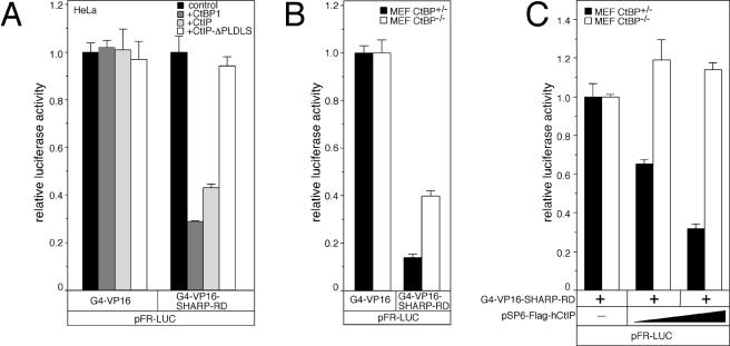
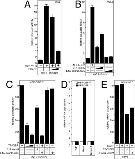
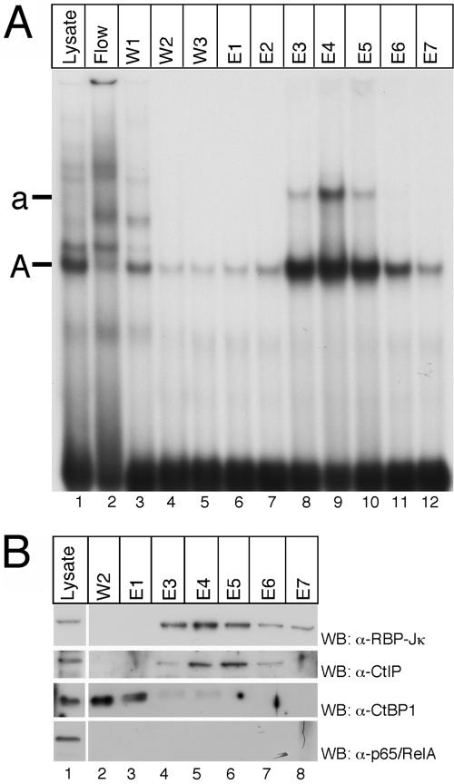
Notch is a transmembrane receptor that determines cell fates and pattern formation in all animal species. After ligand binding, proteolytic cleavage steps occur and the intracellular part of Notch translocates to the nucleus, where it targets the DNA-binding protein RBP-Jkappa/CBF1. In the absence of Notch, RBP-Jkappa represses Notch target genes through the recruitment of a corepressor complex. We and others have identified SHARP as a component of this complex. Here, we functionally demonstrate that the SHARP repression domain is necessary and sufficient to repress transcription and that the absence of this domain causes a dominant negative Notch-like phenotype. We identify the CtIP and CtBP corepressors as novel components of the human RBP-Jkappa/SHARP-corepressor complex and show that CtIP binds directly to the SHARP repression domain. Functionally, CtIP and CtBP augment SHARP-mediated repression. Transcriptional repression of the Notch target gene Hey1 is abolished in CtBP-deficient cells or after the functional knockout of CtBP. Furthermore, the endogenous Hey1 promoter is derepressed in CtBP-deficient cells. We propose that a corepressor complex containing CtIP/CtBP facilitates RBP-Jkappa/SHARP-mediated repression of Notch target genes.


Figures

FIG. 1.
FIG. 1.
Functional characterization of SHARP-RD. (A) Schematic representation of the Gal4-VP16 expression and reporter constructs for SHARP-RD used in the transcriptional repression assay. (B) SHARP-RD is necessary and sufficient to repress Gal4-VP16-mediated transcription. The pFR-LUC reporter construct (2 μg) was transfected alone or together with increasing amounts (5 ng, 10 ng, and 20 ng) of the indicated Gal4-VP16 expression constructs into HeLa cells. (C) Dominant negative effects of SHARP lacking its C-terminal repression domain. In contrast to SHARP, SHARPΔC stimulates RBP-VP16-mediated transcription. The pGA981/6 reporter construct (2 μg) was transfected alone or together with plasmids expressing either Notch-1ΔE (20 ng) or RBP-VP16 (20 ng) as well as with increasing amounts (100 ng and 250 ng) of either SHARP or SHARPΔC expression plasmids into HeLa cells. Luciferase activity was determined in 100-μg portions of total-cell extracts and normalized to the basal promoter activity of the reporter construct. Mean values and standard deviations from four independent experiments are shown. +, with the indicated construct; −, without the indicated construct. (D) Dominant negative effects of SHARP lacking its C-terminal repression domain. SHARPΔC induces a neurogenic phenotype in Xenopus laevis embryos. Embryos were injected with 100 pg GFP expression plasmid alone (panel a) or together with 100 pg mNotch-1ΔE mRNA (panel b), 2.4 ng full-length SHARP mRNA (panel c), or 2.4 ng of SHARPΔC mRNA (panel d) in one cell at the two-cell stage. Whole-mount in situ hybridization for N-tubulin shows primary neurons. The injected sides are marked with an asterisk.
FIG. 2.
FIG. 2.
(A) Endogenous RBP-Jκ proteins bind strongly to GST-SHARP(2002-3664) immobilized on glutathione Sepharose beads. (B through D) Association of CtBP1 with the repression domain of SHARP. (B) Expression plasmids for T7-tagged CtBP1 and FLAG-tagged SHARP-RD were transfected into HEK-293 cells. Expression of transfected SHARP-RD and CtBP1 was detected via Western blotting using antibodies against the T7 (upper blot) or FLAG tags (middle blot). CtBP1 coimmunoprecipitated with SHARP-RD with the antibody against the FLAG epitope from only lysates of cells transfected with both expression constructs (lower blot, lane 3). Coimmunoprecipitated CtBP1 proteins were detected on Western blots by using the anti-T7 antibody. The asterisks indicate antibody heavy and light chains. +, with the indicated construct. IP, immunoprecipitation. (C) Radiolabeled CtBP1 translated in vitro in reticulocyte lysate binds only weakly, if at all, to GST-SHARP-RD (lane 2) but strongly to GST-CtIP immobilized on Sepharose beads (lane 3). (D) CtBP1 proteins from transfected HEK-293 lysates bind GST-SHARP-RD (lanes 4 and 5). Binding proteins in the pull-down assay were separated by SDS-PAGE; RBP-Jκ and FLAG-CtBP1 were detected by Western blotting. WB, Western blot.
FIG. 3.
FIG. 3.
CtIP interacts with the SHARP repression domain. (A) Cell-free synthesized CtIP binds specifically to GST-CtBP1 (lane 2) and GST-SHARP-RD (lane 3) but not to GST-RBP-2N (lane 1). (B) The LXCXE motif (lane 4, middle) and the PLDLS motif (lane 4, lower) within CtIP are dispensable for binding of CtIP to SHARP-RD. (C) CtIP binds to the C terminus of SHARP-RD. Interaction is lost already after the deletion of 36 aa (lane 3). (D and E) SHARP-RD binds to N-terminal (aa 59 to 320) and C-terminal (aa 620 to 897) regions of CtIP. GST proteins were immobilized on Sepharose beads and incubated with in vitro-translated, radiolabeled CtIP proteins. After extensive washing, proteins were eluted and separated on SDS-PAGE. +, with the indicated construct. (F) Schematic representation of CtIP and its interaction domains. SHARP-RD binds, like LMO4, to an N-terminal and a C-terminal domain of CtIP.
FIG. 4.
FIG. 4.
SHARP interacts with CtIP in vivo and links CtIP with RBP-Jκ. (A) Expression of transfected SHARP-RD and CtIP was detected by Western blotting using antibodies against the FLAG (upper panel) or the Myc tags (middle panel). CtIP was coimmunoprecipitated together with SHARP-RD using an antibody directed against the FLAG-epitope exclusively from lysates where both proteins were expressed (lower panel, lane 3). Coimmunoprecipitated CtIP proteins were detected by Western blotting using an anti-Myc antibody. The asterisk indicates the heavy chain of the anti-FLAG antibody. (B, top) Either GST protein or GST-RBP-2N was immobilized on Sepharose beads and incubated with HEK-293 lysates expressing the SHARP(2002-3664) (lanes 1 and 2) or SHARP(2002-3411) (lanes 4 and 5) alone or together with Myc-CtIP protein (lanes 6, 7, 9, and 10). Only when both SHARP(2002-3664) and Myc-CtIP were expressed was a ternary complex formed with GST-RBP-2N (lane 7). This complex was not formed when a C-terminally truncated form of SHARP was expressed together with Myc-CtIP (lane 10). (B, bottom) Expression of the SHARP and CtIP proteins was verified by Western blotting. (C) HEK-293 cells were transiently transfected with an expression plasmid for SHARP(12-3664) and Myc-tagged CtIP. Cells were fixed 24 h after transfection, permeabilized, and immunostained using anti-FLAG and anti-Myc antibodies. The subcellular localization of SHARP (green, panel a) and CtIP (red, panel b) was assayed by fluorescence microscopy. (D) HEK-293 cells were transfected with various SHARP expression constructs together with Myc-CtIP as indicated. Subcellular protein localization was visualized using immunofluorescence staining, as described above. Both, wild-type SHARP (panel a) and CtIP (panel b) proteins are localized predominantly in the nucleus. Transfection of SHARP lacking the nuclear localization signal (aa 2002 to 3664, panel b) resulted in the cytoplasmic localization of CtIP (panel e). Transfection of SHARP(2002-3411) lacking both the nuclear localization signal and the RD (panel c) resulted in a restoration of the nuclear localization of CtIP (panel f).
FIG. 5.
FIG. 5.
CtIP and CtBP act as SHARP corepressors in the SHARP transcriptional repression assay. (A) The pFR-LUC reporter construct (2 μg) was transfected into HeLa cells together with either Gal4-VP16 or Gal4-VP16-SHARP-RD plasmids (20 ng) and expression constructs for CtBP1, CtIP, or CtIP-ΔPLDLS (100 ng). Cotransfection of CtBP1 or CtIP represses SHARP-RD-mediated transcription (right) but not VP16-mediated transcription (left). (B) SHARP-RD-mediated repression is less effective in CtBP-deficient MEFs. The pFR-LUC reporter construct (2 μg) was transfected into MEFs heterozygous (black bars) or CtBP-deficient (white bars) together with G4-VP16 (20 ng) or the G4-VP16-SHARP-RD plasmid (20 ng). Luciferase activity was normalized to the transcriptional activity of Gal4-VP16. Mean values and standard deviations from 12 independent experiments are shown. (C) CtIP corepressor function depends on CtBP in the SHARP repression assay. The pFR-LUC reporter construct (2 μg) was transfected into MEFs heterozygous (black bars) or CtBP-deficient (white bars) together with the G4-VP16-SHARP-RD plasmid (20 ng) and increasing amounts (100 and 200 ng) of CtIP expression plasmids. Luciferase activity was determined from 100-μg portions of total-cell extracts and normalized to the transcriptional activity of the Gal4-VP16 and Gal4-VP16-SHARP-RD constructs alone. Mean values and standard deviations from four independent experiments are shown.
FIG. 6.
FIG. 6.
SHARP and CtBP act as corepressors for Hey1 transcription. (A) The Hey1 reporter construct (2 μg) was transfected into HeLa cells alone or together with RBP-VP16 expression plasmids (60 ng) and increasing amounts of SHARP expression construct (200 and 400 ng). SHARP repressed RBP-VP16-mediated transcription of the human Hey1 promoter. (B) Cotransfections were performed with 40 ng of mNotch-DE expression plasmids or increasing amounts (100 and 200 ng) of E1A-Exon2 expression constructs. Expression of the E1A CtBP-binding motif resulted in the derepression of the Hey1 promoter. (C) CtBP-deficient MEFs were cotransfected with CtBP1 expression plasmids (100 and 200 ng) alone or together with either the E1A-Exon2 expression plasmid (100 and 200 ng) or a construct lacking the CtBP-binding motif (E1Aexon2-ΔCID). Only coexpression of the E1A CtBP-binding motif, together with T7-CtBP1, relieved CtBP1-mediated repression in CtBP-deficient MEFs. Luciferase activity was determined from 100-μg portions of total-cell extracts and normalized to the basal promoter activity of the reporter construct. Mean values and standard deviations from four independent experiments are shown. (D) Hey1 transcription is upregulated in CtBP-deficient MEFs. CP, cyclophilin. Hey1 and Notch-1 mRNA levels were examined using real-time PCR. The mRNA levels were normalized to the endogenous cyclophilin mRNA levels for each cell type. One representative experiment of six is shown. (E) Expression of CtBP1 proteins in CtBP-deficient MEFs represses Hey1 transcription. Cells were transfected with T7-CtBP1 or Flag-CtBP1 together with pEGFPC1. GFP-positive cells were sorted, and Hey1 mRNA levels were examined. The mRNA levels were normalized to the endogenous cyclophilin mRNA levels. One representative experiment of four is shown. +, with the indicated construct; −, without the indicated construct.
FIG. 7.
FIG. 7.
Purification of endogenous RBP-Jκ complexes by DNA affinity chromatography. (A) A DNA fragment containing 12 RBP-Jκ-binding sites was biotinylated and immobilized with streptavidin Sepharose. The column was incubated with cellular extract from Jurkat T cells. After washing, the DNA-binding activity was eluted with increasing NaCl concentrations as indicated in Materials and Methods. RBP-Jκ-specific DNA-binding activity (complexes A [single occupancy] and a [double occupancy] were eluted in fractions E3 to E6 (lanes 8 to 11). (B) Western analysis of lysate (lane 1), washing step 2 (lane 2), and eluted fractions (lanes 3 to 8) using the indicated antibodies. WB, Western blot.

Similar articles

Cited by

References

    1. Ariyoshi, M., and J. W. Schwabe. 2003. A conserved structural motif reveals the essential transcriptional repression function of Spen proteins and their role in developmental signaling. Genes Dev. 17:1909-1920. - PMC - PubMed
    1. Barolo, S., T. Stone, A. G. Bang, and J. W. Posakony. 2002. Default repression and Notch signaling: Hairless acts as an adaptor to recruit the corepressors Groucho and dCtBP to Suppressor of Hairless. Genes Dev. 16:1964-1976. - PMC - PubMed
    1. Chen, P. L., F. Liu, S. Cai, X. Lin, A. Li, Y. Chen, B. Gu, E. Y. Lee, and W. H. Lee. 2005. Inactivation of CtIP leads to early embryonic lethality mediated by G1 restraint and to tumorigenesis by haploid insufficiency. Mol. Cell. Biol. 25:3535-3542. - PMC - PubMed
    1. Chinnadurai, G. 2002. CtBP, an unconventional transcriptional corepressor in development and oncogenesis. Mol. Cell 9:213-224. - PubMed
    1. De Strooper, B., W. Annaert, P. Cupers, P. Saftig, K. Craessaerts, J. S. Mumm, E. H. Schroeter, V. Schrijvers, M. S. Wolfe, W. J. Ray, A. Goate, and R. Kopan. 1999. A presenilin-1-dependent gamma-secretase-like protease mediates release of Notch intracellular domain. Nature 398:518-522. - PubMed

Publication types

MeSH terms