Comparative Study
. 2005 Nov 9;25(45):10546-55.
doi: 10.1523/JNEUROSCI.3275-05.2005.

The role of Snapin in neurosecretion: snapin knock-out mice exhibit impaired calcium-dependent exocytosis of large dense-core vesicles in chromaffin cells

Affiliations
Comparative Study

The role of Snapin in neurosecretion: snapin knock-out mice exhibit impaired calcium-dependent exocytosis of large dense-core vesicles in chromaffin cells

Jin-Hua Tian et al. J Neurosci. .

Abstract

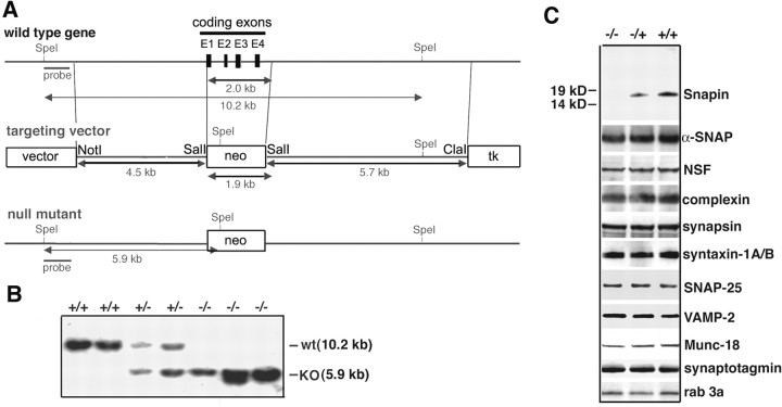
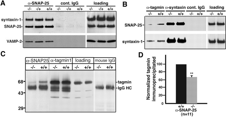
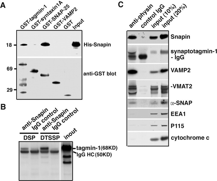
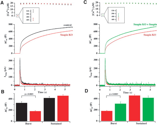
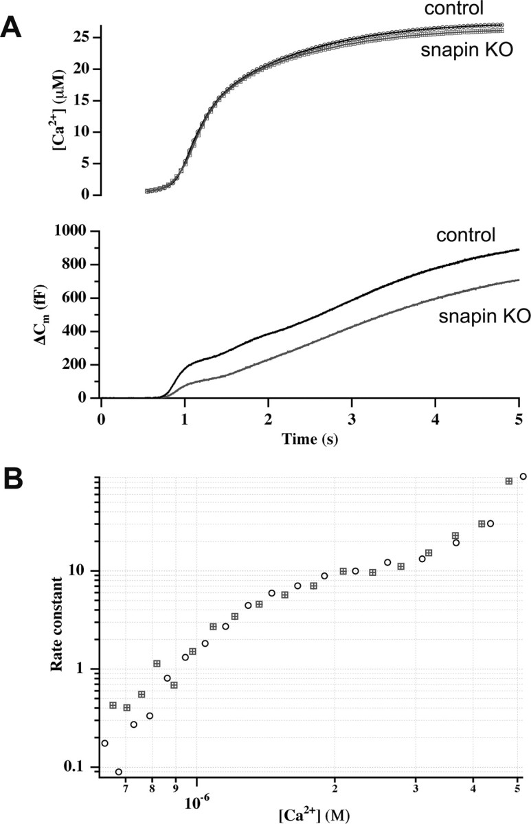
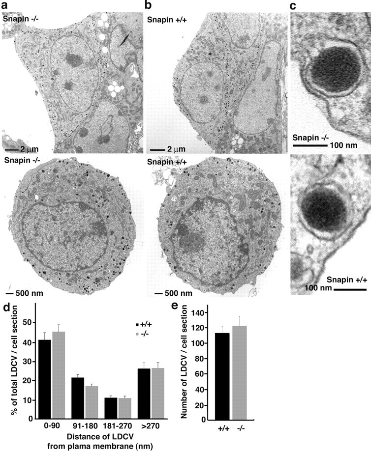
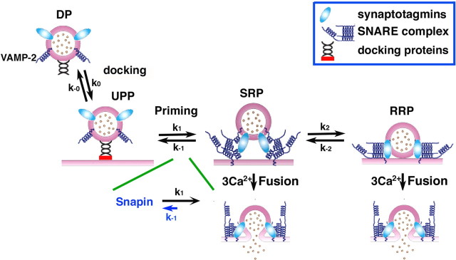
Identification of the molecules that regulate the priming of synaptic vesicles for fusion and the structural coupling of the calcium sensor with the soluble N-ethyl maleimide sensitive factor adaptor protein receptor (SNARE)-based fusion machinery is critical for understanding the mechanisms underlying calcium-dependent neurosecretion. Snapin binds to synaptosomal-associated protein 25 kDa (SNAP-25) and enhances the association of the SNARE complex with synaptotagmin. In the present study, we abolished snapin expression in mice and functionally evaluated the role of Snapin in neuroexocytosis. We found that the association of synaptotagmin-1 with SNAP-25 in brain homogenates of snapin mutant mice is impaired. Consequently, the absence of Snapin in embryonic chromaffin cells leads to a significant reduction of calcium-dependent exocytosis resulting from a decreased number of vesicles in releasable pools. Overexpression of Snapin fully rescued this inhibitory effect in the mutant cells. Furthermore, Snapin is relatively enriched in the purified large dense-core vesicles of chromaffin cells and associated with synaptotagmin-1. Thus, our biochemical and electrophysiological studies using snapin knock-out mice demonstrate that Snapin plays a critical role in modulating neurosecretion by stabilizing the release-ready vesicles.


Figures

Figure 1.
Figure 1.
Generation of mice lacking snapin. A, Maps of the wild-type snapin gene, the respective targeting vector, and the resulting mutant gene. A genomic clone of the snapin gene containing all four coding exons E1-E4 (boxes in top diagram) was used to generate a targeting vector (middle diagram), which was used for homologous recombination in embryonic stem cells (bottom diagram). Restriction enzyme sites (SpeI, NotI, SalI, and ClaI) and position of the probe (bar) used for Southern blot analysis are indicated. Neo, Neomycin resistance gene; TK, thymidine kinase gene. B, Southern blot analysis of the snapin KO mutation in mice. SpeI-digested genomic DNA isolated from mouse tails of wild-type (wt) (+/+), heterozygous (+/-), and homozygous (-/-) animals was hybridized with the probedetecting the DNA fragments at 10.2 and 5.9 kb for the wild-type and mutant alleles, respectively. C, Immunoblot analysis of Snapin and other basic and regulatory components of synaptic vesicle release machinery. Equal amounts of brain homogenates (40 μg) from the E18 embryos of all genotypes were blotted with antibodies as indicated.
Figure 2.
Figure 2.
Effect of snapin knock-out on the association of SNAP-25 with synaptotagmin-1. A, Immunoprecipitation of the native SNARE complex. One microgram of anti-SNAP-25 antibody (α-SNAP-25) coimmunoprecipitated syntaxin-1 and VAMP-2 from solubilized mouse brain homogenates of snapin wild-type (+/+), heterozygous (-/+), and homozygous (-/-) mutants. Coimmunoprecipitates were analyzed by sequential immunoblotting of the same membrane after stripping of previous antibodies. Mouse IgG (cont. IgG) was used as an immunoprecipitation control. B, C, Decreased association of SNAP-25 with synaptotagmin-1 in snapin KO mutant. Brain homogenates (100 μg) from wild-type and homozygous KO littermates were immunoprecipitated by 1 μg of indicated antibodies or control IgG followed by immunoblotting with anti-syntaxin-1 and anti-SNAP-25 (B) or anti-synaptotagmin-1 (C) antibodies. Note that a significant decrease in the association of SNAP-25 with synaptotagmin-1 was observed in the absence of Snapin, whereas the amount of the SNARE complex (syntaxin, SNAP-25, and VAMP2) remains unchanged. Loading lanes represent 15% of homogenates used for each immunoprecipitation. IgGHC, IgG heavy chain. D, Normalized amounts of synaptotagmin-1 coimmunoprecipitated by anti-SNAP-25 antibody from both snapin wild-type (+/+) and KO (-/-) brain homogenates. A 34 ± 3% reduction was observed for the amount of synaptotagmin-1 coprecipitated by anti-SNAP-25 antibody from KO relative to that from wild-type homogenates (mean ± SEM from 11 littermates). A two-tailed Student's t test for paired data was used; **p < 0.01.
Figure 3.
Figure 3.
Snapin is relatively enriched in the purified LDCVs and associated with synaptotagmin-1. A, Snapin binds to synaptotagmin-1 in vitro. GST or GST-tagged fusion proteins (∼1 mg) were immobilized on glutathione-Sepharose beads and then incubated with His-tagged Snapin. Bound protein complexes were sequentially immunoblotted with antibodies against T7-His-tag (top) and GST (bottom). B, Coimmunoprecipitation of synaptotagmin-1 from chemically cross-linked mouse brain homogenates with anti-Snapin antibody followed by immunoblotting with anti-synaptotagmin-1 antibody. C, Snapin is relatively enriched in purified LDCVs. Mouse adrenal gland homogenate was incubated with magnetic beads coated with the antibody against synaptophysin, an LDCV membrane protein, or normal IgG control. The bead-bound fractions were subjected to immunoblotting. Relative purity of the LDCVs was assessed by sequential immunoblotting of LDCV markers including VAMP2, synaptotagmin-1, and VMAT2 and non-LDCV proteins including p115 (Golgi), EEA1 (endosomes), and cytochrome c (mitochondria).
Figure 4.
Figure 4.
Absence of Snapin leads to a reduction in LDCV exocytosis from adrenal chromaffin cells after flash photolysis of caged calcium. A, Averaged calcium concentration ([Ca2+]i; top), capacitance (ΔCm; middle), and amperometric responses (IAmp; bottom) in response to flash-photolysis of NP-EGTA in control cells (47 cells from 7 animals; black line and symbols) and snapin KO cells (112 cells from 12 animals; red line and symbols). Both capacitance and amperometric traces display a rapid, burst-like increase within the first second after the flash, followed by a slower sustained phase of secretion. The inset in the top panel shows the basal [Ca2+]i before the flash, which was not significantly different between the two groups. B, Kinetic analysis of the experiment shown in A. Snapin KO cells show a selective reduction of the exocytotic burst (p < 0.0001; Mann-Whitney U test), whereas the sustained component is unaffected (for details, see Table 1). C, Averaged calcium concentration ([Ca2+]i; top), capacitance (ΔCm; middle), and amperometric responses (IAmp; bottom) in response to flash-photolysis of NP-EGTA in snapin KO cells (112 cells from 12 animals; red line and symbols) and snapin KO cells infected with Snapin-GFP (34 cells from 8 animals; green line and symbols). The inset in the top panel shows the basal [Ca2+]i before the flash, which was not significantly different between the two groups. D, Kinetic analysis of the experiment shown in C. Reintroduction of Snapin into snapin KO cells resulted in a complete recovery of the exocytotic burst size to control values (Table 1) (see A and B for comparison; p < 0.0001), demonstrating the specificity of the observed effect by Snapin. Error bars represent SEM.
Figure 5.
Figure 5.
Reduction in exocytosis in snapin KO cells is not attributable to a change in the Ca2+ dependence of exocytosis. A, Calcium ramp experiments were performed by slow release of Ca2+ from NP-EGTA using weak UV-illumination. Membrane capacitance and Ca2+ concentration were monitored simultaneously during the 5 s of stimulation. In agreement with the flash experiments (Fig. 4), snapin KO cells displayed a smaller secretion than control cells at all calcium concentrations. B, Double logarithmic plot of the rate constant versus calcium for 27 control cells (circles; n = 2) and 26 snapin KO cells (squares; n = 2). The curves are identical. Neither the Ca2+ sensitivity (threshold) nor the Ca2+ cooperativity (slope) of secretion is altered in the absence of Snapin.
Figure 6.
Figure 6.
Morphologically normal docking of LDCVs in the absence of Snapin. A, B, Electron micrographs of isolated chromaffin cells from snapin KO (-/-) (A) and wild-type (+/+) (B) animals. In both cell types, most vesicles were found aligned with the plasma membrane. C, Closeup of a single docked vesicle from both cell types. D, Distribution of vesicles for the distance between the center of chromaffin granules and the plasma membrane in both cell types (16 wild-type cells and 17 KO cells from 4 littermates; mean ± SEM). Note that there is no defect in morphological docking of the LDCVs (0-90 nm between the center of chromaffin granules and the plasma membrane) in the absence of Snapin. E, No significant difference in the total number of vesicles per cell section (mean ± SEM) from both wild-type and KO cells as indicated. Error bars represent SEM.
Figure 7.
Figure 7.
Proposed role of Snapin in promoting the association of SNAP-25 with synaptotagmin during the vesicle maturation/priming process. The illustration shows the kinetic model of vesicle docking, priming, and fusion in chromaffin cells. The priming step corresponds to the assembly of the SNARE complex, and further maturation into the releasable vesicle pool requires synaptotagmins. Fusion from the SRP and RRP is assumed to require three Ca2+ ions. Snapin is proposed to stabilize both releasable pools (SRP and RRP) by reducing the backward rate constant (K-1, in blue) of the priming-unpriming reaction. DP, Depot pool; UPP, pool of docked but unprimed vesicles.

Similar articles

Cited by

References

    1. Ashery U, Varoqueaux F, Voets T, Betz A, Thakur P, Koch H, Neher E, Brose N, Rettig J (2000) Munc13-1 acts as a priming factor for large dense-core vesicles in bovine chromaffin cells. EMBO J 19: 3586-3596. - PMC - PubMed
    1. Augustine GJ (2001) How does calcium trigger neurotransmitter release? Curr Opin Neurobiol 11: 320-326. - PubMed
    1. Bai J, Wang CT, Richards DA, Jackson MB, Chapman ER (2004) Fusion pore dynamics are regulated by synaptotagmin*t-SNARE interactions. Neuron 41: 929-942. - PubMed
    1. Bennett MK, Calakos N, Scheller RH (1992) Syntaxin: a synaptic protein implicated in docking of synaptic vesicles at presynaptic active zones. Science 257: 255-259. - PubMed
    1. Buxton P, Zhang XM, Walsh B, Sriratana A, Schenberg I, Manickam E, Rowe T (2003) Identification and characterization of Snapin as a ubiquitously expressed SNARE-binding protein that interacts with SNAP23 in non-neuronal cells. Biochem J 375: 433-440. - PMC - PubMed

Publication types

MeSH terms

LinkOut - more resources