. 2005 Nov 15;19(22):2668-81.
doi: 10.1101/gad.1360605. Epub 2005 Oct 31.

TAK1, but not TAB1 or TAB2, plays an essential role in multiple signaling pathways in vivo

Affiliations

TAK1, but not TAB1 or TAB2, plays an essential role in multiple signaling pathways in vivo

Jae-Hyuck Shim et al. Genes Dev. .

Abstract

TGF-beta-activated kinase 1 (TAK1), a member of the MAPKKK family, is thought to be a key modulator of the inducible transcription factors NF-kappaB and AP-1 and, therefore, plays a crucial role in regulating the genes that mediate inflammation. Although in vitro biochemical studies have revealed the existence of a TAK1 complex, which includes TAK1 and the adapter proteins TAB1 and TAB2, it remains unclear which members of this complex are essential for signaling. To analyze the function of TAK1 in vivo, we have deleted the Tak1 gene in mice, with the resulting phenotype being early embryonic lethality. Using embryonic fibroblasts lacking TAK1, TAB1, or TAB2, we have found that TNFR1, IL-1R, TLR3, and TLR4-mediated NF-kappaB and AP-1 activation are severely impaired in Tak1(m/m) cells, but they are normal in Tab1(-/-) and Tab2(-/-) cells. In addition, Tak1(m/m) cells are highly sensitive to TNF-induced apoptosis. TAK1 mediates IKK activation in TNF-alpha and IL-1 signaling pathways, where it functions downstream of RIP1-TRAF2 and MyD88-IRAK1-TRAF6, respectively. However, TAK1 is not required for NF-kappaB activation through the alternative pathway following LT-beta signaling. In the TGF-beta signaling pathway, TAK1 deletion leads to impaired NF-kappaB and c-Jun N-terminal kinase (JNK) activation without impacting Smad2 activation or TGF-beta-induced gene expression. Therefore, our studies suggests that TAK1 acts as an upstream activating kinase for IKKbeta and JNK, but not IKKalpha, revealing an unexpectedly specific role of TAK1 in inflammatory signaling pathways.


Figures

Figure 1.
Figure 1.
Targeted disruption of the Tak1 gene. (A) Schematic drawing of the Tak1 gene-trapping strategy. The position of primers (F1, R1, and R2) used in PCR genotyping are indicated by arrowheads. (En2) engrailed 2 gene; (βgeo) a fusion protein between β-galactosidase and neomycin phosphotransferase; (IRES) internal ribosomal entry site; (PLAP) human placental alkaline phosphatase; (SV40 pA) SV40 polyadenylation signal. (B) β-galactosidase staining of E9.5 wild-type (Tak1+/+) and heterozygous embryos (Tak1+/m). Bar, 100 μm. (C) PCR genotyping and immunoblotting analysis of TAK1 in E10 wild-type (+/+), heterozygous (+/m), and homozygous (m/m) mutant embryos. The size of PCR fragments is 599 base pairs (bp) and 195 bp for wild-type and insertional mutant alleles, respectively. (D) Lateral view of the wild type (+/+) and Tak1 mutant (m/m) embryos at E9.5, E10.5, and E11.5. (E) Ventral (left) and Dorsal (middle) view of E10 Tak1 mutant embryos and lateral view of E10.5 Tak1 mutant embryos (right). Whole embryos were used for PCR genotyping. (HF) Head fold; (NT) neural tube.
Figure 2.
Figure 2.
TAK1 deficiency inhibits NF-κB and JNK activation by TNF-α and IL-1. (A,B) Wild-type (WT), Tak1m/m (TAK1), Tab1-/- (TAB1), and Tab2-/- (TAB2) cells were transfected with either pBIIx-luc (left) or AP1-luc (right) reporter together with Renilla luciferase vector. Twenty-four hours after transfection, cells were treated with TNF-α (10 ng/mL; A) or IL-1 (10 ng/mL; B) for 5 h and then analyzed for luciferase activity. Results are expressed as the fold induction in luciferase activity relative to that of untreated cells. Error bars indicate standard deviation. (C,D) IκB-α degradation and JNK phosphorylation by TNF-α (C) or IL-1 (D) were analyzed by immunoblotting with antibodies specific for IκB-α and phospho-JNK, respectively. Immunoblotting with anti-GAPDH4 antibody was performed as a loading control. (E) Cells were treated with TNF-α (10 ng/mL; top) or IL-1 (10 ng/mL; bottom) for the indicated time, nuclear extracts were prepared and immunoblotted with anti-phosho-c-Jun antibody. Immunoblotting with anti-HDAC1 antibody was performed as a loading control. (F,G) Cells were treated with TNF-α (10 ng/mL; E) or IL-1 (10 ng/mL; F) for the indicated times and nuclear extract were prepared. NF-κB DNA-binding activity was analyzed by EMSA.
Figure 3.
Figure 3.
TLR-mediated NF-κB and JNK activation is impaired in Tak1m/m cells. (A) Wild type, Tak1m/m, Tab1-/-, and Tab2-/- cells were cotransfected with CD4/TLR4 and Renilla luciferase vectors together with either pBIIx-luc (left), AP1-luc (middle), or IRF3-luc (right) reporters. Twenty-four hours after transfection, cells were lysed and analyzed for luciferase activity. Results are expressed as fold change in luciferase activity relative to untreated cells. (B) Cells were treated with LPS (1 μg/mL) for the indicated times, nuclear extracts were prepared, and NF-κB DNA-binding activity was analyzed by EMSA. (C) LPS-induced JNK phosphorylation was analyzed by immunoblotting with antiphospho-JNK antibody. Immunoblotting with anti-GAPDH4 antibody was performed to control for gel loading. (D) Cells were transfected with pBIIx-luc (left) or IRF3-luc (right) reporters together with Renilla luciferase vector. Twenty-four hours after transfection, cells were treated with poly(I:C) (5 μg/mL) and luciferase activity was analyzed as in A. (E) Poly(I:C)-induced I-κB-α degradation and JNK phosphorylation were analyzed by immunoblotting with anti-I-κB-α and antiphospho-JNK antibodies, respectively. Immunoblotting with anti-GAPDH4 antibody was performed to control for gel loading.
Figure 4.
Figure 4.
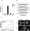
Tak1m/m cells are highly sensitive to TNF-α-induced apoptosis. (A) Wild-type (WT), Tak1m/m, Tab1-/-, and Tab2-/- cells were treated with TNF-α (10 ng/mL) for 8 h. Apoptosis was analyzed by staining with FITC-Annexin V and propidium iodide (PI) staining. Data given is the percentage of cells that stained positive with both PI and Annexin V. (B) Cells were stimulated with TNF-α for the indicated times, lysed, and immunoblotted with anti-caspase 8 antibody. Immunoblotting with anti-GAPDH4 antibody was performed to control for gel loading. (C) Wild-type, p65-/- and Tak1m/m cells were treated with TNF-α (10 ng/mL) for the indicated times. Apoptosis was analyzed by FITC-Annexin V and propidium iodide (PI) staining as in A. (D) Cells were transfected with the GFP expression vector together with pcDNA3, or HA-tagged wild-type TAK1 (TAK1-WT). Twenty-four hours after transfection, cells were treated with TNF-α (10 ng/mL) for 4 h and analyzed by fluorescent microscopy.
Figure 5.
Figure 5.
TAK1 is required for TNF- and IL-1-induced IKK activation. (A) Wild-type (WT), Tak1m/m, Tab1-/-, and Tab2-/- cells were treated with TNF-α (10 ng/mL; top) or IL-1 (10 ng/mL; bottom) for the indicated times. IKK activity was analyzed by in vitro kinase assay using a GST-IκB-α substrate. Protein input was analyzed by immunoblotting analysis with antibodies specific for IKKα and Nemo. (B) Cells were transfected with pcDNA3 or IKKβ (SSEE) together with pBIIx-luc and Renilla luciferase vectors. NF-κB activity was analyzed by luciferase assay and is expressed as fold change compared with untreated control. (C) Wild-type (WT) and Tak1m/m cells were treated with TNF-α (10 ng/mL; top) or IL-1 (10 ng/mL; bottom) for the indicated times, and then immunoblotted with antiphospho-IKKα/β antibody. Immunoblotting analysis with anti-IKKα antibody was performed as a control. (D) Cells were transfected with HA-IKKβ together with either pcDNA3, Flag-tagged TRAF2, or RIP1 expression vector. Cell were harvested 24 h after transfection, and cells were immunoprecipitated with anti-HA antibody and protein A agarose. IKKβ kinase activity was analyzed by in vitro kinase assay as in A.(E) Cells were transfected with HA-IKKβ expression vector together with either pcDNA3, Flag-tagged TRAF6, or MyD88 expression vector. Cells were harvested 24 h after transfection and IKKβ was immunoprecipitated with anti-HA antibody and protein A agarose. IKKβ activity was analyzed by in vitro kinase assay. (F) Cells were treated with IL-1 (10 ng/mL) for the indicated times, and then immunoblotted with anti-IRAK1 antibody. Immunoblotting with anti-GAPDH4 antibody was performed to control for gel loading. (G) Wild-type and Tak1m/m cells were transfected with Flag-IKKβ expression vector together with pcDNA3, or HA-tagged wild-type TAK1 (HA-TAK1-WT). Cells were stimulated 24 h later with TNF-α (10 ng/L) or IL-1 (10 ng/mL) for 20 min, lysed, and immunoprecipitated IKKβ kinase activity was analyzed by in vitro kinase assay.
Figure 6.
Figure 6.
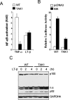
LT-β-induced NF-κB activation is normal in Tak1m/m cells. (A) Wild-type (WT) and Tak1m/m cells were transfected with pBIIx-luc reporter along with Renilla luciferase vector. Twenty-four hours after transfection, cells were treated with TNF-α (10 ng/mL) or anti-LT-βR antibody (2 μg/mL) for 5 or 12 h, respectively, and then analyzed for luciferase activity. Results are expressed as the fold change in luciferase activity relative to untreated cells. (B) Cells were transfected with pcDNA3 or NIK expression vector along with pBIIx-luc reporter and Renilla luciferase vector. NF-κB activity was analyzed by luciferase assay. (C) Cells were stimulated with anti-LT-βR antibody (2 μg/mL) for the indicated times, and then immunoblotted with anti-p100 antibody. Protein amount was analyzed by immunoblotting with anti-GAPDH4 antibody.
Figure 7.
Figure 7.
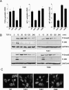
Role of TAK1 complex in TGF-β signaling pathway. (A) Wild-type, Tak1m/m, Tab1-/-, and Tab2-/- cells were transfected with the either 3TP-lux (left), AP-1-luc (middle), or pBIIxluc (right) reporter construct together with Renilla luciferase vector. Twenty-four hours after transfection, cells were treated with TGF-β (1 ng/mL) for 24 h and then analyzed for luciferase activity. Results are expressed as the fold induction in luciferase activity relative to that of untreated cells. (B) Smad2 and JNK phosphorylation by TGF-β (1 ng/mL) were analyzed by immunoblotting with antiphospho-Smad2 antibody and antiphospho-JNK antibody, respectively. Immunoblotting analysis with anti-GAPDH4 antibody was performed as a control. (C) Cells were stimulated with TGF-β (1 ng/mL) for 20 min, stained with antiphospho-Smad2 antibody, and analyzed by immunofluorescence microscopy.
Figure 8.
Figure 8.
Schematic model showing role of TAK1 complex in inflammatory signaling pathways. Red lines indicate TAK1-dependent signaling pathways, while green lines indicate TAK1-independent pathways.

Similar articles

Cited by

References

    1. Austin C.P., Battey, J.F., Bradley, A., Bucan, M., Capecchi, M., Collins, F.S., Dove, W.F., Duyk, G., Dymecki, S., Eppig, J.T., et al. 2004. The knockout mouse project. Nat. Genet. 36: 921–924. - PMC - PubMed
    1. Baud V. and Karin, M. 2001. Signal transduction by tumor necrosis factor and its relatives. Trends Cell. Biol. 11: 372–377. - PubMed
    1. Beg A.A. and Baltimore, D. 1996. An essential role for NF-κBin preventing TNF-α-induced cell death. Science 274: 782–784. - PubMed
    1. Beg A.A., Sha, W.C., Bronson, R.T., Ghosh, S., and Baltimore, D. 1995. Embryonic lethality and liver degeneration in mice lacking the RelA component of NF-κ B. Nature 376: 167–170. - PubMed
    1. Bonizzi G. and Karin, M. 2004. The two NF-κB activation pathways and their role in innate and adaptive immunity. Trends. Immunol. 25: 280–288. - PubMed

Publication types

MeSH terms

Substances