

A role for dual viral hits in causation of subacute sclerosing panencephalitis
- PMID: 16260490
- PMCID: PMC1350947
- DOI: 10.1084/jem.20051376
A role for dual viral hits in causation of subacute sclerosing panencephalitis
Abstract
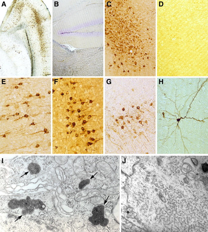
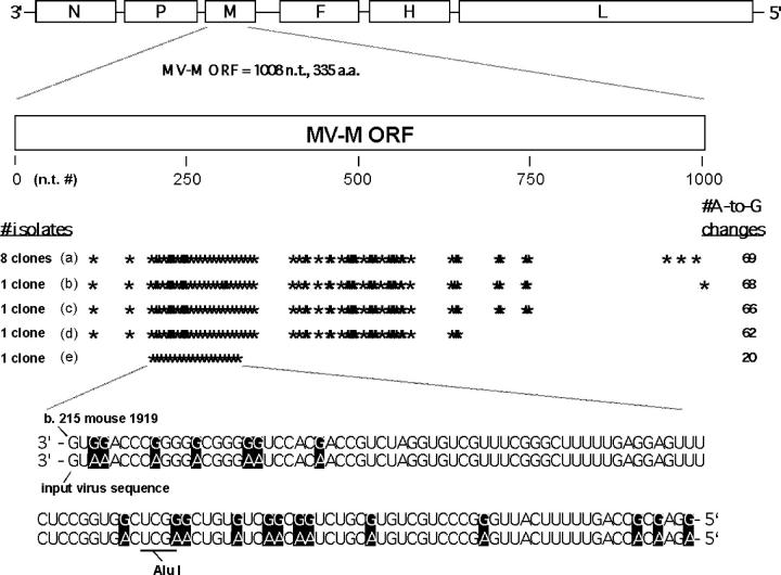
Subacute sclerosing panencephalitis (SSPE) is a progressive fatal neurodegenerative disease associated with persistent infection of the central nervous system (CNS) by measles virus (MV), biased hypermutations of the viral genome affecting primarily the matrix (M) gene with the conversion of U to C and A to G bases, high titers of antibodies to MV, and infiltration of B cells and T cells into the CNS. Neither the precipitating event nor biology underlying the MV infection is understood, nor is their any satisfactory treatment. We report the creation of a transgenic mouse model that mimics the cardinal features of SSPE. This was achieved by initially infecting mice expressing the MV receptor with lymphocytic choriomeningitis virus Cl 13, a virus that transiently suppressed their immune system. Infection by MV 10 days later resulted in persistent MV infection of neurons. Analysis of brains from infected mice showed the biased U to C hypermutations in the MV M gene and T and B lymphocyte infiltration. These sera contained high titers of antibodies to MV. Thus, a small animal model is now available to both molecularly probe the pathogenesis of SSPE and to test a variety of therapies to treat the disease.
Figures
Similar articles
-
Modeling subacute sclerosing panencephalitis in a transgenic mouse system: uncoding pathogenesis of disease and illuminating components of immune control.Curr Top Microbiol Immunol. 2009;330:31-54. doi: 10.1007/978-3-540-70617-5_2. Curr Top Microbiol Immunol. 2009. PMID: 19203103 Review.
-
Evidence that the hypermutated M protein of a subacute sclerosing panencephalitis measles virus actively contributes to the chronic progressive CNS disease.Virology. 2001 Dec 20;291(2):215-25. doi: 10.1006/viro.2001.1182. Virology. 2001. PMID: 11878891
-
Receptor use by vesicular stomatitis virus pseudotypes with glycoproteins of defective variants of measles virus isolated from brains of patients with subacute sclerosing panencephalitis.J Gen Virol. 2003 Aug;84(Pt 8):2133-2143. doi: 10.1099/vir.0.19091-0. J Gen Virol. 2003. PMID: 12867645
-
Effect of the alterations in the fusion protein of measles virus isolated from brains of patients with subacute sclerosing panencephalitis on syncytium formation.Virus Res. 2007 Dec;130(1-2):260-8. doi: 10.1016/j.virusres.2007.07.017. Epub 2007 Sep 7. Virus Res. 2007. PMID: 17825451
-
Pathogenic aspects of measles virus infections.Arch Virol Suppl. 1999;15:139-58. doi: 10.1007/978-3-7091-6425-9_10. Arch Virol Suppl. 1999. PMID: 10470275 Review.
Cited by
-
The Anatomy of a Career in Science.DNA Cell Biol. 2016 Mar;35(3):109-17. doi: 10.1089/dna.2016.3232. Epub 2016 Feb 2. DNA Cell Biol. 2016. PMID: 26836569 Free PMC article. No abstract available.
-
RNA editing enzyme adenosine deaminase is a restriction factor for controlling measles virus replication that also is required for embryogenesis.Proc Natl Acad Sci U S A. 2011 Jan 4;108(1):331-6. doi: 10.1073/pnas.1017241108. Epub 2010 Dec 20. Proc Natl Acad Sci U S A. 2011. PMID: 21173229 Free PMC article.
-
Foxp3+ regulatory T cells control persistence of viral CNS infection.PLoS One. 2012;7(3):e33989. doi: 10.1371/journal.pone.0033989. Epub 2012 Mar 20. PLoS One. 2012. PMID: 22448284 Free PMC article.
-
Measles virus-induced immunosuppression: from effectors to mechanisms.Med Microbiol Immunol. 2010 Aug;199(3):227-37. doi: 10.1007/s00430-010-0152-3. Epub 2010 Apr 8. Med Microbiol Immunol. 2010. PMID: 20376484 Review.
-
"Viral déjà vu" elicits organ-specific immune disease independent of reactivity to self.J Clin Invest. 2006 May;116(5):1254-63. doi: 10.1172/JCI27372. Epub 2006 Apr 6. J Clin Invest. 2006. PMID: 16604192 Free PMC article.
References
-
- Griffin, D. 2001. Measles virus. Fields Virology. B. Fields, D. Knipe, and P. Howley, editors. Lippincott-Raven, Philadelphia. 1401–1442.
-
- Oldstone, M.B.A. 1998. Viruses, Plagues, and History. Oxford University Press, New York, NY. 211 pp.
-
- Centers for Disease Control and Prevention. 2005. Progress in reducing measles mortality—worldwide, 1999-2003. Morb. Mortal. Wkly. Rep. 54:200–203. - PubMed
-
- Centers for Disease Control and Prevention. 1982. Subacute sclerosing panencephalitis surveillance—United States. Morb. Mortal. Wkly. Rep. 31:585–588. - PubMed
-
- Bellini, W.J., J.S. Rota, L.E. Lowe, R.S. Katz, P.R. Dyken, S.R. Zaki, W.-J. Shieh, and P.A. Rota. 2005. Subacute sclerosing panencephalitis: more cases of this fatal disease are prevented by measles immunization than previously recognized. J. Infect. Dis. In press. - PubMed
Publication types
MeSH terms
Substances
Grants and funding
LinkOut - more resources
Full Text Sources
Other Literature Sources
Molecular Biology Databases
