. 2005 Nov;79(21):13538-47.
doi: 10.1128/JVI.79.21.13538-13547.2005.

beta-Adrenoreceptors reactivate Kaposi's sarcoma-associated herpesvirus lytic replication via PKA-dependent control of viral RTA

Affiliations

beta-Adrenoreceptors reactivate Kaposi's sarcoma-associated herpesvirus lytic replication via PKA-dependent control of viral RTA

Margaret Chang et al. J Virol. 2005 Nov.

Abstract

Reactivation of Kaposi's sarcoma-associated herpesvirus (KSHV) lytic replication is mediated by the viral RTA transcription factor, but little is known about the physiological processes controlling its expression or activity. Links between autonomic nervous system activity and AIDS-associated Kaposi's sarcoma led us to examine the potential influence of catecholamine neurotransmitters. Physiological concentrations of epinephrine and norepinephrine efficiently reactivated lytic replication of KSHV in latently infected primary effusion lymphoma cells via beta-adrenergic activation of the cellular cyclic AMP/protein kinase A (PKA) signaling pathway. Effects were blocked by PKA antagonists and mimicked by pharmacological and physiological PKA activators (prostaglandin E2 and histamine) or overexpression of the PKA catalytic subunit. PKA up-regulated RTA gene expression, enhanced activity of the RTA promoter, and posttranslationally enhanced RTA's trans-activating capacity for its own promoter and heterologous lytic promoters (e.g., the viral PAN gene). Mutation of predicted phosphorylation targets at RTA serines 525 and 526 inhibited PKA-mediated enhancement of RTA trans-activating capacity. Given the high catecholamine levels at sites of KSHV latency such as the vasculature and lymphoid organs, these data suggest that beta-adrenergic control of RTA might constitute a significant physiological regulator of KSHV lytic replication. These findings also suggest novel therapeutic strategies for controlling the activity of this oncogenic gammaherpesvirus in vivo.


Figures

FIG. 1.
FIG. 1.
Effect of norepinephrine (NE) on KSHV reactivation from latency. (A) Expression of the KSHV lytic cycle protein ORF59 was assayed by flow cytometry using indirect immunofluorescence in BC3 cells fixed and permeabilized 24 h after exposure to 20 ng/ml PMA, 10 μM norepinephrine, or an equivalent volume of vehicle. (B) Dose dependence of catecholamine effects on KSHV lytic gene expression were also assessed by quantitative real-time RT-PCR detection of KSHV ORF29 mRNA expression (normalized to GAPDH mRNA) 24 h following exposure of BCBL-1 cells to indicated concentrations of epinephrine or norepinephrine. Similar results were observed for ORF50/RTA mRNA (data not shown). (C) Kinetics of norepinephrine-induced lytic gene expression were assessed for immediate-early ORF50/RTA and late lytic ORF29 at the indicated time points by real-time RT-PCR (data represent mean ± standard error fold change in target mRNA concentration in three replicates after normalization to cellular GAPDH). Norepinephrine induction of ORF50/RTA mRNA was statistically significant at all time points (12 h, P < 0.0001; 24 h, P = 0.0011; 36 h, P = 0.0004; 48 h, P = 0.0027; all by t test on log-transformed induction values). Norepinephrine induction of ORF29 was not significant by 12 h (P = 0.2086), but was highly significant at all subsequent time points (24 h, P = 0.0003; 36 h, P = 0.0006; 48 h, P = 0.0003). Similar kinetics were observed in BCBL-1 cells (data not shown). (D) Gardella gel analysis of replicating KSHV DNA was carried out by electrophoresis of KS-1 cell-associated DNA and hybridization with a 32P-labeled PCR amplicon from the KSHV ORF50 locus to distinguish episomal genomic DNA from linear replicating DNA. Data show cells treated for 48 h with vehicle (Control), PMA, norepinephrine, or the PKA activator db-cAMP. (E) KSHV particle-associated DNA was assayed by PCR amplification of KSHV K8.1 sequences in 20 μl of DNase-treated 0.45-μm-filtered supernatant from KS-1 cell cultures treated with vehicle, PMA, or norepinephrine for 48 h (positive control is KS-1 cell-associated DNA). (F) Presence of infectious KSHV in cell culture supernatants from E was assessed by incubating PHA-stimulated PBMC in filtered/DNased supernatants for 1 h followed by washing. 24 h later, PBMC were assayed for KSHV K8.1 DNA and cellular GAPDH by real-time PCR. PBMC not exposed to PEL cell supernatants served as negative controls, and KS-1 cells served as positive controls.
FIG. 2.
FIG. 2.
Role of the β-adrenergic receptor and cAMP/PKA signaling pathway in norepinephrine-induced reactivation of KSHV. (A) To define the role of β-adrenergic receptors in norepinephrine-mediated reactivation of lytic gene expression, BCBL-1 cells were treated with indicated concentrations of the β-antagonist propranalol for 1 h before exposure to 10 μM norepinephrine, and then assayed for KSHV lytic protein expression by Western blot 48 h later (primary antibody: Kaposi's sarcoma patient serum). Results show induction of the major KSHV lytic protein species at MW 49,800 which was the only band diagnostic of reactivation by the PMA positive control. (B) Specificity of adrenergic receptor involvement was tested by treating KS-1 cells with the α-antagonist phentolamine or the β-antagonist propranalol for 1 h prior to norepinephrine (NE) exposure; 24 h later, concentrations of mRNA for the immediate-early ORF50/RTA and late ORF29 were assessed by real-time RT-PCR (values normalized to GAPDH and expressed as a ratio relative to vehicle-treated controls). (C) Expression of mRNA for β1, β2, and β3 adrenergic receptors was assessed in untreated KS-1 and BCBL-1 cells by real-time RT-PCR. Products were resolved on a 3.5% agarose gel and compared to positive control PCR products (parallel amplification of genomic DNA for β1 and β3 adrenergic receptors or a PBMC cDNA library for β2 adrenergic receptors and GAPDH). To verify that RT-PCR results were free of contaminating DNA, BCBL-1 and KS-1 RNA samples were amplified in parallel in the absence of reverse transcriptase (No RT). Data are representative of four independent experiments in which β1 adrenergic receptors were consistently detected at high levels, and β2 and β3 receptors were not significantly expressed. (D) To evaluate the role of PKA and PKC in norepinephrine activation of KSHV lytic gene expression, KS-1 cells were pretreated with the PKA antagonist KT5720 (1 μM) or the PKC antagonists chelerythrine chloride (Chel, 1 μM) or bisindolylmaleimide HCl (300 nM) for 1 h prior to norepinephrine exposure. Expression of mRNA for the late lytic gene ORF29 was assayed by real-time RT-PCR 24 h later. PKA blockade inhibited norepinephrine-induced lytic gene expression by >90%, but PKC inhibitors failed to block norepinephrine effects. Chelerythrine and bisindolylmaleimide (data not shown) efficiently blocked PMA induction of ORF29, verifying that inhibitors were capable of blocking known PKC activators. KT5720 failed to block PMA-mediated ORF29 expression, indicating that norepinephrine/PKA and PMA/PKC signaling pathways are functionally independent in PEL cells. (E) To determine whether cAMP activity was sufficient to induce lytic gene expression, KS-1 cells were treated with graded doses of pharmacological db-cAMP or with indicated concentrations of physiological cAMP inducers prostaglandin E2 (PGE2) and histamine (H2). cAMP/PKA specificity was evaluated using hormones that signal through alternative pathways, such as acetylcholine (ACh) or the glucocorticoid dexamethasone (Dex). (F) To determine if PKA activity alone is sufficient to induce lytic gene expression, KS-1 cells were transduced with a lentiviral vector (34) expressing the catalytic subunit of PKA (PKAc) and a downstream EGFP reporter sequence translated from a single mRNA bearing the PS3 synthetic internal ribosome entry site (73). At 48 h following transduction with an empty vector, or vector bearing bicistronic EGFP and PKAc, EGFP-positive cells were quantified by flow cytometry to assess transduction efficiency. Cells transduced with EGFP alone showed transduction efficiencies comparable to comparable to EGFP plus PKAc (data not shown). (G) Also at 48 h posttransduction, concentrations of mRNA for immediate-early ORF50/RTA and late lytic ORF29 were quantified by real-time RT-PCR (data normalized to GAPDH and expressed as a ratio relative to empty vector controls). Data represent the mean (± standard error) of three independent experiments, with statistical significance evaluated by paired t test. Similar effects were observed with BCBL-1 cells (data not shown).
FIG. 3.
FIG. 3.
Regulation of RTA promoter activity by PKA. (A) Activity of the RTA promoter was assessed by luciferase reporter assays in which pRpluc (firefly luciferase coding sequence controlled by ∼3 kb of KSHV genomic DNA upstream of ORF50) was electroporated into the Ramos or DG75 B cell lines, which are known to be free of KSHV and Epstein-Barr virus, or into BC3 PEL cells containing latent KSHV. Following electroporation, cells were incubated for 18 h in medium supplemented as indicated with the PKA activator db-cAMP (300 μM) or the PKC activator PMA (20 ng/ml). Firefly luciferase activity was normalized to Renilla luciferase activity generated by pRLCMV (Renilla luciferase under control of the CMV promoter), and the statistical significance of triplicate determinations was evaluated by t test. Results showed significant PKA-mediated induction of RTA promoter activity in both cell types, but db-cAMP up-regulated RTA promoter activity more strongly KSHV-containing BC3 cells (as indicated by a cell type × db-cAMP interaction term from a factorial analysis of variance, P < 0.0001). Similar effects were observed when PEL cell lines were treated with 10 μM norepinephrine (data not shown). (B) Bioinformatic analysis of the RTA promoter revealed six potential cAMP response elements (CREs) within 2 kb of the ORF50 transcription start site. To assess their role, we compared db-cAMP effects on the full-length promoter (pRpluc) with effects on a series of truncation mutants omitting the distal 1.05 kb but retaining all six putative CREs (pRp1), omitting the distal four CREs (pRp2), and omitting all predicted CREs (pRp8). Experiments were carried out KS-1 cells as described above (A), with data represented as the mean (+ standard error) fold enhancement in normalized luciferase activity for db-cAMP-treated cells relative to controls. Deletion of the distal four CREs led to a quantitative reduction in PKA-inducibility, but db-cAMP continued to activate the RTA promoter even in constructs lacking all predicted CRE sites (e.g., pRp8). Similar results emerged in BC3 cells (data not shown).
FIG. 4.
FIG. 4.
Posttranslational enhancement of RTA trans-activating capacity by PKA. (A) To determine whether RTA protein might contribute to PKA-mediated activation of RTA promoter activity, FLAG-tagged RTA was expressed in trans (vector: pFLAG/RTA) in uninfected DG-75 cells and supplemented by 1 mM db-cAMP or 20 ng/ml PMA as indicated. Luciferase activity was expressed as the mean fold change above basal promoter activity for triplicate determinations, with statistical significance assessed by t test. Firefly luciferase activity was normalized to Renilla luciferase from pRLCMV, driven by the same CMV immediate-early enhancer-promoter used to express RTA. To ensure that PKA effects were not mediated by altered RTA protein levels, parallel electroporation studies quantified the density of GFP-tagged RTA by flow cytometry. Results of three independent studies are reported as the mean ± standard error of green fluorescence intensity after subtraction of background fluorescence intensity in cells electroporated with vector DNA alone. Neither db-cAMP nor PMA significantly enhanced RTA protein levels. (B) To determine whether PKA-induced enhancement of RTA trans-activating capacity affects heterologous viral promoters, reporter assays were carried out as in A substituting pLUC/-69 (firefly luciferase driven by the KSHV PAN promoter) (64) for the RTA reporter construct. Effects of PKA signaling were tested in uninfected Ramos cells with no ectopic RTA (equivalent quantity of vector DNA) or ectopic RTA as in A. Parallel flow cytometry analyses of cells electroporated with an ORF50-GFP expression vector verified substantial expression of ORF50 protein (P < 0.001 by t test), but db-cAMP and PMA did not significantly alter protein quantity.
FIG. 5.
FIG. 5.
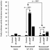
Role of RTA serines 525 and 526 in PKA-mediated enhancement of trans-activation. To evaluate the functional role of predicted phosphorylation targets at S525 and S526 of RTA, both residues were converted to alanines through PCR-based site-directed mutagenesis. RTA's trans-activating capacity was assayed using a luciferase reporter driven by the KSHV PAN promoter as in Fig. 4B. Reporter constructs were electroporated into uninfected Ramos cells accompanied by either 1 μg of pFLAG/RTA (WT RTA), 1 μg of pFLAG/RTA-S525A-S526A (mutant), or 1 μg of empty pFLAG (Vector). Cells were subsequently cultured in the presence of 300 μM db-cAMP or 20 ng/ml PMA as indicated, and Firefly luciferase activity was measured 18 h later as a fold change above basal activity after normalization to control Renilla luciferase levels. Values represent the mean (± standard error) fold increase in PAN promoter activity relative to the vehicle-treated control for each construct (Vector, WT RTA, S525A-S526A mutant RTA). Similar results emerged when data were normalized to a common basal condition (e.g., vehicle-treated cells electroporated with the empty vector). In similar studies using KS-1 cells and norepinephrine, endogenous regulation of RTA expression complicated analyses but data continued to show greater PKA-induced up-regulation of RTA activity on the PAN promoter for wild-type RTA versus the S525A-S526A mutant (results reported in main text).

Similar articles

Cited by

References

    1. Baumann, M., H. Mischak, S. Dammeier, W. Kolch, O. Gires, D. Pich, R. Zeidler, H. J. Delecluse, and W. Hammerschmidt. 1998. Activation of the Epstein-Barr virus transcription factor BZLF1 by 12-O-tetradecanoylphorbol-13-acetate-induced phosphorylation. J. Virol. 72:8105-8114. - PMC - PubMed
    1. Bloom, D. C., J. G. Stevens, J. M. Hill, and R. K. Tran. 1997. Mutagenesis of a cAMP response element within the latency-associated transcript promoter of HSV-1 reduces adrenergic reactivation. Virology 236:202-207. - PubMed
    1. Brown, H. J., M. J. Song, H. Deng, T. T. Wu, G. Cheng, and R. Sun. 2003. NF-κB inhibits gammaherpesvirus lytic replication. J. Virol. 77:8532-8540. - PMC - PubMed
    1. Cacioppo, J. T., J. K. Kiecolt-Glaser, W. B. Malarkey, B. F. Laskowski, L. A. Rozlog, K. M. Poehlmann, M. H. Burleson, and R. Glaser. 2002. Autonomic and glucocorticoid associations with the steady-state expression of latent Epstein-Barr virus. Horm. Behav. 42:32-41. - PubMed
    1. Carey, M., and S. T. Smale. 2000. Transcriptional regulation in eukaryotes: concepts, strategies, and techniques. Cold Spring Harbor Laboratory Press, Cold Spring Harbor, N.Y.

Publication types

MeSH terms

LinkOut - more resources