. 2005 Dec 16;280(50):41732-43.
doi: 10.1074/jbc.M504963200. Epub 2005 Oct 13.

Hypoxia-inducible factor prolyl 4-hydroxylase inhibition. A target for neuroprotection in the central nervous system

Affiliations

Hypoxia-inducible factor prolyl 4-hydroxylase inhibition. A target for neuroprotection in the central nervous system

Ambreena Siddiq et al. J Biol Chem. .

Abstract

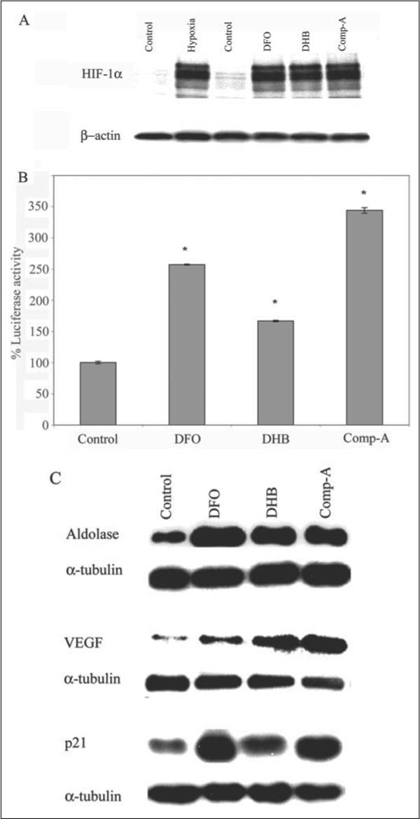
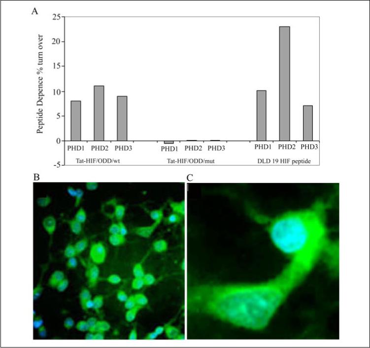
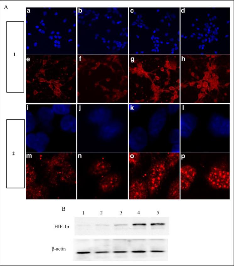
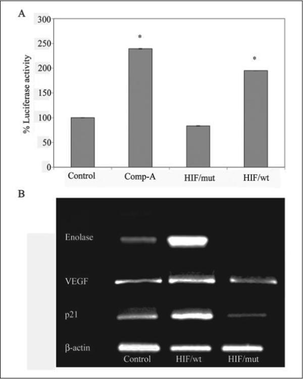
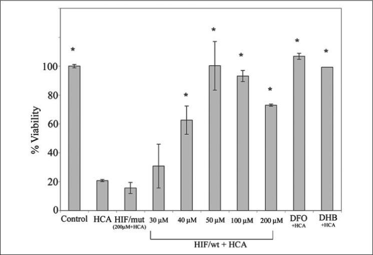
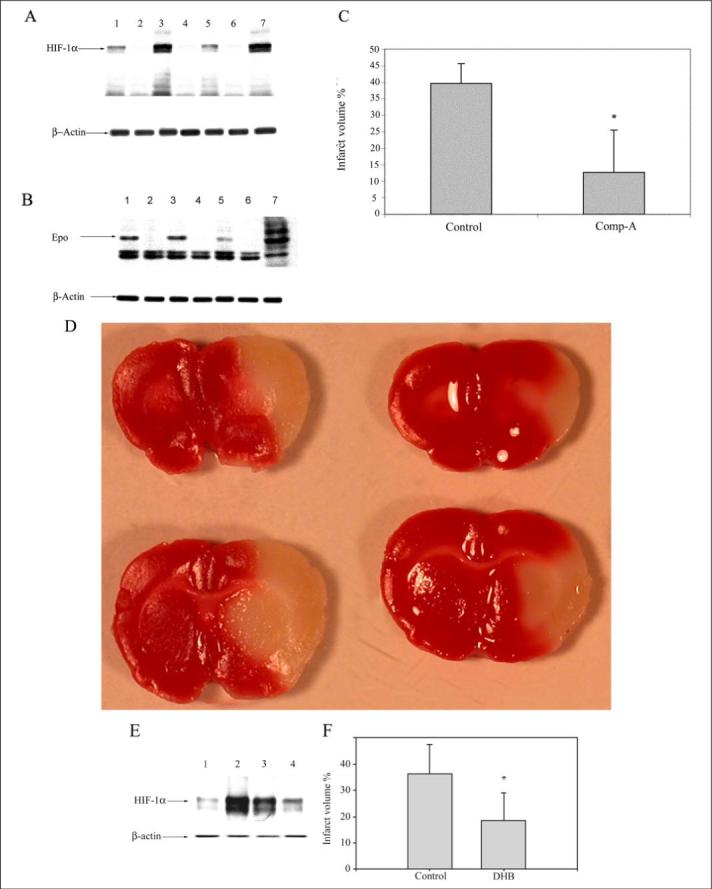
Hypoxia-inducible factor (HIF) prolyl 4-hydroxylases are a family of iron- and 2-oxoglutarate-dependent dioxygenases that negatively regulate the stability of several proteins that have established roles in adaptation to hypoxic or oxidative stress. These proteins include the transcriptional activators HIF-1alpha and HIF-2alpha. The ability of the inhibitors of HIF prolyl 4-hydroxylases to stabilize proteins involved in adaptation in neurons and to prevent neuronal injury remains unclear. We reported that structurally diverse low molecular weight or peptide inhibitors of the HIF prolyl 4-hydroxylases stabilize HIF-1alpha and up-regulate HIF-dependent target genes (e.g. enolase, p21(waf1/cip1), vascular endothelial growth factor, or erythropoietin) in embryonic cortical neurons in vitro or in adult rat brains in vivo. We also showed that structurally diverse HIF prolyl 4-hydroxylase inhibitors prevent oxidative death in vitro and ischemic injury in vivo. Taken together these findings identified low molecular weight and peptide HIF prolyl 4-hydroxylase inhibitors as novel neurological therapeutics for stroke as well as other diseases associated with oxidative stress.


Figures

FIGURE 1
FIGURE 1. Structurally diverse, low molecular weight prolyl 4-hydroxylase inhibitors stabilize HIF-1 protein, enhance activity of a hypoxia-response element-driven luciferase reporter, and increase the expression of established HIF-regulated genes in embryonic cortical neurons
A, cortical neuronal cultures (1 day in vitro (DIV)) were treated with a vehicle control (C, Me2SO (0.01%)), DFO (100 μm), 3,4-DHB (10 μm), or compound A (Comp-A; 40 μm) or exposed to 1% hypoxia (H) overnight. Proteins from cell lysates were separated using gel electrophoresis and immunodetected using an antibody against HIF1-α. β-Actin was monitored as a loading control. B, cortical neuronal cultures (1 DIV) were infected with an adenovirus harboring the hypoxia-response elements from the enolase promoter linked to a luciferase reporter (multiplicity of infection = 100). Parallel infection with adenovirus containing the green fluorescent protein (GFP) indicated reproducible infection efficiencies of ∼30−40%. 24 h following infection with adeno-HRE-luciferase, cortical neuronal cultures were treated with a vehicle control (C), DFO (100 μm), 3,4-DHB (10 μm), or compound A (40 μm). Lysates from each treatment group were generated, and luciferase activity was measured. Graph depicts mean percentage luciferase activity (compared with control) ± S.E. calculated from three separate experiments for each group (* denotes p < 0.05 by ANOVA and Student-Newman-Keuls tests for DFO, 3,4-DHB, or compound A). C, DFO (100 μm), 3,4-DHB (10 μm), and compound A (40 μm) increase expression of established HIF target genes, aldolase, VEGF, or p21waf1/cip1 in cultured cortical neurons as compared with vehicle-treated control. Immunoblots are representative examples of three experiments.
FIGURE 2
FIGURE 2. Structurally diverse, low molecular weight prolyl 4-hydrdoxylase inhibitors abrogate oxidative glutamate toxicity in embryonic cortical neuronal cultures
A, the glutamate analog HCA (5 mm) was added to cortical neuronal cultures (1 DIV). In parallel, DFO (100 μm), 3,4-DHB (10 μm), and compound A (Comp-A) (40 μm) were added with and without HCA. Twenty four hours later, cell viability was determined using the MTT assay. Graph depicts mean ± S.E. for three experiments performed in triplicate (* denotes p < 0.05 from HCA-treated cultures by ANOVA and Student-Newman Keuls tests for control, DFO, 3,4-DHB, or compound A). B–I, live/dead assay of cortical neuronal cultures (2 DIV). Live cells are detected by uptake and trapping of calcein-AM (green fluorescence). Dead cells fail to trap calcein but are freely permeable to the highly charged DNA intercalating dye, ethidium homodimer (red fluorescence). B, control. C, HCA (5 mm). D, DFO (100 μm). E, DFO + HCA. F, 3,4-DHB (10 μm). G, 3,4-DHB + HCA. H, compound A (40 μm). I, compound A + HCA.
FIGURE 3
FIGURE 3. A cell-permeant peptide inhibitor of the HIF prolyl 4-hydroxylases, but not a mutant control, is a substrate for purified, recombinant HIF prolyl 4-hydroxylases 1, 2, and 3 isoforms
A, Tat-HIF/wt peptide (100 μm, composed of the oxygen-dependent domain, including the C-terminal hydroxylation site of human HIF-1α at proline 564 (DDLDEMLAPYIPMDDDFQL; boldface P indicates proline 564) linked to a Tat protein transduction domain YGKKRRQRRR) but not a corresponding peptide with the C-terminal proline hydroxylation site of HIF-1α-mutated (Tat-HIF/mut, DLDLEMLAAYIAMDDDFQL; boldface A indicates the mutation site) acts as a substrate for the HIF prolyl 4-hydroxylases and releases [14C]CO2 from α-ketoglutarate. DLD 19 HIF peptide, composed of the C-terminal hydroxylation site of human HIF-1α at proline 564 without the TAT sequence was used as a positive control. B, representative low magnification image (×40) of cortical neurons treated with a FITC-labeled Tat-HIF/wt peptide (100 μm)(green fluorescence) and the DNA intercalating dye, DAPI, to label nuclei (blue fluorescence). Note all blue nuclei have green fluorescence in the cytoplasm indicating a high efficiency of transduction of the peptide into cells. C, a representative high magnification image (×60) of cortical neurons treated with FITC-Tat-HIF/wt peptide (100 μm) and labeled with the DNA intercalating dye DAPI.
FIGURE 4
FIGURE 4. The peptide inhibitor of the HIF prolyl 4-hydroxylases, but not a mutant control, stabilizes HIF protein in primary cortical neurons
A, Tat-HIF/mut peptide (f and n), Tat-HIF/wt peptide (100 μm) (g and o), DFO (100 μm) (h and p), or vehicle control (e and m) were added to the bathing medium of cortical neurons. Panel 1 represents lower magnification (×20), and panel 2 represents higher magnification (×60). Cells were fixed with 4% paraformaldehyde for 15 min and immunostained for HIF-1α (red) as described under “Experimental Procedures.” DAPI staining was performed to label nuclei (blue) (a–d, ×20; and i–l, ×60). B, immunoblot analysis with an antibody to HIF-1α in cell lysates from nontreated (lane 1) or after treatment with Tat-HIF/mut peptide (100 μm, lane 2), Tat-HIF/wt peptide (20 μm, lane 3; 100 μm, lane 4), or DFO (100 μm, lane 5, positive control).
FIGURE 5
FIGURE 5. A peptide inhibitor of the HIF prolyl 4-hydroxylases, but not a mutant control, induces expression of established HIF-dependent genes
A, Tat-HIF/wt peptide (100 μm) but not a corresponding peptide with the C-terminal proline hydroxylation site of HIF-1α mutated (Tat-HIF/mut, 100 μm) significantly enhances the activity of a hypoxia-response element-driven reporter in cortical neurons (* corresponds to p < 0.05 compared with control by paired t test). Note that the Tat-HIF/wt peptide induces HIF reporter activity to a similar extent as the low molecular weight inhibitor, compound A (Comp-A) (40 μm). B, RT-PCR using specific primers to determine the mRNA level of established HIF target genes, enolase, p21waf1/cip1, or VEGF in vehicle-treated (control) or in neuronal cultures treated with Tat-HIF/wt peptide (100 μm) and Tat-HIF/mut (100 μm) overnight. β-Actin was monitored as a housekeeping gene.
FIGURE 6
FIGURE 6. A cell-permeant peptide inhibitor of the HIF prolyl 4-hydroxylases, but not a mutant control, prevents oxidative glutamate toxicity
The glutamate analog, HCA (5 mm) was added to cortical neurons (1 DIV) with or without Tat-HIF/wt peptide (30, 40, 50, 100, and 200 μm), Tat-HIF/mut peptide (200 μm), DFO (100 μm), and 3,4-DHB (10 μm). Twenty four hours later cell viability was determined using the MTT assay. Graph depicts mean ± S.E. for three experiments performed in triplicate (* denotes p < 0.05 from HCA-treated cultures by ANOVA and Student-Newman Keuls tests for control, Tat-HIF/wt, Tat-HIF/mut, DFO and 3,4-DHB).
FIGURE 7
FIGURE 7. Structurally diverse, low molecular weight prolyl 4-hydroxylase inhibitors stabilize HIF protein, increase the expression of established HIF-regulated genes, and reduce infarct volume induced by permanent focal ischemia in vivo
Three rats were treated in parallel with compound A (lanes 1, 3, and 5) with a dose of 100 mg/kg of body weight (by gavage) 6 h prior to MCAO. As controls, three rats (lanes 2, 4, and 6) received equal volume of vehicle (0.5% carboxymethylcellulose) in a blinded study. Whole brain lysates from individual animals in the treated (n = 9) (lanes 1, 3, and 5) and control group (n = 5) (lanes 2, 4, and 6) were separated using gel electrophoresis and immunodetected using an antibody against HIF1-α (A) and erythropoietin (Epo) (B). Whole brain lysates of 1% hypoxia-exposed animals were used as a positive control for HIF-1α and erythropoietin (lane 7). β-Actin was monitored as a housekeeping gene. C, histologically measured infarct volume of brains of compound A (Comp-A) (n = 9) and vehicle-treated animals (n = 5) after TTC staining. The values are shown as % infarct volume relative to total brain volume (* = p < 0.05 by paired t test). D, representative pictures of vehicle-treated (left) and compound A-treated (right) brains showing infarctions. E, animals were treated with 3,4-DHB (n = 6) (lanes 2 and 3) with a dose of 180 mg/kg of body weight (intraperitoneally) 6 h prior to MCAO. Control animals (n = 5) (lanes 1 and 4) received equal volume of vehicle (ethanol, intraperitoneally) in a blinded study. Whole brain lysates were separated using gel electrophoresis and immunodetected using an antibody against HIF1-α. β-Actin was monitored as a housekeeping gene. F, histologically measured infarct volume of brains of 3,4-DHB (n = 6) and vehicle-treated animals (n = 5) after TTC staining. The values are shown as % infarct volume relative to total brain volume (* = p < 0.05 by paired t test).

Similar articles

Cited by

References

    1. Zecca L, Stroppolo A, Gatti A, Tampellini D, Toscani M, Gallorini M, Giaveri G, Arosio P, Santambrogio P, Fariello RG, Karatekin E, Kleinman MH, Turro N, Hornykiewicz O, Zucca FA. Proc. Natl. Acad. Sci. U. S. A. 2004;101:9843–9848. - PMC - PubMed
    1. Kress GJ, Dineley KE, Reynolds IJ. J. Neurosci. 2002;22:5848–5855. - PMC - PubMed
    1. Connor JR, Menzies SL, Burdo JR, Boyer PJ. Pediatr. Neurol. 2001;25:118–129. - PubMed
    1. Gerlach M, Ben-Shachar D, Riederer P, Youdim MB. J. Neurochem. 1994;63:793–807. - PubMed
    1. Thompson KJ, Shoham S, Connor JR. Brain Res. Bull. 2001;55:155–164. - PubMed

Publication types

MeSH terms

LinkOut - more resources