

Lineage tracing and characterization of insulin-secreting cells generated from adult pancreatic acinar cells
- PMID: 16210247
- PMCID: PMC1257737
- DOI: 10.1073/pnas.0507567102
Lineage tracing and characterization of insulin-secreting cells generated from adult pancreatic acinar cells
Abstract
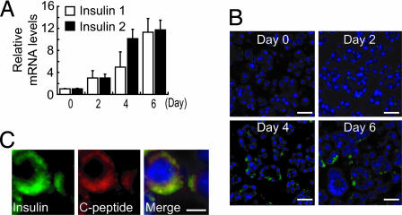
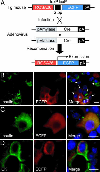
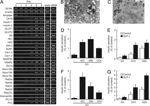
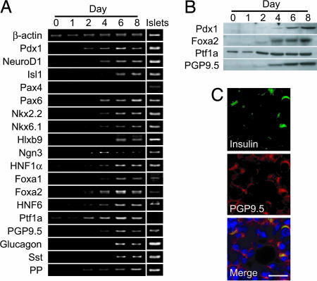
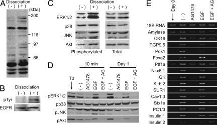
Although several studies have suggested that insulin-secreting cells can be generated in vitro from cells residing in adult exocrine pancreas, neither the origin of these cells nor their precise insulin secretory properties was obtained. We show here that insulin-secreting cells can be derived from adult mouse pancreatic exocrine cells by suspension culture in the presence of EGF and nicotinamide. The frequency of insulin-positive cells was only 0.01% in the initial preparation and increased to approximately 5% in the culture conditions. Analysis by the Cre/loxP-based direct cell lineage tracing system indicates that these newly made cells originate from amylase/elastase-expressing pancreatic acinar cells. Insulin secretion is stimulated by glucose, sulfonylurea, and carbachol, and potentiation by glucagon-like peptide-1 also occurs. Insulin-containing secretory granules are present in these cells. In addition, we found that the enzymatic dissociation of pancreatic acini itself leads to activation of EGF signaling, and that inhibition of EGF receptor kinase blocks the transdifferentiation. These data demonstrate that pancreatic acinar cells can transdifferentiate into insulin-secreting cells with secretory properties similar to those of native pancreatic beta cells, and that activation of EGF signaling is required in such transdifferentiation.
Figures
Similar articles
-
Generation of insulin-secreting cells from pancreatic acinar cells of animal models of type 1 diabetes.Am J Physiol Endocrinol Metab. 2007 Jan;292(1):E158-65. doi: 10.1152/ajpendo.00180.2006. Epub 2006 Aug 22. Am J Physiol Endocrinol Metab. 2007. PMID: 16926384
-
Differentiation of pancreatic acinar cells to hepatocytes requires an intermediate cell type.Gastroenterology. 2010 Jun;138(7):2519-30. doi: 10.1053/j.gastro.2010.02.011. Epub 2010 Feb 20. Gastroenterology. 2010. PMID: 20178796
-
Lineage tracing evidence for transdifferentiation of acinar to duct cells and plasticity of human pancreas.Gastroenterology. 2011 Aug;141(2):731-41, 741.e1-4. doi: 10.1053/j.gastro.2011.04.050. Epub 2011 May 4. Gastroenterology. 2011. PMID: 21703267
-
Can beta-cells be derived from exocrine pancreas?Diabetes Obes Metab. 2008 Nov;10 Suppl 4:170-8. doi: 10.1111/j.1463-1326.2008.00949.x. Diabetes Obes Metab. 2008. PMID: 18834444 Review.
-
Pancreatic acinar-to-beta cell transdifferentiation in vitro.Front Biosci. 2008 May 1;13:5824-37. doi: 10.2741/3119. Front Biosci. 2008. PMID: 18508625 Review.
Cited by
-
Expression of the Notch signaling pathway and effect on exocrine cell proliferation in adult rat pancreas.Am J Pathol. 2006 Oct;169(4):1206-14. doi: 10.2353/ajpath.2006.050926. Am J Pathol. 2006. PMID: 17003479 Free PMC article.
-
Pancreatic regenerating gene I and acinar cell differentiation: influence on cellular lineage.Pancreas. 2009 Jul;38(5):572-7. doi: 10.1097/mpa.0b013e3181a1d9f9. Pancreas. 2009. PMID: 19557902 Free PMC article.
-
In vivo reprogramming of pancreatic acinar cells to three islet endocrine subtypes.Elife. 2014 Jan 1;3:e01846. doi: 10.7554/eLife.01846. Elife. 2014. PMID: 24714494 Free PMC article.
-
Exploiting Single-Cell Tools in Gene and Cell Therapy.Front Immunol. 2021 Jul 12;12:702636. doi: 10.3389/fimmu.2021.702636. eCollection 2021. Front Immunol. 2021. PMID: 34322133 Free PMC article. Review.
-
Oreocnide integrifolia Flavonoids Augment Reprogramming for Islet Neogenesis and β-Cell Regeneration in Pancreatectomized BALB/c Mice.Evid Based Complement Alternat Med. 2012;2012:260467. doi: 10.1155/2012/260467. Epub 2012 Feb 27. Evid Based Complement Alternat Med. 2012. PMID: 22474495 Free PMC article.
References
-
- Bonner-Weir, S. (2000) Trends Endocrinol. Metab. 11, 375–378. - PubMed
-
- Dor, Y., Brown, J., Martinez, O. I. & Melton, D. A. (2004) Nature 429, 41–46. - PubMed
-
- Bouwens, L. (1998) Microsc. Res. Technol. 43, 332–336. - PubMed
-
- Wang, R. N., Kloppel, G. & Bouwens, L. (1995) Diabetologia 38, 1405–1411. - PubMed
Publication types
MeSH terms
Substances
LinkOut - more resources
Full Text Sources
Other Literature Sources
Medical
