. 2005 Sep 2;309(5740):1534-9.
doi: 10.1126/science.1113150.

Inositol hexakisphosphate is bound in the ADAR2 core and required for RNA editing

Affiliations

Inositol hexakisphosphate is bound in the ADAR2 core and required for RNA editing

Mark R Macbeth et al. Science. .

Abstract

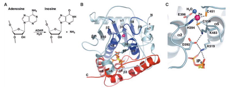
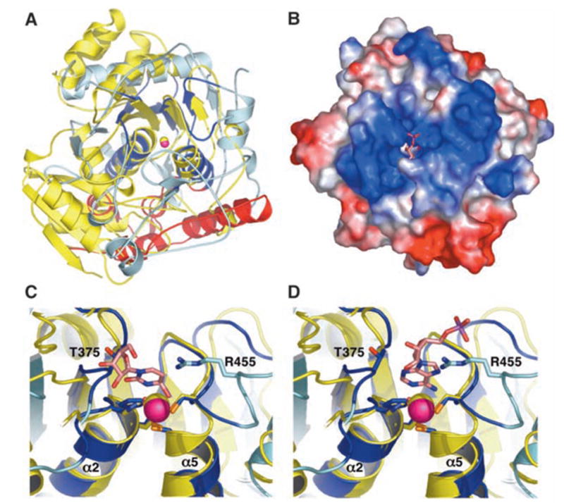
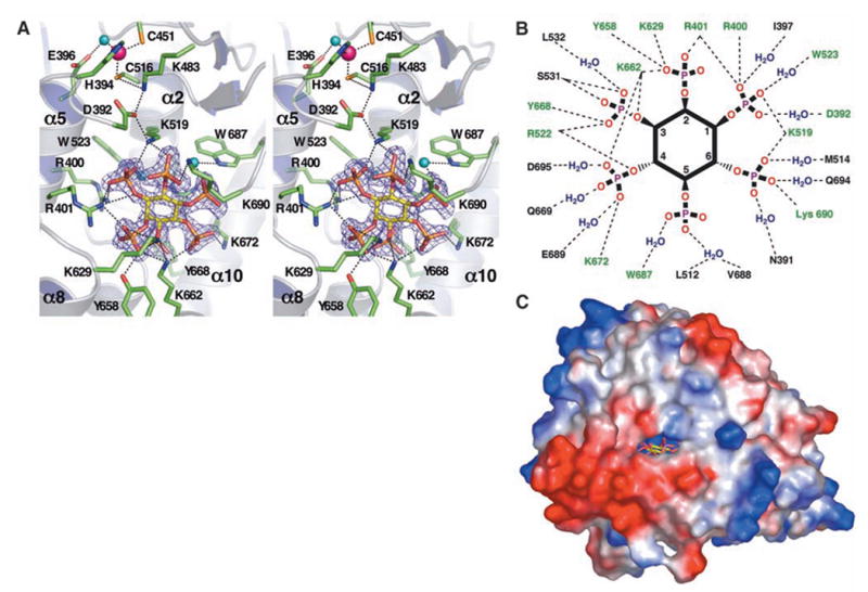
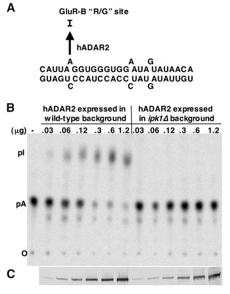
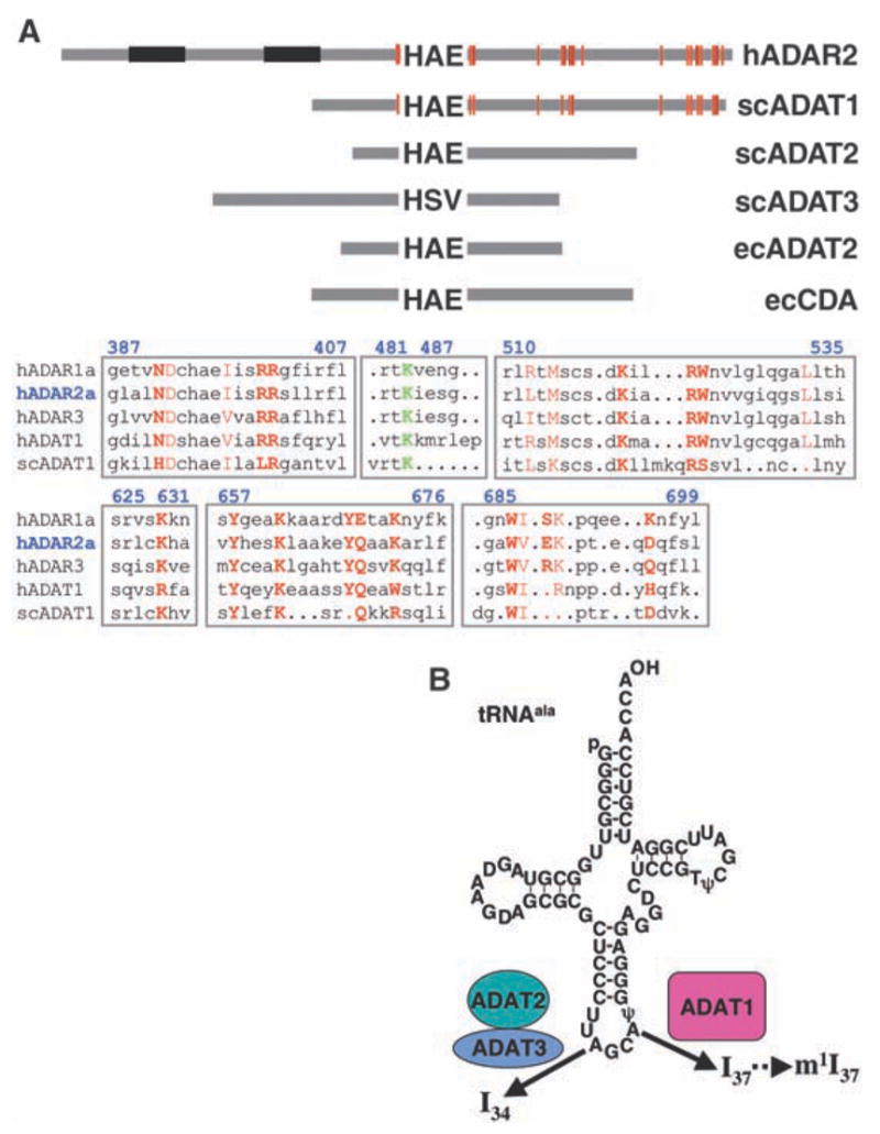
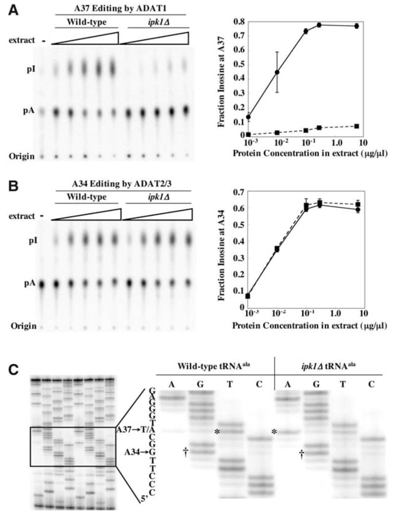
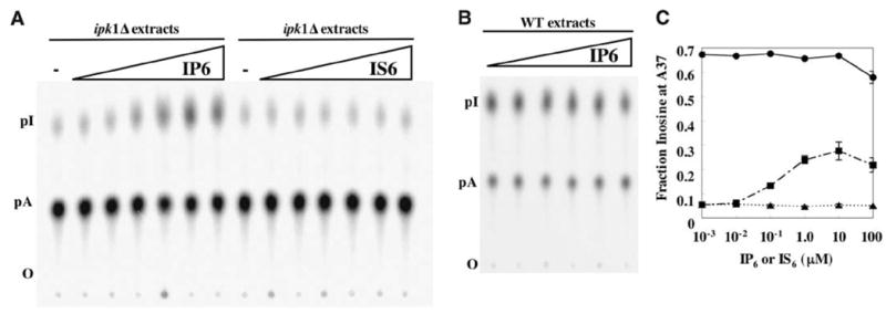
We report the crystal structure of the catalytic domain of human ADAR2, an RNA editing enzyme, at 1.7 angstrom resolution. The structure reveals a zinc ion in the active site and suggests how the substrate adenosine is recognized. Unexpectedly, inositol hexakisphosphate (IP6) is buried within the enzyme core, contributing to the protein fold. Although there are no reports that adenosine deaminases that act on RNA (ADARs) require a cofactor, we show that IP6 is required for activity. Amino acids that coordinate IP6 in the crystal structure are conserved in some adenosine deaminases that act on transfer RNA (tRNA) (ADATs), related enzymes that edit tRNA. Indeed, IP6 is also essential for in vivo and in vitro deamination of adenosine 37 of tRNAala by ADAT1.


Figures

Fig. 1
Fig. 1
(A) ADAR catalyzed hydrolytic deamination of adenosine to inosine in dsRNA. (B) Ribbon model of hADAR2-D. The active-site zinc atom is represented by a magenta sphere. The N-terminal α/β domain (residues 306 to 620) is colored cyan, with the region that shares structural similarity with CDA and TadA colored dark blue (deamination motif; residues 350 to 375, 392 to 416, 439 to 455, 514 to 525, and 542 to 551). The C-terminal helical domain (residues 621 to 700), which with contributions from the deamination motif makes the major contacts to IP6 (ball and stick), is colored red. Ends of the disordered segment (residues 462 to 473) are indicated with asterisks. (C) Residue interactions at the active site. Shown are the zinc ion, coordinating residues (H394, C451, and C516), the nucleophilic water (blue sphere), and the proposed proton-shuttling residue, E396. The hydrogen-bond relay that connects the active site to the IP6 is also indicated. Single-letter abbreviations for amino acid residues are defined in (42).
Fig. 2
Fig. 2
(A) Superposition of Escherichia coli CDA (yellow) and hADAR2-D (color scheme as in Fig. 1B) shows that these structures are highly diverged. View direction is similar to Fig. 1B. The only hADAR2-D residues that have structural equivalents in CDA are dark blue. (B) Electrostatic surface potential reveals a highly basic (blue) region flanking the active site. View direction is from the top of (A). The modeled AMP (pink) and catalytic zinc ion (magenta, partially occluded) are visible in the active site cleft. (C) Superposition of the hADAR2 and CDA active sites. Zebularine (pink) bound to CDA would clash with the loop containing T375 of hADAR2-D. (D) Docking of AMP (pink) in the same chemically equivalent orientation as zebularine would not clash with the T375 loop. Single-letter abbreviations for amino acid residues are defined in (42).
Fig. 3
Fig. 3
(A) Stereo image of the active site and IP6 binding site in hADAR2-D. The zinc ion (magenta sphere) and E396 are coordinating the nucleophilic water (aqua sphere). The hydrogen bond relay between the zinc and IP6 (yellow sticks) is shown as dashes, as are hydrogen bonds between conserved residues (green sticks) and IP6. IP6 interactions with W523 and W687 are mediated by water (aqua spheres). The 2FoFc (where Fo is the observed structure factor and Fc is the calculated structure factor) difference electron density map demonstrating the presence of the bound IP6 is contoured at 4σ. Because IP6 is nearly buried, part of the protein molecule in the foreground has been cut away for clarity. (B) Schematic diagram of residues that hydrogen-bond with IP6 directly or through one water molecule. H bonds, dashed lines; conserved residues, green. For clarity, the inositol ring is depicted as planar, although it actually binds in the ‘‘chair’’ conformation. (C) Only view from which IP6 (yellow) is visible from the protein exterior in a surface representation. The ‘‘window’’ measures 8.4 Å by 4.6 Å between van der Waal’s surfaces. Single-letter abbreviations for amino acid residues are defined in (42).
Fig. 4
Fig. 4
(A) The 27-mer R/G site RNA substrate used to assay hADAR2 editing activity. (B) Editing of the R/G site RNA by hADAR2 expressed in wild-type or ipk1Δ yeast. The R/G site adenosine was labeled with 32P and incubated with increasing concentrations of expressed hADAR2 in extracts. Reacted RNA was treated with nuclease P1, the resulting 5 nucleotide monophosphates separated by thin-layer chromatography (TLC), and the plate exposed to a PhosphorImager screen. The amount of hADAR2 in each extract was determined by Western blotting, and extract was added to give the final ADAR2 concentrations as indicated. (C) Western blot showing the amount of hADAR2 in each reaction. Single-letter abbreviations for amino acid residues are defined in (42).
Fig. 5
Fig. 5
(A) Schematic diagram showing the relative lengths and domain structures of hADAR2 and family members from S. cerevisiae (sc) and E. coli (ec). Proteins are anchored at the invariant zinc-coordinating histidine (H). Residues that coordinate IP6 are red lines; double-stranded RNA binding motifs are in black. Alignments for regions surrounding the residues that coordinate IP6 in hADAR2 are shown below, with blue numbering corresponding to hADAR2. IP6 coordinating residues are in red, with side-chain contacts in bold. Residues N391, W523, Q669, W687, E689, and D695 are water-mediated contacts. The conserved K483, which is part of a hydrogen-bond relay from IP6 to the active site zinc, is shown in green. Sequences diverge considerably in the region surrounding K483; the alignment shown was chosen because the conserved lysine of various subfamilies is aligned with K483 of hADAR2. Notably, the IP6 coordinating residues are found in ADAR3, which suggests that inefficient IP6 binding is not the reason this enzyme lacks deaminase activity (43). (B) The tRNAala substrate used in this study showing the sites of modification by the ADAT proteins. Single-letter abbreviations for amino acid residues are defined in (42).
Fig. 6
Fig. 6
(A) Editing of tRNAala A37 in vitro by extracts of wild-type or ipk1Δ yeast strains. tRNAala, labeled with 32P at A37, was incubated with increasing amounts of yeast extract protein, as indicated (14). Reacted RNA was processed as in Fig. 4B, and nuclease P1 products were separated by TLC (left). The fraction of inosine in each lane was quantified, and the average of three determinations was plotted as a function of protein concentration (right; error bars, standard deviation; when error was very small, error bars are obscured by data point symbols). Solid line, editing by ADAT1 from wild-type extracts; dashed line, editing by ADAT1 from ipk1Δ extracts. (B) As in (A) but showing editing of A34-labeled tRNAala. Solid line, editing by ADAT2/3 from wild-type extracts; dashed line, editing by ADAT2/3 from ipk1Δ extracts. (C) Editing of endogenous tRNA in vivo. tRNA was prepared from wild-type or ipk1Δ strains, reverse transcribed, and amplified by PCR. PCR products were sequenced using dideoxy nucleotide triphosphates and a 32P-labeled primer that anneals to the nontemplate strand at the 5 end of the gene. The right panel shows an expanded view of the sequencing gel shown on the left. The dideoxy sequencing lanes are indicated at the top of each lane, and the 5 to 3 sequence to the left of the gel is read from bottom to top. Bands corresponding to A34 to G editing in the wild-type and ipk1Δ tRNAs are labeled with daggers, and the band representing the A37 site that is not edited in the ipk1Δ tRNA is labeled with an asterisk. Consistent with the observation of residual activity in the mutant extract in vitro (A), some editing of A37 in the mutant extract occurs.
Fig. 7
Fig. 7
(A) Addition of IP6, but not IS6, rescued ADAT1 activity in extracts prepared from ipk1Δ yeast. Wild-type or ipk1Δ protein extract (0.1 μg/μl) was incubated with IP6 or IS6 for 15 min at 30°C before the addition of A37-labeled tRNAala. IP6 and IS6 concentrations were 10-fold dilutions from 100 μM to 10−3 μM. The tRNA was processed as described in Fig. 4B. (B) Addition of IP6 had no effect on ADAT1 activity in wild-type extracts using the reaction conditions of (A). Without the addition of IP6, wild-type extracts gave 70% A to I conversion. (C) The average fraction of inosine produced in three experiments each of (A) and (B) plotted as a function of IP6 or IS6 concentration; error bars show the standard deviation (small error bars are obscured by the data point symbol). Circles, ADAT1 activity from wild-type extracts with IP6 added; squares, activity of ipk1Δ extracts with IP6; triangles, activity of ipk1Δ extracts with IS6.

Similar articles

Cited by

References

    1. Bass BL. Annu Rev Biochem. 2002;71:817. - PMC - PubMed
    1. Higuchi M, et al. Nature. 2000;406:78. - PubMed
    1. Palladino MJ, Keegan LP, O’Connell MA, Reenan RA. Cell. 2000;102:437. - PubMed
    1. Tonkin LA, et al. EMBO J. 2002;21:6025. - PMC - PubMed
    1. Knight SW, Bass BL. Mol Cell. 2002;10:809. - PubMed

Publication types