. 2005 Sep;11(9):944-51.
doi: 10.1038/nm1280. Epub 2005 Aug 21.

Using siRNA in prophylactic and therapeutic regimens against SARS coronavirus in Rhesus macaque

Affiliations

Using siRNA in prophylactic and therapeutic regimens against SARS coronavirus in Rhesus macaque

Bao-jian Li et al. Nat Med. 2005 Sep.

Abstract

Development of therapeutic agents for severe acute respiratory syndrome (SARS) viral infection using short interfering RNA (siRNA) inhibitors exemplifies a powerful new means to combat emerging infectious diseases. Potent siRNA inhibitors of SARS coronavirus (SCV) in vitro were further evaluated for efficacy and safety in a rhesus macaque (Macaca mulatta) SARS model using clinically viable delivery while comparing three dosing regimens. Observations of SARS-like symptoms, measurements of SCV RNA presence and lung histopathology and immunohistochemistry consistently showed siRNA-mediated anti-SARS efficacy by either prophylactic or therapeutic regimens. The siRNAs used provided relief from SCV infection-induced fever, diminished SCV viral levels and reduced acute diffuse alveoli damage. The 10-40 mg/kg accumulated dosages of siRNA did not show any sign of siRNA-induced toxicity. These results suggest that a clinical investigation is warranted and illustrate the prospects for siRNA to enable a massive reduction in development time for new targeted therapeutic agents.


Conflict of interest statement

Bao-jian Li and Frank Y Xie are consultants for and Du Cheng is an employee of Guangzhou Top Genomics, Ltd. Qingquan Tang, Frank Y. Xie, Yijia Liu, Martin C. Woodle and Patrick Y. Liu are employees of Intradigm Corporation. Both Top Genomics, Ltd. and Intradigm Corporation are biopharmaceutical companies that are developing RNAi therapeutics for the treatment of human disease.

Figures

Figure 1
Figure 1. Selection and validation of siRNA duplexes targeting SCV sequence.
(a) The RT-PCR–amplified region is marked at the most upstream region of open reading frame 1 (ORF1). The siSC2- and siSC5-targeted regions (red dashes) were also marked within the Spike- and NSP12-coding regions of the SCV genome, respectively. Black arrows indicate the locations of the two targeted sequences within the viral RNA genome and gray arrowheads indicate mutation sites. Electron microscopy images of SCV particles are indicated by arrows within SCV-infected FRhK-4 cell (b) and the SCV-infected FRhK-4 cell treated with siSC2-5 (c). Scale bar in b and c, 200 nm. (d) Luciferase expression (measured in relative luciferase units, RLU) in mouse lungs after co-delivery of the expression plasmid pCI-scLuc and either siSC2-5 or siCONc-d, in either D5W or Infasurf solution. *P < 0.05, n = 5.
Figure 2
Figure 2. Severity of lung histopathology in SCV-challenged macaques.
All lung histology sections were stained with H&E (original magnification, × 100). (a) Normal lung section from macaque #921 without SCV infection “−”. (b) Minor inflammation “±”, from macaque #138, slight broadening of alveolar septa and sparse monocyte infiltration. (c) Apparent inflammation “+”, from macaque #077, hemorrhage in septa, elastic fibers of alveolar wall distorted as shown by silver staining. (d) Early symptom of acute DAD “++”, from macaque # 015, alveolus septa broadening with increasing infiltration of inflammatory cells. (e) Typical symptom of acute DAD “+++”, from macaque #212, extensive exudation and septa broadening, shrinking of alveoli caused by pressure, restricted fusion of the thick septa, obvious septa hemorrhage, ruptured elastic fiber of alveolar wall and slight cell infiltration in alveolar cavities. (f) Severe acute DAD “++++”, from macaque #202, massive cell filtration and alveoli shrinking, sheets of septa fusion, necrotic lesions at the hemorrhagic septa and massive cell infiltration in alveolar cavities.
Figure 3
Figure 3. Histopathological characteristics of SCV-infected macaque lungs.
(a,b) Lung sections were stained with H&E (original magnification, × 100). (a) Section from macaque #214 shows the alveolar walls collapsed and acute diffuse interstitial injury with interstitial edema (arrows) at an early stage of the disease. (b) Section from macaque #015 shows hyaline membrane formation (arrows) along the alveoli and pneumocyte desquamation. (c,d) Lung sections stained with H&E (original magnification, × 200). (c) Section from macaque #166 shows that damaged alveoli were filled with hemorrhage and inflammatory cells (upper arrows) and pneumocytes with nuclear enlargement, prominent nucleolus and amphophilic granular cytoplasm resulting in focal giant-cell formation (lower arrows). (d) Section from macaque #202 shows inflammatory cells, including neutrophils, lymphocytes, macrophages and monocytes, were present in the damaged alveoli. (e) IHC staining of SCV-infected monkey lung section with keratin-specific monoclonal antibody indicates the epithelial origin of the pneumocytes (arrows) (original magnification, × 200). (f) IHC staining of SCV-infected monkey lung section with CD68 monoclonal antibody indicates microphage infiltrates (arrows) (original magnification, × 500).
Figure 4
Figure 4. siSC2-5 relieved SARS symptoms.
(a) Comparison of mean body temperature. Mean body temperature represents mean value of average body temperature of each group during the 20-d period. **P < 0.01 for t-test compared to viral infection control group, n = 20. IC, viral infection control; NS, nonspecific siRNA control; PL, prophylactic treatment; CD, co-delivery treatment; PE, postexposure treatment. (b) Distribution of the average body temperatures throughout the 20-d period. The regression analysis was conducted based on the average body temperature of each group on each day. The average body temperature of each group was calculated with n = 4 before 7 d.p.i. and n = 2 afterward. (c) IgG titers of SCV-specific antibody in serum samples were measured at 10 d.p.i. (left) and 19 d.p.i. (right). (d) The average histopathological scores of each group, including lung samples collected at both 7 d.p.i and 20 d.p.i, were compared against the viral infection control group. *P < 0.05, n = 4.
Figure 5
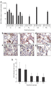
Figure 5. siSC2-5 inhibits SCV replication.
(a) Q-RT-PCR detection of SCV RNA (both genomic and RNA from open reading frame 1) from oropharyngeal swab specimens collected at 4 d.p.i. IC, viral infection control; NS, nonspecific siRNA control; PL, prophylactic treatment; CD, co-delivery treatment; PE, postexposure treatment. Numbers on x-axis refer to animal identification number. (bg) The SCV-specific antigen was detected in alveoli of the deep lungs including various cell types (original magnification, × 200), confirmed by the specific staining with monoclonal antibodies. (b) The upper arrow indicates an SCV-infected type II pneumocyte and the lower arrow indicates an infected alveolar macrophage. (c) Arrows indicate SCV-infected epithelium-originated type I pneumocytes. (d) Arrows indicate SCV antigen–positive alveolar macrophages. (eg) Arrows indicate SCV-infected cells scattered within the siSC2-5–treated lungs. (h) Comparison of average SCV-infected cell counts of each group with that of viral infection control group. IC, viral infection control; NS, nonspecific siRNA control; PL, prophylactic treatment; CD, co-delivery treatment; PE, postexposure treatment. *P < 0.05, n = 4.

Similar articles

Cited by

References

    1. Fouchier RA, et al. Aetiology: Koch's postulates fulfilled for SARS virus. Nature. 2003;423:240. doi: 10.1038/423240a. - DOI - PMC - PubMed
    1. Kuiken T, et al. Newly discovered coronavirus as the primary cause of severe acute respiratory syndrome. Lancet. 2003;362:263–270. doi: 10.1016/S0140-6736(03)13967-0. - DOI - PMC - PubMed
    1. Ksiazek TG, et al. A novel coronavirus associated with severe acute respiratory syndrome. N. Engl. J. Med. 2003;348:1953–1966. doi: 10.1056/NEJMoa030781. - DOI - PubMed
    1. Peiris JS, et al. Coronavirus as a possible cause of severe acute respiratory syndrome. Lancet. 2003;361:1319–1325. doi: 10.1016/S0140-6736(03)13077-2. - DOI - PMC - PubMed
    1. Guan Y, et al. Isolation and characterization of viruses related to the SARS coronavirus from animals in southern China. Science. 2003;302:276–278. doi: 10.1126/science.1087139. - DOI - PubMed

Publication types

MeSH terms

Associated data