Induction and maintenance of late-phase long-term potentiation in isolated dendrites of rat hippocampal CA1 pyramidal neurones
- PMID: 16109729
- PMCID: PMC1464174
- DOI: 10.1113/jphysiol.2005.092924
Induction and maintenance of late-phase long-term potentiation in isolated dendrites of rat hippocampal CA1 pyramidal neurones
Abstract
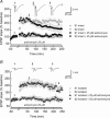
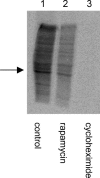
Expression of N-methyl-d-aspartate (NMDA) receptor-dependent long-term potentiation (LTP) in the CA1 region of the hippocampus can be divided into an early (1-2 h), protein synthesis-independent phase and a late (>4 h), protein synthesis-dependent phase. In this study we have addressed whether the de novo protein synthesis required for the expression of late-LTP can be sustained solely from the translation of mRNAs located in the dendrites of CA1 pyramidal neurones. Our results show that late-LTP, lasting at least 5 h, can be maintained in hippocampal slices where the dendrites located in stratum radiatum have been isolated from their cell bodies by a microsurgical cut. The magnitude of the potentiation of the slope of field EPSPs in these 'isolated' slices was similar to that recorded in 'intact' slices. Incubation of the slices with the mRNA translation inhibitor cycloheximide or the mammalian target of rapamycin (mTOR) inhibitor rapamycin blocked late-LTP in both 'intact' and 'isolated' slice preparations. In contrast, incubation of slices with the transcription inhibitor, actinomycin D, resulted in a reduction of sustained potentiation, at 4 h, in 'intact' slices while in 'isolated' slices the magnitude of potentiation was similar to that seen in untreated slices. These results indicate that late-LTP can be induced and maintained in 'isolated' dendritic preparations via translation of pre-existing mRNAs.
Figures


Similar articles
-
Late-phase, protein synthesis-dependent long-term potentiation in hippocampal CA1 pyramidal neurones with destabilized microtubule networks.Br J Pharmacol. 2007 Aug;151(7):1071-7. doi: 10.1038/sj.bjp.0707314. Epub 2007 Jun 4. Br J Pharmacol. 2007. PMID: 17549044 Free PMC article.
-
The development of synaptic plasticity induction rules and the requirement for postsynaptic spikes in rat hippocampal CA1 pyramidal neurones.J Physiol. 2007 Dec 1;585(Pt 2):429-45. doi: 10.1113/jphysiol.2007.142984. Epub 2007 Oct 11. J Physiol. 2007. PMID: 17932146 Free PMC article.
-
Long-term plasticity at excitatory synapses on aspinous interneurons in area CA1 lacks synaptic specificity.J Neurophysiol. 1998 Jan;79(1):13-20. doi: 10.1152/jn.1998.79.1.13. J Neurophysiol. 1998. PMID: 9425172
-
Continuous blockade of GABA-ergic inhibition induces novel forms of long-lasting plastic changes in apical dendrites of the hippocampal cornu ammonis 1 (CA1) in vitro.Neuroscience. 2010 Jan 13;165(1):188-97. doi: 10.1016/j.neuroscience.2009.10.015. Epub 2009 Oct 24. Neuroscience. 2010. PMID: 19837134
-
Excitation-transcription coupling, neuronal gene expression and synaptic plasticity.Nat Rev Neurosci. 2023 Nov;24(11):672-692. doi: 10.1038/s41583-023-00742-5. Epub 2023 Sep 29. Nat Rev Neurosci. 2023. PMID: 37773070 Review.
Cited by
-
Persistent organic pollutants at the synapse: Shared phenotypes and converging mechanisms of developmental neurotoxicity.Dev Neurobiol. 2021 Jul;81(5):623-652. doi: 10.1002/dneu.22825. Epub 2021 May 2. Dev Neurobiol. 2021. PMID: 33851516 Free PMC article. Review.
-
Long-lasting LTP requires neither repeated trains for its induction nor protein synthesis for its development.PLoS One. 2012;7(7):e40823. doi: 10.1371/journal.pone.0040823. Epub 2012 Jul 11. PLoS One. 2012. PMID: 22792408 Free PMC article.
-
Targeting metaplasticity mechanisms to promote sustained antidepressant actions.Mol Psychiatry. 2024 Apr;29(4):1114-1127. doi: 10.1038/s41380-023-02397-1. Epub 2024 Jan 4. Mol Psychiatry. 2024. PMID: 38177353 Free PMC article. Review.
-
Neuronal activation increases the density of eukaryotic translation initiation factor 4E mRNA clusters in dendrites of cultured hippocampal neurons.Exp Mol Med. 2009 Aug 31;41(8):601-10. doi: 10.3858/emm.2009.41.8.066. Exp Mol Med. 2009. PMID: 19381064 Free PMC article.
-
Demonstration of ion channel synthesis by isolated squid giant axon provides functional evidence for localized axonal membrane protein translation.Sci Rep. 2018 Feb 2;8(1):2207. doi: 10.1038/s41598-018-20684-8. Sci Rep. 2018. PMID: 29396520 Free PMC article.
References
-
- Anderson WW, Collingridge GL. The LTP Program: a data acquisition program for on-line analysis of long-term potentiation and other synaptic events. J Neurosci Meth. 2001;108:71–83. - PubMed
-
- Banko JL, Hou L, Klann E. NMDA receptor activation results in PKA- and ERK-dependent Mnk1 activation and increased eIF4E phosphorylation in hippocampal area CA1. J Neurochem. 2004;91:462–470. - PubMed
-
- Bradshaw KD, Emptage NJ, Bliss TV. A role for dendritic protein synthesis in hippocampal late LTP. Eur J Neurosci. 2003;18:3150–3152. - PubMed
Publication types
MeSH terms
Substances
LinkOut - more resources
Full Text Sources
Miscellaneous

