. 2005 Sep 7;24(17):3093-103.
doi: 10.1038/sj.emboj.7600769. Epub 2005 Aug 18.

G1 checkpoint failure and increased tumor susceptibility in mice lacking the novel p53 target Ptprv

Affiliations

G1 checkpoint failure and increased tumor susceptibility in mice lacking the novel p53 target Ptprv

Gilles Doumont et al. EMBO J. .

Abstract

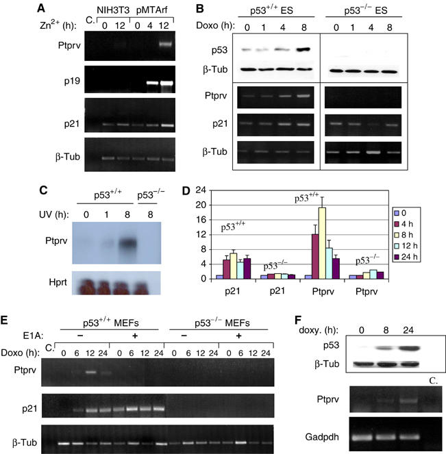
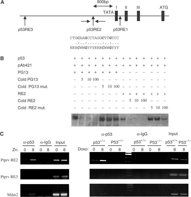
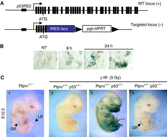
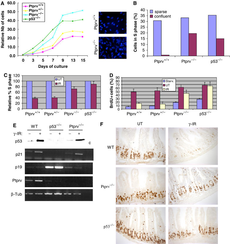
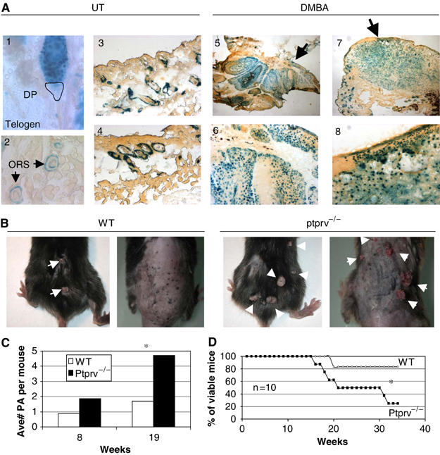
In response to DNA damage, p53 activates a G1 cell cycle checkpoint, in part through induction of the cyclin-dependent kinase inhibitor p21(Waf1/Cip1). Here we report the identification of a new direct p53 target, Ptprv (or ESP), encoding a transmembrane tyrosine phosphatase. Ptprv transcription is dramatically and preferentially increased in cultured cells undergoing p53-dependent cell cycle arrest, but not in cells undergoing p53-mediated apoptosis. This observation was further confirmed in vivo using a Ptprv null-reporter mouse line. A p53-responsive element is present in the Ptprv promoter and p53 is recruited to this site in vivo. Importantly, while p53-dependent apoptosis is intact in mice lacking Ptprv, Ptprv-null fibroblasts and epithelial cells of the small intestine are defective in G1 checkpoint control. Thus, Ptprv is a new direct p53 target and a key mediator of p53-induced cell cycle arrest. Finally, Ptprv loss enhances the formation of epidermal papillomas after exposure to chemical carcinogens, suggesting that Ptprv acts to suppress tumor formation in vivo.


Figures

Figure 1
Figure 1
Ptprv expression is p53-inducible. (A) Semiquantitative PCR analysis shows expression of Ptprv in p19ARF-inducible pMTArf cells exposed to zinc for 4 and 12 h. Expression of p19ARF, p21, and Ptprv was detected only in the pMTArf cells, not in the NIH-3T3 parental cell line. β-Tub served here as a normalization control. (B) Western analysis (top panel) and semiquantitative PCR (lower panel) show levels of p53 protein and Ptprv mRNA, respectively, in p53 WT and p53-deficient ES cells, exposed to doxorubicin (Doxo, 0.2 μg/ml). Induction of p21 and Ptprv mRNA was concomitant with p53 protein stabilization, and was detected only in WT cells. β-Tub served here as a normalization control. (C) RNAse protection assay shows induction of Ptprv expression only in p53 WT and not in p53-deficient ES cells, exposed to UVC (30 J/m2). (D) Q-PCR analysis shows the induction of expression of Ptrpv in WT and not in p53-null MEFs following γ-IR (10 Gy). The data represent the mean (±the standard deviation) of three independent experiments. (E) Semiquantitative PCR analysis shows Ptprv expression in p53 WT and p53-deficient E1A-expressing MEFs, exposed to doxorubicin (Doxo, 0.2 μg/ml). Induction of Ptprv expression was only detected in WT E1A-negative cells and not in WT E1A-expressing or p53-deficient cells. β-Tub served here as a normalization control. (F) Western analysis (top panels) and semiquantitative PCR (lower panels) show expression of p53 protein levels and Ptprv mRNA levels, respectively, in p53-inducible SAOS-2 cells following doxycycline (Doxy) treatment.
Figure 2
Figure 2
Identification of a p53-binding site in the Ptprv promoter. (A) Partial genomic structure of mouse Ptprv showing exon–intron organization at the 5′ end of the locus. Three potential p53-binding sites were identified (RE1–RE3) in this region. The complete sequence of RE2 is compared to the consensus p53-binding site and positions mutated in p53RE2 mut. indicated in bold. (B) DNA-binding activity of in vitro-translated p53 to oligonucleotides containing the consensus p53-binding site (PG13) and Ptprv RE2. DNA binding was activated using the C-terminal anti-p53 antibody pAb421. Competitive inhibition of p53 binding to RE2 was observed with unlabeled cold RE2, but not mutant p53RE2 (p53RE2 mut.), when added at five-, 10-, and 100-fold molar excess over the labeled RE2 oligonucleotide. (C) ChIP assay of p53 DNA-binding activity in zinc-inducible p19ARF pMTArf cells (left panel) and doxorubicin (Doxo)-treated WT and p53-null ES cells (right panel). A rabbit polyclonal antibody to p53 (FL-393) or control rabbit IgG was used. PCR analysis using primers described in Materials and methods is shown using input DNA (1/20 of ChIP) or DNA after ChIP.
Figure 3
Figure 3
Ptprv induction in vivo. (A) Schematic representation of the WT and the targeted Ptprv loci. An internal robosome entry site-nLacZ casette was knocked in the Ptprv locus, leading to deletion of the majority of the Ptprv coding sequence (Dacquin et al, 2004). (B, C) Cells and embryos of the Ptprv-nLacZ mice were sujected to in situ and whole-mount staining, respectively, with X-gal substrate to visualize the activity of the lacZ reporter gene. (B) Ptprv+/− ES cells were left untreated (NT) or exposed to UVC, staining was performed 8 and 24 h after exposure. (C) Whole-mount lacZ staining of E12.5 embryos performed either without (embryo 1) or with prior exposure to 5 Gy of γ-IR (embryos 2–4). Staining was performed 8 H postirradiation. GR, gonadal ridge (staining as an arc in the center of the embryo); LB, limb bud.
Figure 4
Figure 4
Ptprv is dispensible for p53-induced apoptosis (A) MEFs with the indicated genotypes were infected with a retrovirus expressing E1A and exposed to doxorubicin (Doxo; left panel) or serum deprived (right panel). Apoptotic cells were measured with annexin V staining and FACS analysis. The graphs represent the average percentages±s.e.m. of viable cells in three independent experiments. (B) E13.5 Embryos with the indicated genotypes were treated in utero with γ-IR (10 Gy). Sections through the neuroepithelium were assayed for apoptosis. The sections were stained with antibody directed against the activated form of caspase-3. (C) Freshly isolated mouse thymocytes were γ-irradiated (IR, 10 Gy) and apoptosis was measured 10 h later by FACS analysis. The graph represents the average percentages±s.e.m. of viable cells in two independent experiments.
Figure 5
Figure 5
Loss of Ptprv results in defective G1 checkpoint control. (A) To measure cell proliferation, passage 2 MEFs of different genotypes were plated at day 0. Cultures were harvested at daily intervals, and the total number of cells were determined and normalized to the number of cells at day 0 (4 h after plating) (relative number of cells). The right panel shows a Hoechst staining (5 μg/ml) of WT and Ptprv-null cultures at day 9. (B) Sparse or confluent MEF cultures were labeled with BrdU and analyzed by FACS. The graph displays the relative reduction in the number of S-phase cells (BrdU-positive) compared with sparse cultures of the same genotype. FACS analysis also revealed that the WT and Ptprv-null cells have equivalent cell volumes (data not shown). (C) Effect of γ-IR (10 Gy) on the cell cycle of asynchronously growing MEFs of different genotypes. The percentage of S-phase cells is shown relative to the percentage of S-phase cells in UT cultures. Three independent experiments were performed, and the mean values with standard deviations (error bars) are presented. (D) S-phase entry following serum stimulation and γ-IR of synchronized MEFs. The percentages of BrdU-positive cells immediately after starvation (Starv.), cells grown for 24 h in growth medium (UT), or cells that had undergone γ-IR are shown. Three independent experiments were performed, and the mean values with standard deviations (error bars) are presented. (E) Western analysis (top panel) and semiquantitative PCR (lower panel) show expression of p53 protein levels and p21 mRNA levels, respectively, in p53 WT, p53-deficient, and Ptprv-null MEFs, exposed to γ-IR. Induction of p21 mRNA was concomitant with p53 protein stabilization, and was detected in WT and in Ptprv-null cells. β-Tub served here as a normalization control. (F) Cell proliferation in the epithelium of small intestine of WT, Ptprv-null and p53-null mice after γ-IR (15 Gy). Comparison of BrdU incorporation in UT mice and irradiated mice (γ-IR) 8 h after irradiation.
Figure 6
Figure 6
Ptprv-deficient mice display increased papilloma yield after chemical carcinogenesis. (A) Expression of Ptprv in the skin of UT or DMBA-treated Ptprv-null/reporter mice as measured by X-gal staining on cryosections. LacZ expression in UT mice is only detected in hair follicles, particularly in resting hair follicles (telogen, see 1). Within the hair follicles, expression is confined to the ORS (see 2–4), and is excluded from the dermal papilla (DP). No expression was detected in the normal epidermis. In contrast, strong staining was observed in the hyperplastic epidermis (see 6 and 8) of DMBA-treated mice. Two different lesions isolated from DMBA-treated mice are shown (see images 5 and 7, acquired with an × 2 objective). Images 6 and 8 are the same lesions showed at higher magnification (acquired with an × 10 objective). Strong expression was evident in most tumor cells of epithelial origin in both lesions (see 5 and 7). (B) Gross morphology of DMBA-induced skin tumors at 16 weeks of treatment. Two representative WT and Ptprv-null mice are shown. (C) Average number of papillomas (more than 2 mm in diameter) per mouse after 8 and 19 weeks of treatment. (D) Survival curves of DMBA-treated WT (n=10) and Ptprv-null mice (n=10) with respect to the number of weeks. In (C) and (D), asterisks denote statistical significance.

Similar articles

Cited by

References

    1. Alonso A, Sasin J, Bottini N, Friedberg I, Friedberg I, Osterman A, Godzik A, Hunter T, Dixon J, Mustelin T (2004) Protein tyrosine phosphatases in the human genome. Cell 117: 699–711 - PubMed
    1. Alonso L, Fuchs E (2003) Stem cells of the skin epithelium. Proc Natl Acad Sci USA 100 (Suppl 1): 11830–11835 - PMC - PubMed
    1. Ben-Ze'ev A, Shtutman M, Zhurinsky J (2000) The integration of cell adhesion with gene expression: the role of beta-catenin. Exp Cell Res 261: 75–82 - PubMed
    1. Brugarolas J, Chandrasekaran C, Gordon JI, Beach D, Jacks T, Hannon GJ (1995) Radiation-induced cell cycle arrest compromised by p21 deficiency. Nature 377: 552–557 - PubMed
    1. Capozza F, Williams TM, Schubert W, McClain S, Bouzahzah B, Sotgia F, Lisanti MP (2003) Absence of caveolin-1 sensitizes mouse skin to carcinogen-induced epidermal hyperplasia and tumor formation. Am J Pathol 162: 2029–2039 - PMC - PubMed

Publication types

MeSH terms

Substances