. 2005 Aug 16;102(33):11681-6.
doi: 10.1073/pnas.0501494102. Epub 2005 Aug 4.

Hyperphosphorylation regulates the activity of SREBP1 during mitosis

Affiliations

Hyperphosphorylation regulates the activity of SREBP1 during mitosis

Maria T Bengoechea-Alonso et al. Proc Natl Acad Sci U S A. .

Abstract

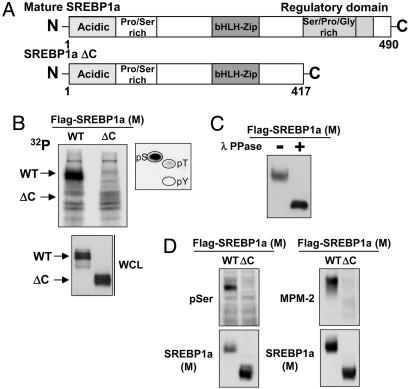
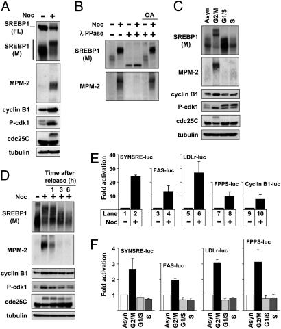
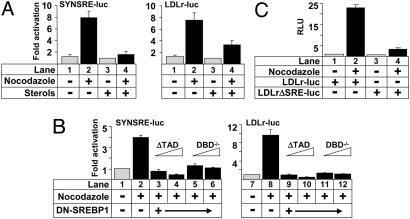
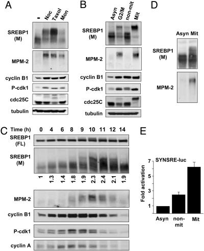
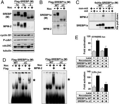
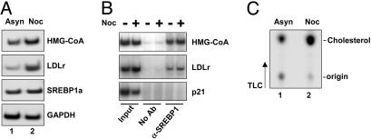
The sterol regulatory element-binding protein (SREBP) family of transcription factors controls the biosynthesis of cholesterol and other lipids, and lipid synthesis is critical for cell growth and proliferation. We were, therefore, interested in the expression and activity of SREBPs during the cell cycle. We found that the expression of a number of SREBP-responsive promoter-reporter genes were induced in a SREBP-dependent manner in cells arrested in G2/M. In addition, the mature forms of SREBP1a and SREBP1c were hyperphosphorylated in mitotic cells, giving rise to a phosphoepitope recognized by the mitotic protein monoclonal-2 (MPM-2) antibody. In contrast, SREBP2 was not hyperphosphorylated in mitotic cells and was not recognized by the MPM-2 antibody. The MPM-2 epitope was mapped to the C terminus of mature SREBP1, and the mitosis-specific hyperphosphorylation of SREBP1 depended on this domain of the protein. The transcriptional and DNA-binding activity of SREBP1 was enhanced in cells arrested in G2/M, and these effects depended on the C-terminal domain of the protein. In part, these effects could be explained by our observation that mature SREBP1 was stabilized in G2/M. In agreement with these observations, we found that the synthesis of cholesterol was increased in G2/M-arrested cells. Thus, our results demonstrate that the activity of mature SREBP1 is regulated by phosphorylation during the cell cycle, suggesting that SREBP1 may provide a link between lipid synthesis, proliferation, and cell growth.


Figures

Fig. 1.
Fig. 1.
Mature SREBP1 is highly phosphorylated. (A) Schematic illustration of mature SREBP1a and the ΔC mutant, lacking the C-terminal domain. bHLH, basic helix–loop–helix. (B) 293T cells were transfected with Flag-tagged mature (M) SREBP1a, either WT or ΔC, followed by metabolic labeling with 32P and immunoprecipitation. The migration of the WT and ΔC proteins is indicated. The band corresponding to WT SREBP1a was excised and used for phosphoamino acid analysis (Right). The levels of SREBP1a in whole-cell lysates (WCL) were detected by Western blotting. (C) Mature SREBP1a was immunoprecipitated from transfected 293T cells and incubated in the absence or presence of λ-phosphatase, separated on SDS/PAGE, and visualized by Western blotting. (D) 293T cells were transfected with mature SREBP1a, either WT or ΔC, followed by immunoprecipitation of the Flag-tagged proteins and separation on SDS/PAGE. The phosphorylation of SREBP1a was monitored with phospho-serine (pSer, Left) or MPM-2 (Right) antibodies.
Fig. 2.
Fig. 2.
Mature SREBP1 is hyperphosphorylated in cells arrested in G2/M. (A) HeLa cells were left untreated or treated with nocodazole (Noc) to induce G2/M arrest. After immunoprecipitation of SREBP1, the levels and phosphorylation (MPM-2) of SREBP1 were determined by Western blotting. The migration of the precursor (FL) and mature (M) forms of SREBP1 is indicated. (B) HeLa cells were treated as in A, incubated in the absence or presence of λ-phosphatase in the absence or presence of the phosphatase inhibitor okadaic acid (OA), and separated by SDS/PAGE, and the levels and phosphorylation (MPM-2) of SREBP1 were determined by Western blotting. (C) HeLa cells were left untreated or arrested in G2/M, G1/S, or S as described in Materials and Methods. After immunoprecipitation of SREBP1, the levels and phosphorylation (MPM-2) of SREBP1 were determined by Western blotting. Asyn, asynchronous. (D) HeLa cells were left untreated or treated with nocodazole to induce G2/M arrest. Where indicated, the nocodazole-treated cells were washed with PBS and released from the G2/M arrest in normal media for the indicated times. After immunoprecipitation of SREBP1, the levels and phosphorylation (MPM-2) of SREBP1 were determined by Western blotting. (E) HepG2 cells were transfected with SYNSRE-luc, LDLr-luc, FAS-luc, FPPS-luc, and CyclinB1-luc. Twenty-four hours after transfection, cells were treated with nocodazole, and luciferase activity was measured. (F) HeLa cells were transfected with SYNSRE-luc, FAS-luc, LDLr-luc, and FPPS-luc. Twenty-four hours after transfection, cells were treated as in C, and luciferase activity was measured.
Fig. 3.
Fig. 3.
Transcriptional activation of SREBP-responsive promoter-reporter genes in G2/M. (A) HeLa cells were transfected with SYNSRE-luc and LDLr-luc. Twenty-four hours after transfection, cells were treated with nocodazole in the absence or presence of sterols (cholesterol and 25-hydroxycholesterol; 50 and 5.0 μg/ml, respectively) to suppress the activation of endogenous SREBPs, and luciferase activity was measured. (B) HepG2 cells were transfected with SYNSRE-luc and LDLr-luc in the absence or presence of empty expression vector or dominant-negative SREBP1 (ΔTAD and DBD–/–). Twenty-four hours after transfection, cells were treated with nocodazole, and luciferase activity was measured. (C) HepG2 cells were transfected with LDLr-luc or LDLrΔSRE-luc. Twenty-four hours after transfection, cells were treated with nocodazole, and luciferase activity was measured. RLU, relative light units.
Fig. 4.
Fig. 4.
The hyperphosphorylation and activation of SREBP1 is specific for mitotic cells. (A) HeLa cells were left untreated or treated with nocodazole (Noc), taxol, or monastrol (Mon) to induce G2/M arrest. After immunoprecipitation of SREBP1, the levels and phosphorylation (MPM-2) of mature (M) SREBP1 were determined by Western blotting. (B) HeLa cells were left untreated or treated with nocodazole to induce G2/M arrest (lanes 1 and 2), and M-phase cells were separated by mitotic shake-off (lanes 3 and 4). After immunoprecipitation of SREBP1, the levels and phosphorylation (MPM-2) of mature SREBP1 were determined by Western blotting. Asyn, asynchronous; non-mit, non-mitotic; Mit, mitotic. (C) HeLa cells were synchronized at the G1/S transition by a double-thymidine treatment. Cells were collected at the indicated time points after release from the second thymidine block. Cell lysates were generated and analyzed by Western blotting with antibodies to the indicated proteins. The migration of the precursor (FL) and mature (M) forms of SREBP1 is indicated. The relative expression of mature SREBP1 is indicated at the bottom of Upper. After immunoprecipitation of SREBP1, the phosphorylation (MPM-2) of mature SREBP1 was determined by Western blotting. (D) Asynchronous HeLa cells were left untreated, and M-phase cells were separated by mitotic shake-off. After immunoprecipitation of SREBP1, the levels and phosphorylation (MPM-2) of mature SREBP1 were determined by Western blotting. (E) HeLa cells were transfected with the SYNSRE-luc promoter-reporter gene and treated with nocodazole to induce G2/M arrest, and M-phase cells were separated by mitotic shake-off, and luciferase activity was measured.
Fig. 5.
Fig. 5.
Phosphorylation of the C terminus of mature SREBP1 in mitotic cells. (A) 293T cells were transfected with mature (M) SREBP1a, SREBP1c, or SREBP2 and left untreated or treated with nocodazole (Noc). After immunoprecipitation, the levels and phosphorylation (MPM-2) of the SREBPs were determined by Western blotting. (B) 293T cells were transfected with mature SREBP1a, either WT or ΔC, and left untreated or treated with nocodazole. After immunoprecipitation, the levels and phosphorylation (MPM-2) of SREBP1 were determined by Western blotting. (C) Recombinant 6×His-SREBP1a, either WT or ΔC, was incubated with whole-cell lysates from asynchronous HeLa cells or cells arrested in G2/M in kinase buffer (10 mM Hepes, pH 7.4/3.5 mM MgCl2/0.2 mM DTT/1 mM ATP/10 mM β-glycerophosphate). The 6×His-tagged proteins were captured on NiTA-agarose, washed, and resolved by SDS/PAGE. The phosphorylation of SREBP1a was monitored with the MPM-2 antibody. non-mit, non-mitotic; mit, mitotic. (D) 293T cells were transfected with mature SREBP1a, either WT or ΔC, and left untreated or treated with nocodazole. Whole-cell lysates were used in EMSAs with a 32P-labeled probe containing the SRE-1 sequence from the LDLr promoter. Where indicated, anti-Flag (Left) or MPM-2 (Right) antibodies were included in the assay. *, Supershifted complexes. (E) HepG2 cells were transfected with SYNSRE-luc or LDLr-luc in the presence of mature SREBP1a, either WT or ΔC. Twenty-four hours after transfection, cells were treated with nocodazole, and luciferase activity was measured.
Fig. 6.
Fig. 6.
The expression of SREBP target genes and cholesterol synthesis are enhanced in G2/M. (A) RNA was isolated from asynchronous (Asyn, lane 1) or nocodazole (Noc)-treated (lane 2) HepG2 cells. Total RNA was used to determine the expression of the LDLr, HMG-CoA synthase (HMG-CoA), SREBP1a, and GAPDH genes by semiquantitative RT-PCR. (B) HeLa cells were left untreated or treated with nocodazole to induce G2/M arrest. Cells were processed for chromatin immunoprecipitation analysis of the LDLr, HMG-CoA synthase, and p21 (negative control) genes, using anti-SREBP1 antibodies for immunoprecipitation. (C) HeLa cells were left untreated or treated with nocodazole to induce G2/M arrest. Two hours before the end of the experiment, cells were placed in fresh media supplemented with [14C]acetate. Lipids were extracted and resolved by TLC. Radioactive products were visualized by PhosphorImage analysis.

Similar articles

Cited by

References

    1. Brown, M. S. & Goldstein, J. L. (1999) Proc. Natl. Acad. Sci. USA 96, 11041–11048. - PMC - PubMed
    1. Edwards, P. A., Tabor, D., Kast, H. R. & Venkateswaran, A. (2000) Biochim. Biophys. Acta 1529, 103–113. - PubMed
    1. Rawson, R. B., Zelenski, N. G., Nijhawan, D., Ye, J., Sakai, J., Hasan, M. T., Chang, T. Y., Brown, M. S. & Goldstein, J. L. (1997) Mol. Cell 1, 47–57. - PubMed
    1. Sakai, J., Rawson, R. B., Espenshade, P. J., Cheng, D., Seegmiller, A. C., Goldstein, J. L. & Brown, M. S. (1998) Mol. Cell 2, 505–514. - PubMed
    1. Horton, J. D., Goldstein, J. L. & Brown, M. S. (2002) J. Clin. Invest. 109, 1125–1131. - PMC - PubMed

Publication types

MeSH terms

LinkOut - more resources