. 2005 Aug 5;19(3):405-19.
doi: 10.1016/j.molcel.2005.07.011.

Crystal structure of A. aeolicus argonaute, a site-specific DNA-guided endoribonuclease, provides insights into RISC-mediated mRNA cleavage

Affiliations

Crystal structure of A. aeolicus argonaute, a site-specific DNA-guided endoribonuclease, provides insights into RISC-mediated mRNA cleavage

Yu-Ren Yuan et al. Mol Cell. .

Abstract

Argonaute (Ago) proteins constitute a key component of the RNA-induced silencing complex (RISC). We report the crystal structure of Aquifex aeolicus Ago (Aa-Ago) together with binding and cleavage studies, which establish this eubacterial Ago as a bona fide guide DNA strand-mediated site-specific RNA endonuclease. We have generated a stereochemically robust model of the complex, where the guide DNA-mRNA duplex is positioned within a basic channel spanning the bilobal interface, such that the 5' phosphate of the guide strand can be anchored in a basic pocket, and the mRNA can be positioned for site-specific cleavage by RNase H-type divalent cation-coordinated catalytic Asp residues of the PIWI domain. Domain swap experiments involving chimeras of human Ago (hAgo1) and cleavage-competent hAgo2 reinforce the role of the PIWI domain in "slicer" activity. We propose a four-step Ago-mediated catalytic cleavage cycle model, which provides distinct perspectives into the mechanism of guide strand-mediated mRNA cleavage within the RISC.


Figures
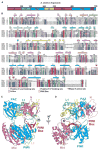
Figure 1
Figure 1. Sequence Alignment and Crystal Structure of Aa-Ago
(A) Domain and linker topology of the Aa-Ago. (B) Sequence alignment of Ago domains. The aligned sequences (Genebank ID, designated gi) are in the order of Aquifex aeolicus-1147 (AaAgo, gi:5606619), Pyrococcus furiosus-0537 (PfAgo, gi:18976909), Drosophila PIWI (DmPIWI, gi:17136736), human PIWI (HsPIWI, gi:18098558), Drosophila Argonaute1 (DmAgo1, gi:17647145), human Argonaute1 (HsAgo1, gi:6912352), and human Argonaute2 (HsAgo2, gi:29171734). The secondary structure diagram for Aa-Ago is shown on the top, color coded by domains and linkers. Conserved residues are shaded in blue (60% conservation) and green (80% conservation), whereas essentially invariant residues are shaded in red. (C) A view of the overall bilobal architecture of Aa-Ago. The domains and linkers are color coded as in (A). (D) An alternate view of Aa-Ago rotated counter-clockwise by 90° along the z axis.
Figure 2
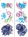
Figure 2. Domain Alignments within the PAZ-Containing (1–311) and PIWI-Containing (312–706) Lobes of Aa-Ago
(A) The PAZ-containing lobe is oriented as in Figure 1C with the N (1–108) and PAZ (166–262) domains in magenta and cyan, respectively, whereas the linkers L1 (residues 108–166) and L2 (262–311) are in green and yellow, respectively. A conserved tryptophan (W226) within the PAZ pocket is shown in orange. (B) Clockwise rotation of (A) along the y axis showing the face of the PAZ-containing lobe positioned to interact with its counterpart on the face of the PIWI-containing module lobe. (C) GRASP-based electrostatics view of (B) highlighting surface basic patches (colored blue). (D) The PIWI-containing module is oriented as it is in Figure 1C with the linker L2 (312–334) in yellow and the Mid (335–488) and PIWI (488–706) domains in magenta and cyan, respectively, whereas the PIWI box (622–650) is colored red. Conserved acidic amino acids (D502, D571, and E578) within the RNase H fold of the PIWI domain are shown in red with a coordinated divalent Ca2+ cation in orange. Conserved basic amino acids (K443, R444, and K480) within the Mid domain, which are candidates for anchoring the 5′-phosphate of the guide strand, are shown in blue. (E) Counter-clockwise rotation of (D) along the y axis showing the face of the PIWI-containing lobe positioned to interact with its counterpart on the face of the PAZ-containing lobe. (F) GRASP-based electrostatics view of (E) highlighting basic patches (colored blue), with the most basic segment highlighted by a red arrow.
Figure 3
Figure 3. Positioning of Key Residues in the N, PAZ, Mid, and PIWI Domains of Aa-Ago
(A) Relative positions of basic K34, K37, and R41 residues on the surface of the N domain across from the PAZ domain. (B) Conserved aromatic residues F209, H219, and W226 lining the 2 nt 3′ OH binding pocket and conserved basic residues R195 and K246 on the RNA binding surface of the PAZ domain. (C) Relative positioning of conserved basic K443, R444, and K480 and polar Q454 and Q476 residues, candidates for anchoring the 5′ phosphate of the guide strand, within a pocket on the Mid domain. Also shown are aromatic residues Y439 and Y681 that line this potential recognition pocket. (D) Relative positioning of invariant catalytic acidic D502, D571, and E578 residues and bound Ca2+ cation on the surface of the RNase H fold of the PIWI domain. Invariant basic R570 is also positioned in the catalytic pocket, whereas conserved basic K600 is directed toward the catalytic pocket. The Ca cation is also coordinated by D683, which is an Arg residue in hAgo1 and a His residue in hAgo2.
Figure 4
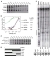
Figure 4. Nucleic Acid Double Filter Assays of Aa-Ago and Cleavage Assays of Chimeric hAgo1 and hAgo2 Constructs
(A) X-ray images of the nitrocellulose Protran BA-85 membrane (binds protein) and Nylon HyBond-N+ membrane (binds nucleic acids) in the double filter binding assay where various 32P-labeled nucleic acids bound Aa-Ago with concentrations increasing from left (1 × 10⁻⁹ M) to right (1 × 10⁻⁵ M). (B) The binding curves for Aa-Ago with various nonself-complementary nucleic acid ligands (Table S1), which are 32P-labeled at their 5′ ends. The deduced apparent KD values are listed on the right. (C) X-ray images of the nitrocellulose Protran BA-85 membrane and Nylon HyBond-N+ membrane in the competitive binding assay starting from Aa-Ago bound to a 5′-phosphorylated 21-mer RNA. The concentration of competitor decreased from left (1 × 10⁻⁵ M) to right (1 × 10⁻⁹ M). (D) Schematic showing design of chimerical constructs associated with domain swapping experiments between hAgo1 and hAgo2. Chi-a and Chi-b are the chimerical proteins resulting from swapping both Mid and PIWI domains of hAgo1 and hAgo2; Chi-c and Chi-d are the chimerical proteins resulting from swapping of only the PIWI domain of the hAgo1 and hAgo2. (E) FLAG/HA-tagged hAgo1, hAgo2, and chimerical proteins purified from HEK 293 cells were reconstituted by using single-stranded siRNA and subsequently incubated with 32P cap-labeled target RNA. The cleavage products were resolved by 8% denaturing RNA-PAGE, and radioactivity was detected by phosphorimaging. The black bar to the left side of the image represents the region of the target RNA complementary to utilized guide RNA. The expression levels of the proteins used in the assays were assessed by Western blotting using anti-HA antibody and are shown in the lower panel.
Figure 5
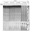
Figure 5. Target RNA Cleavage Activity of Aa-Ago
Recombinant Aa-Ago was assayed for cleavage of 32P cap-labeled target RNA after reconstituted with ssRNA (r) or ssDNA (d) of variant length (18-, 21- and 24-mer) in the presence of indicated divalent cation cofactor at either 35°C or 55°C. As a control, human recombinant Ago2 was also assayed for cleavage activity at 35°C in the presence of magnesium after reconstituted with 21-mer ssRNA. After treatment with proteinase K, the cleavage products were resolved on an 8% denaturing PAGE gel and radioactivity was detected by phosphorimaging. The black bar to the right side of the image represents the region of the target RNA complementary to the 21 nt guide strand.
Figure 6
Figure 6. Stereochemically Robust Model of Aa-Ago Bound to Guide DNA-mRNA
(A) A view of the model of the complex with the same perspective as the free Aa-Ago shown in Figure 1C. The hybrid duplex between the guide DNA strand (colored red) and the mRNA strand (colored blue) is shown in a tubular representation, with a thicker diameter for the sugar-phosphate backbone and thinner diameter for the bases. (B) A view of the model of the complex with the same perspective as the free Aa-Ago shown in Figure 1D. (C) The phosphodiester backbone corresponding to positions 2–8 from the 5′ end of the guide strand are positioned within a trough-like segment of the Mid and PIWI domains in the model of the complex. The guide strand is shown in red, with phosphorus atoms as yellow balls. The trough is shown in a surface representation and exhibits surface complementarity with the sugar-phosphate backbone of the 5′ end region of the guide strand.
Figure 7
Figure 7. A Schematic of the Reaction Cycle Involving Guide RNA-Dictated mRNA Loading, Cleavage, and Product Release within the Context of the Ago Scaffold
Conformers I, II, III, and IV are as described in the text. Transition I to II corresponds to the mRNA nucleation step, transition II to III to the mRNA propagation step, transition III to IV corresponds to the cleavage step, and transition IV to I to the product release step.

Similar articles

Cited by

References

    1. Brunger AT. X-PLOR: A System for Crystallography and NMR, Version 3.1. New Haven, CT: Yale University Press; 1992.
    1. Campos-Olivas R, Louis JM, Clerot D, Gronenborn B, Gronenborn AM. The structure of replication initiator unites diverse aspects of nucleic acid metabolism. Proc Natl Acad Sci USA. 2002;99:10310–10315. - PMC - PubMed
    1. Carmell MA, Xuan Z, Zhang MQ, Hannon GJ. The Argonaute family: tentacles that reach into RNAi, developmental control, stem cell maintenance, and tumorigenesis. Genes Dev. 2002;16:2733–2742. - PubMed
    1. Cerutti L, Mian N, Bateman A. Domains in gene silencing and cell differentiation proteins: the novel PAZ domain and redefinition of the Piwi domain. Trends Biochem Sci. 2000;25:481–482. - PubMed
    1. Chiu YL, Rana TM. siRNA function in RNAi: a chemical modification analysis. RNA. 2003;9:1034–1048. - PMC - PubMed

Publication types

MeSH terms