. 2005 Aug;25(16):7092-106.
doi: 10.1128/MCB.25.16.7092-7106.2005.

WW domains provide a platform for the assembly of multiprotein networks

Affiliations

WW domains provide a platform for the assembly of multiprotein networks

Robert J Ingham et al. Mol Cell Biol. 2005 Aug.

Abstract

WW domains are protein modules that mediate protein-protein interactions through recognition of proline-rich peptide motifs and phosphorylated serine/threonine-proline sites. To pursue the functional properties of WW domains, we employed mass spectrometry to identify 148 proteins that associate with 10 human WW domains. Many of these proteins represent novel WW domain-binding partners and are components of multiprotein complexes involved in molecular processes, such as transcription, RNA processing, and cytoskeletal regulation. We validated one complex in detail, showing that WW domains of the AIP4 E3 protein-ubiquitin ligase bind directly to a PPXY motif in the p68 subunit of pre-mRNA cleavage and polyadenylation factor Im in a manner that promotes p68 ubiquitylation. The tested WW domains fall into three broad groups on the basis of hierarchical clustering with respect to their associated proteins; each such cluster of bound proteins displayed a distinct set of WW domain-binding motifs. We also found that separate WW domains from the same protein or closely related proteins can have different specificities for protein ligands and also demonstrated that a single polypeptide can bind multiple classes of WW domains through separate proline-rich motifs. These data suggest that WW domains provide a versatile platform to link individual proteins into physiologically important networks.


Figures

FIG. 1.
FIG. 1.
Domain organization and alternate names of WW domain-containing proteins analyzed in this study. WW domain-containing proteins examined in this study, their unique GeneIDs, as well as alternate names are indicated. The relative domain organization of each of the proteins was determined by searching the National Center for Biotechnology Information conserved domain database (CDD) (51) and is indicated. WW domains specifically analyzed in this study are indicated with an asterisk. a.a., amino acids; PTB, phosphotyrosine binding; PH, pleckstrin homology; SH3, Src homology 3.
FIG. 2.
FIG. 2.
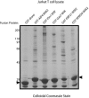
Purification of WW domain-binding proteins. Proteins were precipitated from Jurkat cell lysate with the indicated fusion proteins as outlined in Materials and Methods. Precipitated proteins were separated on SDS-PAGs, and gels were stained with colloidal Coomassie blue to visualize proteins. One representative gel is shown here. The positions of molecular mass markers (in kilodaltons) are indicated to the left. The positions of bait proteins are indicated by arrows.
FIG.3.
FIG.3.
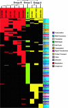
Hierarchical clustering of WW domains with precipitated proteins. The GST-WW domains (abscissa) and precipitated proteins (ordinate) were hierarchically clustered as outlined in Materials and Methods. The tree at the top indicates the relatedness of the WW domains with respect to precipitated proteins. This clustering distinguishes three groups of WW domains: group A (AIP4 WW2, NEDD4-1 WW2, and WWOX WW1) in yellow, group B (FBP11 WW1 and WW2, FBP21 WW2, FE65 WW, CA150 WW2, and Gas7 WW) in red, and group C (Pin1 WW) in green. The color of the precipitated protein indicates cellular function and is shown to the right.
FIG. 4.
FIG. 4.
Examination of precipitated proteins for known proline-based WW domain-binding motifs. The proteins precipitated with each of the GST-WW pulldowns were analyzed for the presence of the indicated proline-containing motifs. Note that the pie charts do not equal 100%, since individual proteins can have more than one motif. The number of proteins precipitated by each WW domain and the percentage of proteins with at least one of the proline-rich motifs are indicated below the corresponding pie charts. The ∼24,000 reference proteins with unique GeneIDs were downloaded from the National Center for Biotechnology Information database and searched locally for the indicated motifs.
FIG. 5.
FIG. 5.
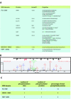
Analysis of GST-WW domain pulldowns for phosphopeptides. (A) The MS data from the GST-Pin1 WW, NEDD4-1 WW2, and FBP11 WW2 pulldowns were analyzed for the presence of phosphopeptides as described in Materials and Methods. Identified phosphopeptides and their host proteins are indicated. Definitively phosphorylated residues are indicated in red, and possible phosphorylation sites where the identification was ambiguous are indicated in green. A lowercase “m” denotes an oxidized methionine, while a lowercase “q” denotes a pyroglutamine residue. Amino acids in parentheses are residues N and C terminal to the peptide in the protein sequence. Peptides indicated by an asterisk are doubly phosphorylated. (B) The MS/MS spectra for the THRAP3 phosphopeptide is shown. Note that only b (blue), y (red), and parental ion minus phosphoric acid (P(m/z) − H3PO4) or minus phosphoric acid and water (P(m/z) − H3PO4-H2O) (green) are indicated. The number associated with each ion is the mass/charge ratio (m/z) of that ion. (C) Statistics showing the number of phosphorylation sites, number of phosphorylated proteins, and number of phosphorylated proteins as a percentage of total proteins precipitated with each WW domain (from Fig. 3).
FIG. 6.
FIG. 6.
Identified proteins are direct, in vivo, binding partners. (A) Proteins were precipitated from 293T cell lysate (anti-RNA Pol II LS immunoprecipitate [ippt]) or Jurkat cell lysate (anti-p68 ippts) with the indicated antibodies, separated on SDS-PAGs, transferred to PVDF membranes, and blotted with antiserum against AIP4 or Diaphanous 1 (top blots). Blots were stripped and reprobed with the indicated antibodies (bottom blots). (B) 293T cells were transfected with cDNAs coding for a doubly Myc-tagged AIP4 (Myc-AIP4), enzymatically inactive AIP4 mutant (Myc-C830A AIP4), an AIP4 construct lacking the entire C2 domain (Myc-ΔC2 AIP4), or a construct in which the second tryptophan (tyrosine in the case of the fourth WW domain) residue in each WW domain had been mutated to alanine (Myc-WWmut AIP4). Cell lysates were immunoprecipitated with the anti-Myc MAb 9E10, and immunoprecipitates were separated on SDS-PAGs, transferred to PVDF membranes, and blotted for the large subunit of RNA polymerase II (anti-RNA Pol II LS) or the p68 and p25 subunits of CFIm (anti-p68 and anti-p25). The blots were stripped and reprobed with the 9E10 anti-Myc MAb (bottom blots). Cell lysates are included to show that the levels of RNA Pol II LS, p68, and p25 are equivalent in each of the lysates. Note that the dot in the top blot of the Myc-WWmut AIP4 pulldown is an artifact. (C) Baculovirus-expressed, purified recombinant His-tagged p68 or p25 protein was incubated with GST alone or a GST fusion protein of the first WW domain of AIP4 (GST-AIP4 WW1). GST precipitates were separated on SDS-PAGs, transferred to nitrocellulose, and blotted with either anti-p68 antiserum (top blot) or anti-p25 antiserum (bottom blot). The positions of molecular mass markers (in kilodaltons) are indicated to the left. The positions of p68 and p25 proteins are indicated by the arrows. The asterisk in the top blot indicates a p68 degradation product, while the asterisk in the bottom blot indicates the GST-AIP4 WW1 fusion protein. The input is one/fifth of the material used for the pulldown experiment. (D) 293T cells were transfected as indicated with cDNAs coding for a doubly Myc-tagged AIP4 (Myc-AIP4), Flag-tagged p68 protein (FLAG-p68), or Flag-tagged p68 protein in which the tyrosine residue in the PY motif had been mutated to alanine (FLAG-p68 Y/A). Cell lysates were immunoprecipitated with the anti-FLAG M2 MAb, and immunoprecipitates were separated on SDS-PAGs, transferred to PVDF membranes, and blotted with the anti-Myc MAb (top blot). The blot was stripped and reprobed with the anti-Flag M2 MAb (middle blot). Cell lysate is included to show that equivalent amounts of Myc-tagged AIP4 were expressed in the appropriate transfections (bottom blot). (E) 293T cells were transfected as indicated with cDNAs coding for a Flag-tagged p68 protein (FLAG-p68), a doubly Myc-tagged AIP4 (Myc-AIP4), an enzymatically inactive AIP4 mutant (Myc-C830A AIP4), or an HA-tagged ubiquitin (HA-Ub) construct. Cell lysates were immunoprecipitated with the anti-FLAG M2 MAb, and immunoprecipitates were separated on SDS-PAGs, transferred to PVDF membranes, and blotted with an anti-HA antiserum (top blot). The blot was stripped and reprobed with the anti-Myc 9E10 MAb (middle blot). Cell lysate is included to show that equivalent amounts of FLAG protein were expressed in the appropriate transfections (bottom blot). The positions of ubiquitylated p68 proteins are indicated by arrows. The positions of molecular mass standards (in kilodaltons) are indicated to the left.
FIG. 7.
FIG. 7.
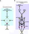
Protein complexes precipitated by GST-WW domains. (A) The transcriptional complex precipitated by the AIP4 WW2 domain is shown. The WW domain is indicated in yellow, direct WW domain-binding proteins in red, and likely indirectly precipitating proteins in green. Gene symbols and common names (in parentheses) are provided. Black lines indicate a direct physical interaction, while gray lines indicate a physical association, but not necessarily direct. (B) Actin cytoskeleton-regulating complex precipitated by the FE65 WW domain (as well as other group B [II/III] WW domains) is shown and labeled as described above for panel A. Although not found in the FE65 pulldowns, NAP1 (NCKAP1) was included (in blue) as it has been shown to bridge ABI1 with CYFIP1/2 (24, 35) and was found in association with other group B (II/III) WW domains (Fig. 3). Note that this model represents one possible cytoskeleton-regulating complex based on published work. Since the WASP proteins (WAS and WASL), WIPIP, WIRE, WASF2, and ABI1 are extremely proline rich and contain motifs that are predicted to interact with group B WW domains, they may interact directly with group B WW domains.
FIG. 8.
FIG. 8.
Specificity of binding between the different WW domains of AIP4 and the second WW domains of AIP4 and NEDD4-1. (A) 293T cells were transfected with a cDNA coding for a Flag-tagged KIAA0144 protein (FLAG-KIAA0144 blot) or left untransfected (other blots). Cell lysates were prepared, and an equivalent amount of lysate was precipitated with GST alone or with the indicated GST-AIP4 WW domain. Half of each precipitate was separated on an SDS-PAG, transferred to a PVDF membrane, and blotted with the indicated antibody. The other half of the GST pulldown was separated on an SDS-PAG and stained with Coomassie blue to show that equivalent amounts of GST fusion protein were used for the pulldowns (bottom blot). Cell lysate is included to show the electrophoretic mobilities of the indicated proteins. (B) Osprey diagram (7) showing the proteins identified by MS to precipitate with the second WW domains of NEDD4-1 and AIP4. Proteins with PY motifs are indicated in red. Note that the large subunit of RNA polymerase II (POL2A) does not have a PY motif but does possess several copies of the YSPTSPS heptamer sequence in its C-terminal domain (CTD), which has been shown to bind the WW domains of NEDD4 family proteins (12). (C) 293T cells were transfected with a cDNA coding for a Flag-tagged KIAA0144 protein (FLAG-KIAA0144 blot) or left untransfected (other blots). Cell lysates were prepared, and proteins were precipitated with the indicated GST fusion proteins from either 2 mg or 500 μg of cell lysate. Half of each precipitate was separated on an SDS-PAG, transferred to a PVDF membrane, and blotted with the indicated antibody. The other half of the GST pulldown was separated on an SDS-PAG and stained with Coomassie blue to show that equivalent amounts of GST fusion protein were used for the pulldowns (bottom blot). Cell lysate (10 μg) was included to show the electrophoretic mobility of the immunoblotted protein and to give an indication of the amount of protein precipitated by the GST fusion proteins. (D) 293T cells were transfected with a cDNA coding for a Flag-tagged KIAA0144 protein (wild type [wt]) or a Flag-tagged KIAA0144 protein in which the tyrosine residue in the PY motif had been mutated to alanine (Y/A). Cell lysates were prepared, and proteins were precipitated with the indicated fusion proteins from equivalent amounts of cell lysate. Half of each precipitate was separated on an SDS-PAG, transferred to a PVDF membrane, and blotted with the anti-FLAG M2 MAb. The other half of the GST pulldown was separated on an SDS-PAG and stained with Coomassie blue to show that equivalent amounts of GST fusion protein were used for the pulldowns (bottom blot). Cell lysate was included to show the electrophoretic mobility of the FLAG-KIAA0144 protein. (E) SPOTS membranes were prepared as outlined in Materials and Methods. Proteins (12-mers) comprising the entire coding sequence of KIAA0144 were generated, with a moving window of four amino acids. SPOTS membranes were probed with 1 μΜ of either GST-AIP4 WW2, GST-NEDD4-1 WW2, or GST alone. Spots specific to GST-AIP4 WW2 and NEDD4-1 WW2 are numbered and indicated in red. The sequences of these peptides (with the PY motif and LPXY motifs in red) are shown below the blots. Spots marked by an asterisk were recognized by GST alone in other experiments.
FIG. 9.
FIG. 9.
Recognition of the p68 CFIm protein by multiple WW domains. (A) The coding sequence of the p68 protein is shown. The proline-rich region used for the SPOTS blots in panel C is indicated in pink. The PY (red), PPPPP stretch (yellow), and three PPGPPP motifs (green) are also indicated. The RNA recognition motif is underlined. (B) 293T cells were transfected with cDNAs coding for either a Flag-tagged p68 protein (wild type [wt]) or a Flag-tagged p68 protein in which the tyrosine residue in the PY motif had been mutated to alanine (Y/A). Cell lysates were prepared, and an equivalent amount of lysate was precipitated with GST alone or the indicated GST-WW domain fusion protein. Half of each precipitate was separated on an SDS-PAG, transferred to a PVDF membrane, and blotted with the anti-FLAG M2 MAb. The other half of the GST pulldown was separated on an SDS-PAG and stained with Coomassie blue to show that equivalent amounts of GST fusion protein were used for the pulldowns (bottom blot). Cell lysate is included to show the electrophoretic mobilities of the indicated proteins; note that the p68 Y/A was consistently expressed at higher levels than the wt protein. (C) Peptides (12-mers), with a moving window of five amino acids, were generated for the proline-rich region of p68 (panel A, pink region), and SPOTS membranes were probed with 1 μM of GST alone or the indicated GST-WW domain fusion protein. Peptides recognized by one group of WW domains over another are boxed and numbered, and the peptide sequences are provided below the blots. In the boxed sequences, the PY (red), PPPPP (yellow), and PPGPPP (green) motifs in the peptides are highlighted.

Similar articles

Cited by

References

    1. Beaudenon, S. L., M. R. Huacani, G. Wang, D. P. McDonnell, and J. M. Huibregtse. 1999. Rsp5 ubiquitin-protein ligase mediates DNA damage-induced degradation of the large subunit of RNA polymerase II in Saccharomyces cerevisiae. Mol. Cell. Biol. 19:6972-6979. - PMC - PubMed
    1. Bedford, M. T., D. C. Chan, and P. Leder. 1997. FBP WW domains and the Abl SH3 domain bind to a specific class of proline-rich ligands. EMBO J. 16:2376-2383. - PMC - PubMed
    1. Bedford, M. T., D. Sarbassova, J. Xu, P. Leder, and M. B. Yaffe. 2000. A novel pro-Arg motif recognized by WW domains. J. Biol. Chem. 275:10359-10369. - PubMed
    1. Bednarek, A. K., K. J. Laflin, R. L. Daniel, Q. Liao, K. A. Hawkins, and C. M. Aldaz. 2000. WWOX, a novel WW domain-containing protein mapping to human chromosome 16q23.3-24.1, a region frequently affected in breast cancer. Cancer Res. 60:2140-2145. - PubMed
    1. Bompard, G., and E. Caron. 2004. Regulation of WASP/WAVE proteins: making a long story short. J. Cell Biol. 166:957-962. - PMC - PubMed

Publication types

MeSH terms