. 2005 Dec 15;392(Pt 3):589-99.
doi: 10.1042/BJ20050542.

Regulation of ERK1 gene expression by coactivator proteins

Affiliations

Regulation of ERK1 gene expression by coactivator proteins

Beanca Y Chu et al. Biochem J. .

Abstract

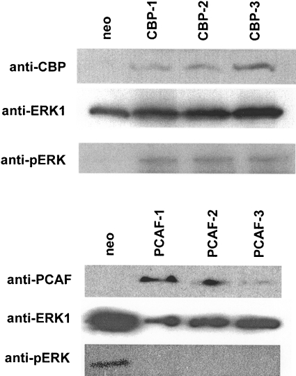
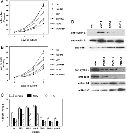
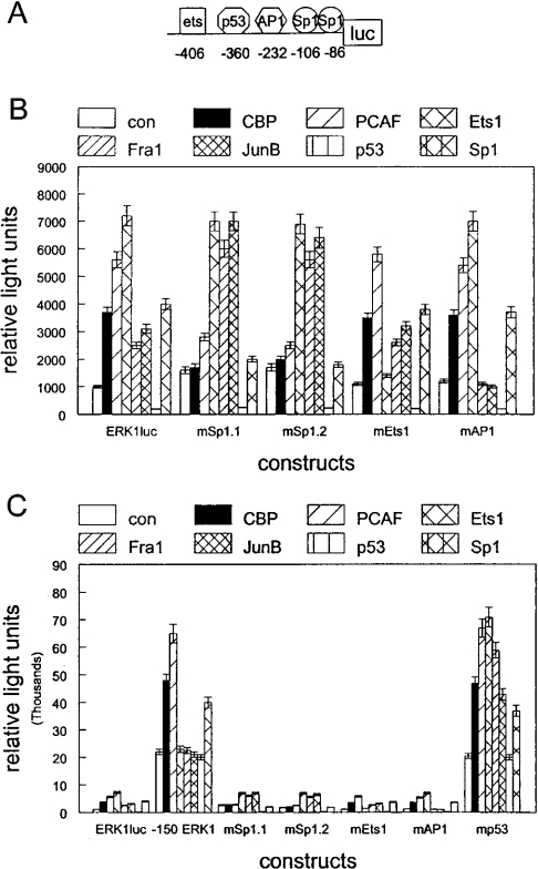
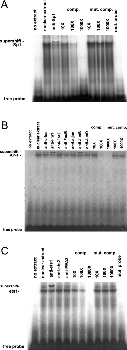
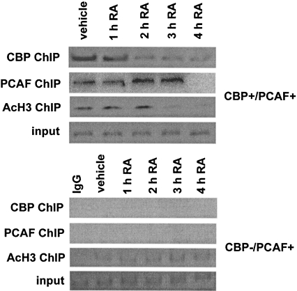
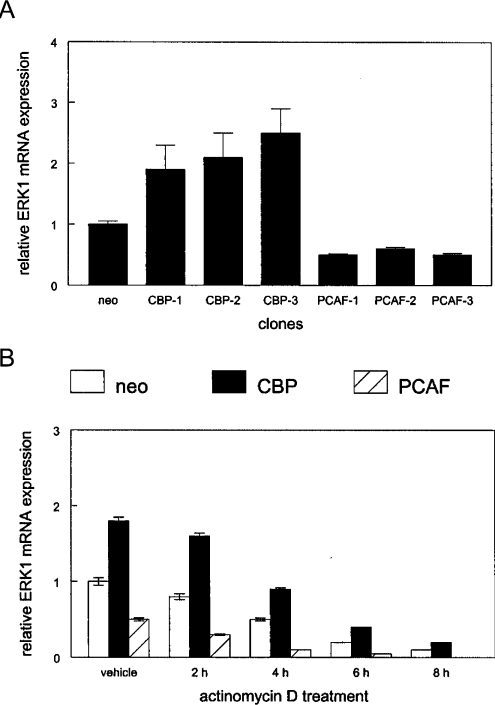
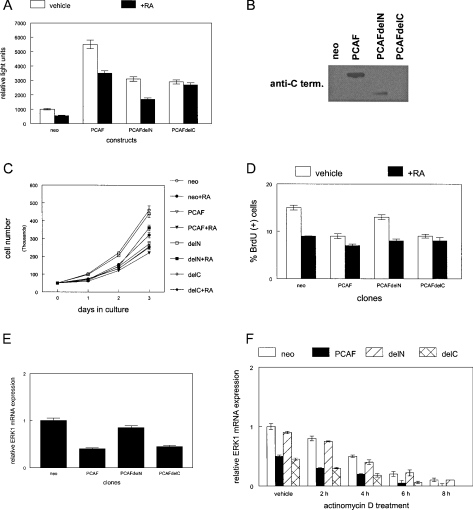
RARs (retinoic acid receptors) mediate the effect of their ligand RA (retinoic acid) on gene expression. We previously showed that RA inhibited cellular proliferation in part by decreasing expression of the mitogen activated protein kinase ERK1 (extracellular signal regulated kinase 1). However, the mechanism by which RA regulates ERK1 expression is largely uncharacterized. The present study characterizes coactivator-mediated regulation of RA target gene expression by analysing ERK1 promoter activation. CBP (CREB-binding protein) and PCAF (p300/CBP associated factor) are transcriptional coactivators that interact with nuclear hormone receptors such as RARs. CBP and PCAF differentially regulated ERK1 expression in stable clones. CBP clones expressed higher ERK1 protein levels, proliferated faster in culture and were resistant to RA-mediated growth inhibition. PCAF clones expressed lower levels of ERK1 protein and cells grew more slowly than controls. CBP and PCAF regulation of the ERK1 promoter was dependent on two Sp1 (specificity protein 1) sites located between -86 and -115 bp. Immunoprecipitation and yeast two-hybrid analysis revealed that PCAF interacted with Sp1 via CBP. A putative p53 binding site at -360 bp functioned as a major repressor of ERK1 promoter activity even in the absence of exogenous p53 expression. CBP and PCAF occupancy of the proximal ERK1 promoter was dramatically decreased by RA treatment. PCAF mediated inhibition of ERK1 expression was due to decreased stability of the kinase mRNA. We conclude that CBP and PCAF coactivators mediate ERK1 gene expression at both the transcriptional and post-transcriptional level.


Figures

Figure 1
Figure 1. ERK1 expression is induced in CBP-expressing clones and decreased in PCAF-expressing clones
Expression of CBP, PCAF and ERK1 proteins was determined by Western blotting using anti-CBP, anti-PCAF, anti-phospho ERK (anti-pERK) and anti-ERK1 antibodies in stable clones transfected with CBP or PCAF expression vectors (CBP-1, -2 and -3; PCAF-1, 2 and 3) as described in the Materials and Methods section. Lack of expression in a neomycin resistant control clone is shown. These experiments were performed 3 times with similar results. Representative blots are shown.
Figure 2
Figure 2. CBP but not PCAF expression increased cellular proliferation in an ERK-dependent manner which was not inhibited by RA treatment
(A) CBP- and PCAF-expressing clones and neomycin resistant control cells (neo) were plated in triplicate and treated with 1 μM RA (+RA) or vehicle for up to 3 days. Cultures were trypsinized and counted with a haemocytometer at 1 day intervals. These experiments were performed 3 times with similar results. Error bars indicate S.E.M. (B) CBP- and PCAF-expressing clones and neomycin resistant control cells (neo) were plated in triplicate and treated with 10 μM PD98059 (+PD; MEK inhibitor) or vehicle for up to 3 days. Cultures were trypsinized and counted with a haemocytometer at 1 day intervals. These experiments were performed 3 times with similar results. Error bars indicate S.E.M. (C) PD98059 but not RA treatment inhibited BrdU incorporation in CBP stable clones. CBP- (CBP 1, 2 and 3), and PCAF-expressing clones (PCAF 1, 2 and 3) and neomycin resistant control cells (neo) were plated in triplicate and treated with RA, PD98059 or vehicle for 16 h. Cells were labelled with BrdU for 1 h and processed as described in the Materials and Methods section. The number of BrdU-positive cells was expressed as a percentage of total cells counted by fluorescence microscopy. These experiments were performed 3 times with similar results. Error bars indicate S.E.M. (D) CBP but not PCAF clones expressed higher levels of G2 phase cell cycle regulatory proteins. Cyclin E, cyclin B and cdk1 protein expression is shown by Western blot of three stable clones (upper panel) transfected with CBP expression vector (CBP-1, 2 and 3) or neomycin resistant control cells (neo) as described in the Materials and Methods section. PCAF-expressing clones (PCAF 1, 2 and 3) expressed lower levels of cyclin B and cdk1 and higher relative-amounts of G1 phase proteins (cdk4 and cdk6, lower panel). These experiments were performed three times with similar results. Representative blots are shown.
Figure 3
Figure 3. CBP- and PCAF-mediated induction of the ERK1 promoter is dependent on two Sp1 sites
(A) Diagram of the ERK1 proximal promoter region showing the positions of two Sp1 sites at −86 and −106 bp, the AP1 site at −232 bp, the p53 site at −360 bp and the ets site at −406 bp. (B, C) The ERK1 promoter fused to the luciferase reporter gene (ERK1luc) was transiently transfected into triplicate cultures of SCC12 cells as described in the Materials and Methods section. A second reporter construct (−150 ERK1) was created, in which the ERK1 promoter was truncated at −150 bp, which deleted putative ets, AP1 and p53 recognition sequences. Inactivating double point-mutations in the Sp1 (mSp1.1, mSp1.2), ets (mEts1), AP1 (mAP1) and p53 (mp53) sites were created by site-directed mutagenesis. Expression vectors for CBP, PCAF, Ets1, Fra1, JunB, p53 or control plasmid were cotransfected with the ERK1 promoter/reporter constructs. ERK1 promoter activity was measured in relative light units by luciferase assay. These experiments were performed three times with similar results. Error bars indicate S.E.M.
Figure 4
Figure 4. Sp1, Fra1, JunB and Ets1 proteins bind to their recognition sequences in the ERK1 promoter region
EMSA was performed using SCC12 nuclear extract as described in the Materials and Methods section. (A) Anti-Sp1 antibody was used to determine the presence of Sp1 protein in the shifted complex (supershift). Molar excess (10–1000 fold) of unlabelled competitor oligonucleotides containing intact (comp) or mutant (mut) Sp1 sites was included in some reactions. No binding to the labelled mutant (mut) probe was observed, nor was binding detected in the absence of nuclear extract (no extract). The position of the free probe is shown. (B) Fra1 and JunB proteins bind to a putative AP1 site in the ERK1 promoter. EMSA using SCC12 nuclear extract was performed as described in the Materials and Methods section. Anti-AP1 antibodies were used to determine the presence of fos and jun proteins in the shifted complexes (supershift). Molar excess (10–1000 fold) of unlabelled competitor oligonucleotides containing intact (comp) or mutant (mut) AP1 sites was included in some reactions. No binding was observed in the absence of nuclear extract (no extract). The position of the free probe is shown. (C) Ets1 protein binds to a putative ets site in the ERK1 promoter. EMSA was performed using SCC12 nuclear extract as described in the Materials and Methods section. Anti-ets1, ets2, or PEA3 antibodies were used to determine the presence of ets proteins in the shifted complex (supershift). Molar excess (10–1000 fold) of unlabelled competitor oligonucleotides containing intact (comp) or mutant (mut) ets sites was included in some reactions. No binding to the labelled mutant (mut) probe was observed, nor was binding detected in the absence of nuclear extract (no extract). These experiments were performed three times with similar results. Representative gels are shown.
Figure 5
Figure 5. RA treatment induces loss of coactivator proteins and histone deacetylation of the proximal ERK1 promoter in genomic DNA
Cells expressing both CBP and PCAF (CBP+/PCAF+) or PCAF alone (CBP/PCAF+) were treated with 1 μM RA or vehicle for up to 4 h. CBP/PCAF occupancy and histone acetylation (AcH3) of the proximal ERK1 promoter was determined by ChIP as described in the Materials and Methods section. Genomic DNA before CBP or PCAF immunoprecipitation was used as the PCR amplification control (input). These experiments were performed three times with similar results. Representative blots are shown.
Figure 6
Figure 6. Sp1 forms a transcriptional complex with PCAF via CBP in SCC12 clones
(A) Sp1 protein was immunoprecipitated (IP Sp1) from SCC12 clones followed by Western blotting with anti-CBP and anti-PCAF antibodies to detect interaction with these coactivators. Preimmune IgG (IP IgG) did not immunoprecipitate Sp1 protein (anti-Sp1). These experiments were performed three times with similar results. Representative blots are shown. (B) Yeast two-hybrid analysis was used to detect interaction of Sp1 with coactivators as described in the Materials and Methods section. CBP, PCAF or Sp1 did not individually activate the GAL4/lacZ reporter in yeast strain Y190. Cotransformation of CBP and Sp1 (C/Sp) or CBP and PCAF (C/P) but not PCAF and Sp1 (P/Sp) produced reporter gene activity in this yeast strain. The full-length GAL4 transcription factor encoded by plasmid pCL was used as a positive control. The control plasmids pVA3 and pTD1 were cotransformed as the positive control for protein interaction. The interaction deficient plasmid pLAM5′ 1 (L) was cotransformed with pTD1 as a negative control. Untransformed yeast extract also was used as a negative control (con). LacZ activity was measured in relative light units by luminometry. These experiments were performed three times with similar results. Error bars indicate S.E.M.
Figure 7
Figure 7. Decreased ERK1 expression in PCAF clones is due to reduced mRNA stability
(A) Real-time RT-PCR analysis of relative ERK1 mRNA levels in neomycin resistant control (neo), CBP (CBP 1, 2, 3) and PCAF (PCAF 1, 2, 3) clones was performed as described in the Materials and Methods section. These experiments were performed three times with similar results. Error bars indicate S.E.M. (B) Real-time RT-PCR analysis of relative ERK1 mRNA levels in neomycin resistant (neo) or PCAF clones treated with actinomycin D or vehicle for up to 8 h was performed as described in the Materials and Methods section. These experiments were performed three times with similar results. Error bars indicate S.E.M.
Figure 8
Figure 8. The PCAF amino terminus mediates post-transcriptional regulation of ERK1 mRNA levels
(A) The ERK1 promoter fused to the luciferase reporter gene was transiently transfected into triplicate cultures of SCC12 cells as described in the Materials and Methods section. Expression vectors for PCAF, an amino-terminal truncation mutant (PCAFdelN), a carboxyl-terminal truncation mutant (PCAFdelC) or control plasmid (neo) were cotransfected with the ERK1 promoter/reporter constructs. Transfected cells were treated with 1 μM RA (+RA) or vehicle for 24 h. ERK1 promoter activity was measured in relative light units by luciferase assay. These experiments were performed 3 times with similar results. Error bars indicate S.E.M. (B) Expression of full-length and mutant PCAF (PCAFdelN) in stable clones is shown by Western blot. Lack of expression in neomycin resistant control clones is shown. The anti-carboxyl terminal PCAF antibody (anti-C term.) does not detect the carboxyl-terminal truncation mutant (PCAFdelC). These experiments were performed three times with similar results. A representative blot is shown. (C) The PCAF amino terminus mediates the growth inhibitory properties of PCAF. Triplicate cultures of stable clones expressing PCAF, the amino-terminal truncation-mutant (delN), the carboxyl-terminal truncation mutant (delC), or neomycin resistance plasmid (neo) were grown for three days in 1 μM RA (+RA) or vehicle. Cultures were trypsinized daily and counted using a haemocytometer. These experiments were performed three times with similar results. Error bars indicate S.E.M. (D) The PCAF carboxyl-terminal truncation mutant mediates cellular growth inhibition by RA. Clones expressing PCAF, the amino-terminal truncation-mutant PCAFdelN, the carboxyl-terminal truncation-mutant PCAFdelC, and neomycin resistance (neo) were plated in triplicate and treated with 1 μM RA (+RA) or vehicle for 16 h. Cells were labelled with BrdU for 1 h and processed as described in the Materials and Methods section. The number of BrdU-positive cells was expressed as a percentage of total cells counted by fluorescence microscopy. These experiments were performed three times with similar results. Error bars indicate S.E.M. (E) The PCAF amino terminus mediates post-transcriptional regulation of ERK1 mRNA levels. Total RNA was isolated from stable clones expressing PCAF, the amino-terminal truncation mutant PCAFdelN, the carboxyl-terminal truncation mutant PCAFdelC or neomycin resistance. Relative ERK1 mRNA levels were quantified by real-time RT-PCR as described in the Materials and Methods section. These experiments were performed three times with similar results. Error bars indicate S.E.M. (F) Real-time RT PCR analysis of relative ERK1 mRNA levels in neomycin resistant (neo) or PCAF clones (PCAF, delN, delC) treated with actinomycin D or vehicle for up to 8 h was performed as described in the Materials and Methods section. These experiments were performed three times with similar results. Error bars indicate S.E.M.

Similar articles

Cited by

References

    1. Kastner P., Mark M., Chambon P. Nonsteroid nuclear receptors: what are genetic studies telling us about their role in real life? Cell. 1995;83:859–869. - PubMed
    1. Mangelsdorf D. J., Thummel C., Beato M., Herrlich P., Schutz G., Umesono K., Blumberg B., Kastner P., Mark M., Chambon P., Evans R. M. The nuclear receptor superfamily: the second decade. Cell. 1995;83:835–839. - PMC - PubMed
    1. Mangelsdorf D. J., Evans R. M. The RXR heterodimers and orphan receptors. Cell. 1995;83:841–850. - PubMed
    1. Leblanc B. P., Stunnenberg H. G. 9 cis retinoic acid signaling: changing partners causes some excitement. Genes Dev. 1995;9:1811–1816. - PubMed
    1. Chakravarti D., LaMorte V. J., Nelson M. C., Nakajima T., Schulman I. G., Juguilon H., Montminy M., Evans R. M. Role of CBP/p300 in nuclear receptor signaling. Nature (London) 1996;383:99–103. - PubMed

Publication types

MeSH terms