Pneumonitis and multi-organ system disease in common marmosets (Callithrix jacchus) infected with the severe acute respiratory syndrome-associated coronavirus
- PMID: 16049331
- PMCID: PMC1603565
- DOI: 10.1016/S0002-9440(10)62989-6
Pneumonitis and multi-organ system disease in common marmosets (Callithrix jacchus) infected with the severe acute respiratory syndrome-associated coronavirus
Abstract
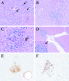
Severe acute respiratory syndrome (SARS) is a significant emerging infectious disease. Humans infected with the etiological agent, SARS-associated coronavirus (SARS-CoV), primarily present with pneumonitis but may also develop hepatic, gastrointestinal, and renal pathology. We inoculated common marmosets (Callithrix jacchus) with the objective of developing a small nonhuman primate model of SARS. Two groups of C. jacchus were inoculated intratracheally with cell culture supernatant containing SARS-CoV. In a time course pathogenesis study, animals were evaluated at 2, 4, and 7 days after infection for morphological changes and evidence of viral replication. All animals developed a multifocal mononuclear cell interstitial pneumonitis, accompanied by multinucleated syncytial cells, edema, and bronchiolitis in most animals. Viral antigen localized primarily to infected alveolar macrophages and type-1 pneumocytes by immunohistochemistry. Viral RNA was detected in all animals from pulmonary tissue extracts obtained at necropsy. Viral RNA was also detected in tracheobronchial lymph node and myocardium, together with inflammatory changes, in some animals. Hepatic inflammation was observed in most animals, predominantly as a multifocal lymphocytic hepatitis accompanied by necrosis of individual hepatocytes. These findings identify the common marmoset as a promising nonhuman primate to study SARS-CoV pathogenesis.
Figures


Similar articles
-
Pathological changes in masked palm civets experimentally infected by severe acute respiratory syndrome (SARS) coronavirus.J Comp Pathol. 2008 May;138(4):171-9. doi: 10.1016/j.jcpa.2007.12.005. Epub 2008 Mar 17. J Comp Pathol. 2008. PMID: 18343398 Free PMC article.
-
[Clinical pathology and pathogenesis of severe acute respiratory syndrome].Zhonghua Shi Yan He Lin Chuang Bing Du Xue Za Zhi. 2003 Sep;17(3):217-21. Zhonghua Shi Yan He Lin Chuang Bing Du Xue Za Zhi. 2003. PMID: 15340561 Chinese.
-
[Expression of SARS-CoV in various types of cells in lung tissues].Beijing Da Xue Xue Bao Yi Xue Ban. 2005 Oct 18;37(5):453-7. Beijing Da Xue Xue Bao Yi Xue Ban. 2005. PMID: 16224511 Chinese.
-
Histologic pulmonary lesions of SARS-CoV-2 in 4 nonhuman primate species: An institutional comparative review.Vet Pathol. 2022 Jul;59(4):673-680. doi: 10.1177/03009858211067468. Epub 2021 Dec 29. Vet Pathol. 2022. PMID: 34963391 Review.
-
SARS-CoV replication and pathogenesis in an in vitro model of the human conducting airway epithelium.Virus Res. 2008 Apr;133(1):33-44. doi: 10.1016/j.virusres.2007.03.013. Epub 2007 Apr 23. Virus Res. 2008. PMID: 17451829 Free PMC article. Review.
Cited by
-
Detached epithelial cell plugs from the upper respiratory tract favour distal lung injury in Golden Syrian hamsters (Mesocricetus auratus) when experimentally infected with the A.2 Brazilian SARS-CoV-2 strain.Mem Inst Oswaldo Cruz. 2024 Oct 21;119:e240100. doi: 10.1590/0074-02760240100. eCollection 2024. Mem Inst Oswaldo Cruz. 2024. PMID: 39442103 Free PMC article.
-
Marmosets as models of infectious diseases.Front Cell Infect Microbiol. 2024 Feb 23;14:1340017. doi: 10.3389/fcimb.2024.1340017. eCollection 2024. Front Cell Infect Microbiol. 2024. PMID: 38465237 Free PMC article. Review.
-
Ontology-based taxonomical analysis of experimentally verified natural and laboratory human coronavirus hosts and its implication for COVID-19 virus origination and transmission.PLoS One. 2024 Jan 22;19(1):e0295541. doi: 10.1371/journal.pone.0295541. eCollection 2024. PLoS One. 2024. PMID: 38252647 Free PMC article.
-
Association between acute liver injury & severity and mortality of COVID-19 patients: A systematic review and meta-analysis.Heliyon. 2023 Sep 20;9(9):e20338. doi: 10.1016/j.heliyon.2023.e20338. eCollection 2023 Sep. Heliyon. 2023. PMID: 37809564 Free PMC article.
-
Functional Cardiovascular Characterization of the Common Marmoset (Callithrix jacchus).Biology (Basel). 2023 Aug 11;12(8):1123. doi: 10.3390/biology12081123. Biology (Basel). 2023. PMID: 37627007 Free PMC article.
References
Publication types
MeSH terms
Substances
Grants and funding
LinkOut - more resources
Full Text Sources
Medical
Miscellaneous

