. 2005 Aug 1;202(3):353-61.
doi: 10.1084/jem.20050778. Epub 2005 Jul 25.

Calcium-sensing soluble adenylyl cyclase mediates TNF signal transduction in human neutrophils

Affiliations

Calcium-sensing soluble adenylyl cyclase mediates TNF signal transduction in human neutrophils

Hyunsil Han et al. J Exp Med. .

Abstract

Through chemical screening, we identified a pyrazolone that reversibly blocked the activation of phagocyte oxidase (phox) in human neutrophils in response to tumor necrosis factor (TNF) or formylated peptide. The pyrazolone spared activation of phox by phorbol ester or bacteria, bacterial killing, TNF-induced granule exocytosis and phox assembly, and endothelial transmigration. We traced the pyrazolone's mechanism of action to inhibition of TNF-induced intracellular Ca2+ elevations, and identified a nontransmembrane ("soluble") adenylyl cyclase (sAC) in neutrophils as a Ca2+-sensing source of cAMP. A sAC inhibitor mimicked the pyrazolone's effect on phox. Both compounds blocked TNF-induced activation of Rap1A, a phox-associated guanosine triphosphatase that is regulated by cAMP. Thus, TNF turns on phox through a Ca2+-triggered, sAC-dependent process that may involve activation of Rap1A. This pathway may offer opportunities to suppress oxidative damage during inflammation without blocking antimicrobial function.


Figures
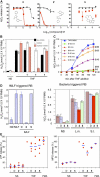
Figure 1.
Figure 1.
Identification of compounds that inhibit the neutrophil respiratory burst in response to TNF. (A) Chemical structures and concentration-response curves. Compounds were added at 37°C for 30 min before stimulation with TNF (red squares) or PMA (black triangles). H2O2 release at 90 min is displayed as percentage of H2O2 release seen with TNF or PMA in the presence of the vehicle, DMSO. (B) Reversibility. Neutrophils incubated with DMSO (D), 2, or 5 were left unmanipulated (black bars) or washed (open bars) with buffer before both sets were plated and stimulated with TNF. Other neutrophils from which the compounds had been washed off were reexposed to the compounds (red hatched bars). (C) Influence of 2 after the onset of respiratory burst. At indicated times, 2 was added to neutrophils already undergoing a TNF-triggered respiratory burst (RB). (D) Inhibition by 2 of the respiratory burst triggered by fMLF but not by bacteria. Neutrophils were preincubated with DMSO (D), 2, or 5 and not stimulated (NS) or stimulated with fMLF (100 nM), L. monocytogenes (L.m.) or S. enterica. (S.t.) (S.H2O2 release is depicted at the time it reached plateau. (A–C) means ± SEM for triplicates. (E) Impact on degranulation. Neutrophils were stimulated and their supernates were assayed for lactoferrin (LTF) and myeloperoxidase (MPO) as a percent of that released in the absence of compound. Each dot is the mean of duplicates in one experiment. Horizontal bars are group means. TNF and PMA were used at 100 ng/ml in all figures.
Figure 2.
Figure 2.
Impact of inhibitors on neutrophil spreading. Neutrophils were plated on FBS-coated glass cover slips, incubated or not with each compound at 37°C for 30 min before stimulation with TNF, PMA, or an equal volume of buffer (no stimulus, NS). After 30 min, the cells were fixed and photographed with phase-contrast microscopy (100×).
Figure 3.
Figure 3.
Impact of inhibitors on components of the phox complex. (A) Translocation of p47phox to membranes. Neutrophils were incubated with DMSO (D) or 2 at 37°C for 30 min and stimulated with TNF (T), PMA (P), or buffer (NS). After 40 min, cells were lysed and membrane fractions collected by ultracentrifugation, separated by SDS-PAGE, and Western-blotted with anti-p47phox antibody. (B) Activation of Rap1A. Lysates of neutrophils that had been pretreated for 30 min with DMSO (D), 2, KH7 (25 μM), or the tmAC inhibitor ddAdo (25 μM), and then stimulated for 30 min with TNF or buffer (NS) alone were incubated with agarose beads coupled to recombinant RalGDS-Rap binding domain to affinity purify GTP-bound Rap1A. Beads were boiled in SDS sample buffer and the supernate subjected to SDS-PAGE and Western blot with anti-Rap1A antibody (top row). Western blot of total cell lysates (bottom row) served as a control.
Figure 4.
Figure 4.
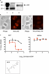
sAC as a critical element of TNF signaling in neutrophils. (A) Western blot. Neutrophils were lysed and subjected to SDS-PAGE/Western blot with anti-sAC mAb R21. Lane 1 (control): lysate of Chinese hamster ovary cells transfected with sperm isoform of sAC. Lanes 2, 3: neutrophil lysates treated without (lane 2) or with (lane 3) protease inhibitor diisopropylfluorophosphate. (B) Immunofluorescence microscopy. Neutrophils were stained with or without biotinylated anti-sAC mAb R41 followed by streptavidin-Alexafluor 594. (C) Effects of sAC inhibitor KH7 and tmAC inhibitor ddAdo on the respiratory burst. Compounds were added to adherent neutrophils at 37°C for 30 min before stimulation with TNF (red squares) or PMA (black triangles). (D) sAC activity in neutrophils (5 × 10⁶/well) preincubated 30 min with the phosphodiesterase inhibitor, isobutylmethylxanthine (100 μM) and DMSO (D), 2, or KH7, then stimulated with ionomycin (1 μM). cAMP accumulation was measured at 0 min and 2 min after stimulation. (E) Impact on TNF-triggered Ca2+ elevation in Fluo-3/AM-loaded neutrophils. Left panels are pseudocolored images of adherent neutrophils pretreated with DMSO (D) or 2 for 30–60 min and then treated with TNF or no stimulus (NS). Average relative fluorescence intensity (RFI) for all cells in similar microscopic fields is plotted as a function of time on the right. Arrow marks addition of TNF. Some cells were pretreated with KH7 (25 μM) as indicated. (F) Speculative model.
Figure 4.
Figure 4.
sAC as a critical element of TNF signaling in neutrophils. (A) Western blot. Neutrophils were lysed and subjected to SDS-PAGE/Western blot with anti-sAC mAb R21. Lane 1 (control): lysate of Chinese hamster ovary cells transfected with sperm isoform of sAC. Lanes 2, 3: neutrophil lysates treated without (lane 2) or with (lane 3) protease inhibitor diisopropylfluorophosphate. (B) Immunofluorescence microscopy. Neutrophils were stained with or without biotinylated anti-sAC mAb R41 followed by streptavidin-Alexafluor 594. (C) Effects of sAC inhibitor KH7 and tmAC inhibitor ddAdo on the respiratory burst. Compounds were added to adherent neutrophils at 37°C for 30 min before stimulation with TNF (red squares) or PMA (black triangles). (D) sAC activity in neutrophils (5 × 10⁶/well) preincubated 30 min with the phosphodiesterase inhibitor, isobutylmethylxanthine (100 μM) and DMSO (D), 2, or KH7, then stimulated with ionomycin (1 μM). cAMP accumulation was measured at 0 min and 2 min after stimulation. (E) Impact on TNF-triggered Ca2+ elevation in Fluo-3/AM-loaded neutrophils. Left panels are pseudocolored images of adherent neutrophils pretreated with DMSO (D) or 2 for 30–60 min and then treated with TNF or no stimulus (NS). Average relative fluorescence intensity (RFI) for all cells in similar microscopic fields is plotted as a function of time on the right. Arrow marks addition of TNF. Some cells were pretreated with KH7 (25 μM) as indicated. (F) Speculative model.

Similar articles

Cited by

References

    1. Weiss, S.J. 1989. Tissue destruction by neutrophils. N. Engl. J. Med. 320:365–376. - PubMed
    1. Fang, F.C. 2004. Antimicrobial reactive oxygen and nitrogen species: concepts and controversies. Nat. Rev. Microbiol. 2:820–832. - PubMed
    1. Reeves, E.P., H. Lu, H.L. Jacobs, C.G. Messina, S. Bolsover, G. Gabella, E.O. Potma, A. Warley, J. Roes, and A.W. Segal. 2002. Killing activity of neutrophils is mediated through activation of proteases by K+ flux. Nature. 416:291–297. - PubMed
    1. Weiss, S.J., G. Peppin, X. Ortiz, C. Ragsdale, and S.T. Test. 1985. Oxidative autoactivation of latent collagenase by human neutrophils. Science. 227:747–749. - PubMed
    1. Peppin, G.J., and S.J. Weiss. 1986. Activation of the endogenous metalloproteinase, gelatinase, by triggered human neutrophils. Proc. Natl. Acad. Sci. USA. 83:4322–4326. - PMC - PubMed

Publication types

MeSH terms