. 2005 Aug 1;170(3):349-55.
doi: 10.1083/jcb.200504028. Epub 2005 Jul 25.

A local mechanism mediates NAD-dependent protection of axon degeneration

Affiliations

A local mechanism mediates NAD-dependent protection of axon degeneration

Jing Wang et al. J Cell Biol. .

Abstract

Axon degeneration occurs frequently in neurodegenerative diseases and peripheral neuropathies. Important insight into the mechanisms of axon degeneration arose from findings that the degeneration of transected axons is delayed in Wallerian degeneration slow (Wlds) mice with the overexpression of a fusion protein with the nicotinamide adenine dinucleotide (NAD) synthetic enzyme, nicotinamide mononucleotide adenylyltransferase (Nmnat1). Although both Wld(s) and Nmnat1 themselves are functional in preventing axon degeneration in neuronal cultures, the underlying mechanism for Nmnat1- and NAD-mediated axon protection remains largely unclear. We demonstrate that NAD levels decrease in degenerating axons and that preventing this axonal NAD decline efficiently protects axons from degeneration. In support of a local protective mechanism, we show that the degeneration of axonal segments that have been separated from their soma could be prevented by the exogenous application of NAD or its precursor nicotinamide. Furthermore, we provide evidence that such Nmnat1/NAD-mediated protection is primarily mediated by their effects on local bioenergetics. Together, our results suggest a novel molecular pathway for axon degeneration.


Figures

Figure 1.
Figure 1.
Reduced NAD levels in degenerating axons. (A) HPLC chromatograms of the axonal segments undergoing Wallerian degeneration. DRG explants cultured for 5 d were subjected to axonal transection, and the acidic extracts of axonal segments were prepared at the indicated time-points after transection and analyzed by HPLC. The chromatograms containing NAD and a few other pyridine compounds, including IMP, AMP, and nicotinic acid (Na) plus GDP from the same amounts of axonal extracts are shown. The absorbance (mAU) was monitored at 260 nm. (B) Quantification of NAD levels in control and transected axons. NAD levels were normalized against total axonal protein contents and expressed as nmol/mg. The results are presented from six independent experiments. Statistical analysis was done by t test. ***, P < 0.001. (C) Representative phase images of axons undergoing Wallerian degeneration taken at indicated time-points after transection. Bar, 50 μm. (D) Quantification of axon degeneration results. Statistical analysis was done by t test. ***, P < 0.001.
Figure 2.
Figure 2.
Nmnat1 and Wlds delay both NAD decrease and Wallerian degeneration. (A and B) Wallerian degeneration is delayed by overexpression of Nmnat1 or Wlds protein. DRG explants were cultured for 5 d and infected with HSVs expressing YFP, Nmnat1, or Wlds 24 h before axotomy. Representative phase images (A) were taken from transected axonal segments at indicated time-points after transection, and the extents of axon degeneration were quantified base on phase images (B). Statistical analysis was done by two-way ANOVA using GraphPad Prism software. P < 0.001 for both Nmnat1 and Wlds vs. YFP. Bar, 50 μm. (C) Localization of overexpressed Nmnat1 and Wlds proteins. Dissociated DRG neurons were cultured for 5 d and infected with HSVs expressing YFP or myc-tagged Nmnat1 or Wlds protein (MOI = 0.1). 24 h after infection the neurons were fixed and stained with antibodies against myc (left panels) and Hoechst (middle panels). Bar, 100 μm. (D) Both Wlds and Nmnat1 overexpression delay the decline of NAD levels in degenerating axons. The DRG explants were infected with HSVs expressing YFP, Nmnat1, or Wlds 24 h before axonal transection. Axonal segments collected at the indicated time-points were analyzed by HPLC to measure NAD levels. Statistical analysis was done by two-way ANOVA using GraphPad Prism software. P < 0.001 for both Nmnat1 and Wlds vs. YFP.
Figure 3.
Figure 3.
Exogenously provided NAD and nicotinamide delay Wallerian degeneration in soma-free axonal segments. (A) Wallerian degeneration of soma-separated axonal segments is delayed by NAD and nicotinamide (NAm) without pretreatment. DRG explants cultured for 5 d were subjected to axonal transection and the ganglia were removed. At the same time, indicated concentrations of NAD or nicotinamide were added to the medium of the axonal segments only. Quantification of axon degeneration was performed from phase images taken at different time-points after transection. Statistical analysis was done by two-way ANOVA using GraphPad Prism software. P < 0.001 for NAD (5 mM, 20 mM), NAm (5 mM, 25 mM) vs. control. (B and C) Quantification of the protective effects of 1 mM (B) and 5 mM (C) NAD added at indicated time-points. Pre 24h: NAD added 24 h before transection; pre 0h: NAD added at the same time as transection; post 3, 5, 7 h: NAD added 3, 5, and 7 h post-transection, respectively. Quantification was performed from phase images taken at indicated time-points (B) or at 12 h after transection (C). Statistical analysis: two-way ANOVA in B: P < 0.001 for NAD 1 mM pre 0 h and 1 mM pre 24 h vs. control; P > 0.05 for NAD 1 mM pre 0 h vs. pre 24 h; t test in C: ***, P < 0.001. (D) Nicotinamide (NAm) attenuates NAD decrease in soma-free axonal segments. DRG explants cultured for 5 d were subjected to axonal transection and the ganglia were removed. At the same time, indicated concentrations of nicotinamide were added to the medium with axonal segments only. The axonal samples were collected at indicated time-points after transection and subjected to HPLC analysis. The results from three independent experiments are presented and statistical analysis was done by two-way ANOVA using GraphPad Prism software. P < 0.001 for 5 mM NAm and 25 mM NAm vs. control.
Figure 4.
Figure 4.
Impaired ATP metabolism in degenerating axons. (A) Both Wlds and Nmnat1 delay the decrease of ATP levels in transected axons. 24 h before axonal transection, DRG explant cultures were infected with HSVs expressing YFP, Wlds, or Nmnat1. The axonal segments were collected at the indicated times-points after transection and subjected to HPLC analysis. Axonal ATP levels in the axons expressing Wlds and Nmnat1 are expressed as the percentages of that in control YFP-expressing axons (also for B and C). The results from three independent experiments are presented and statistical analysis was done by two-way ANOVA using GraphPad Prism software. P < 0.001 for Wlds and Nmnat1 vs. YFP controls. (B and C) Exogenously provided NAD (B) and nicotinamide (C) delay the decrease of ATP levels in soma-free axonal segments. Indicated concentration of NAD (B) or nicotinamide (NAm) (C) was added to the cultures at the same time when the axonal transection was performed. The axonal segments were collected at the indicated time-points after transection and subjected to HPLC analysis. The results from three independent experiments are presented, and statistical analysis was done by two-way ANOVA using GraphPad Prism software. P < 0.001 for 5 and 20 mM NAD and 25 mM NAm vs. controls; P < 0.01 for 5 mM NAm vs. control. (D) Methyl-pyruvate slows down Wallerian degeneration. 10 mM of methyl-pyruvate was added to the cultures at the same time when the axonal transection was performed. Quantification of axon degeneration was performed from phase images taken at indicated time-points after transection. Statistical analysis was done by two-way ANOVA using GraphPad Prism software. P < 0.001 for methyl-pyruvate vs. control. (E) Methyl-pyruvate delays the decline of axonal ATP levels. 10 mM methyl-pyruvate (Py) was added to the cultures at the time of axon transection. The axonal segments were collected at the indicated time-points after transection and subjected to HPLC analysis. The results from three independent experiments are presented and statistical analysis was done by two-way ANOVA using GraphPad Prism software. P < 0.001 for 10 mM methyl-pyruvate vs. controls.
Figure 5.
Figure 5.
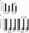
Sirt1 is not required for NAD-dependent axon protection. (A) Degeneration of DRG axons from Sirt1 (+/+) and Sirt1 (−/−) mice explant cultures. DRG explants cultured for 5 d were subjected to axonal transection. At the time of transection, 0 or 5 mM NAD was added to the medium. Quantification was performed from phase images taken 0 or 10 h after transection from duplicate experiments from at least three mice for each genotype. Statistical analysis was done by two-way ANOVA. There is no significant difference in all groups between Sirt1 (+/+) and Sirt1 (−/−). (B) The protective effects of overexpressing Nmnat1 or Wlds on Wallerian degeneration of DRG neurons from Sirt1 (+/+) and Sirt1 (−/−) mice. Virally infected DRG explants were subjected to axonal transection. Quantification was performed from phase images taken at indicated time-points after transection. Statistical analysis was done by two-way ANOVA using GraphPad Prism software. There is no significant difference in all groups between Sirt1 (+/+) and Sirt1 (−/−).

Similar articles

Cited by

References

    1. Araki, T., Y. Sasaki, and J. Milbrandt. 2004. Increased nuclear NAD biosynthesis and SIRT1 activation prevent axonal degeneration. Science. 305:1010–1013. - PubMed
    1. Blander, G., and L. Guarente. 2004. The Sir2 family of protein deacetylases. Annu. Rev. Biochem. 73:417–435. - PubMed
    1. Brunet, A., L.B. Sweeney, J.F. Sturgill, K.F. Chua, P.L. Greer, Y. Lin, H. Tran, S.E. Ross, R. Mostoslavsky, H.Y. Cohen, et al. 2004. Stress-dependent regulation of FOXO transcription factors by the SIRT1 deacetylase. Science. 303:2011–2015. - PubMed
    1. Coleman, M.P., and V.H. Perry. 2002. Axon pathology in neurological disease: a neglected therapeutic target. Trends Neurosci. 25:532–537. - PubMed
    1. Conforti, L., A. Tarlton, T.G. Mack, W. Mi, E.A. Buckmaster, D. Wagner, V.H. Perry, and M.P. Coleman. 2000. A Ufd2/D4Cole1e chimeric protein and overexpression of Rbp7 in the slow Wallerian degeneration (WldS) mouse. Proc. Natl. Acad. Sci. USA. 97:11377–11382. - PMC - PubMed

Publication types

MeSH terms