G-CSF potently inhibits osteoblast activity and CXCL12 mRNA expression in the bone marrow
- PMID: 16037394
- PMCID: PMC1895331
- DOI: 10.1182/blood-2004-01-0272
G-CSF potently inhibits osteoblast activity and CXCL12 mRNA expression in the bone marrow
Abstract
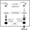
Accumulating evidence indicates that interaction of stromal cell-derived factor 1 (SDF-1/CXCL12 [CXC motif, ligand 12]) with its cognate receptor, CXCR4 (CXC motif, receptor 4), generates signals that regulate hematopoietic progenitor cell (HPC) trafficking in the bone marrow. During granulocyte colony-stimulating factor (G-CSF)-induced HPC mobilization, CXCL12 protein expression in the bone marrow decreases. Herein, we show that in a series of transgenic mice carrying targeted mutations of their G-CSF receptor and displaying markedly different G-CSF-induced HPC mobilization responses, the decrease in bone marrow CXCL12 protein expression closely correlates with the degree of HPC mobilization. G-CSF treatment induced a decrease in bone marrow CXCL12 mRNA that closely mirrored the fall in CXCL12 protein. Cell sorting experiments showed that osteoblasts and to a lesser degree endothelial cells are the major sources of CXCL12 production in the bone marrow. Interestingly, osteoblast activity, as measured by histomorphometry and osteocalcin expression, is strongly down-regulated during G-CSF treatment. However, the G-CSF receptor is not expressed on osteoblasts; accordingly, G-CSF had no direct effect on osteoblast function. Collectively, these data suggest a model in which G-CSF, through an indirect mechanism, potently inhibits osteoblast activity resulting in decreased CXCL12 expression in the bone marrow. The consequent attenuation of CXCR4 signaling ultimately leads to HPC mobilization.
Figures


Similar articles
-
G-CSF induces stem cell mobilization by decreasing bone marrow SDF-1 and up-regulating CXCR4.Nat Immunol. 2002 Jul;3(7):687-94. doi: 10.1038/ni813. Epub 2002 Jun 17. Nat Immunol. 2002. PMID: 12068293
-
Characterization of hematopoietic progenitor mobilization in protease-deficient mice.Blood. 2004 Jul 1;104(1):65-72. doi: 10.1182/blood-2003-05-1589. Epub 2004 Mar 9. Blood. 2004. PMID: 15010367
-
Suppression of CXCL12 production by bone marrow osteoblasts is a common and critical pathway for cytokine-induced mobilization.Blood. 2009 Aug 13;114(7):1331-9. doi: 10.1182/blood-2008-10-184754. Epub 2009 Jan 13. Blood. 2009. PMID: 19141863 Free PMC article.
-
[The role of the SDF-1-CXCR4 axis in hematopoiesis and the mobilization of hematopoietic stem cells to peripheral blood].Postepy Hig Med Dosw (Online). 2007 Jun 13;61:369-83. Postepy Hig Med Dosw (Online). 2007. PMID: 17572657 Review. Polish.
-
Comparison between granulocyte colony-stimulating factor and granulocyte-macrophage colony-stimulating factor in the mobilization of peripheral blood stem cells.Curr Opin Hematol. 2002 May;9(3):190-8. doi: 10.1097/00062752-200205000-00003. Curr Opin Hematol. 2002. PMID: 11953663 Review.
Cited by
-
The role of chemokines in mediating graft versus host disease: opportunities for novel therapeutics.Front Pharmacol. 2012 Feb 24;3:23. doi: 10.3389/fphar.2012.00023. eCollection 2012. Front Pharmacol. 2012. PMID: 22375119 Free PMC article.
-
BET inhibitors induce apoptosis through a MYC independent mechanism and synergise with CDK inhibitors to kill osteosarcoma cells.Sci Rep. 2015 May 6;5:10120. doi: 10.1038/srep10120. Sci Rep. 2015. PMID: 25944566 Free PMC article.
-
Elevating body temperature enhances hematopoiesis and neutrophil recovery after total body irradiation in an IL-1-, IL-17-, and G-CSF-dependent manner.Blood. 2012 Sep 27;120(13):2600-9. doi: 10.1182/blood-2012-02-409805. Epub 2012 Jul 17. Blood. 2012. PMID: 22806894 Free PMC article.
-
Nitrogen-containing bisphosphonate induces a newly discovered hematopoietic structure in the omentum of an anemic mouse model by stimulating G-CSF production.Cell Tissue Res. 2017 Feb;367(2):297-309. doi: 10.1007/s00441-016-2525-4. Epub 2016 Nov 5. Cell Tissue Res. 2017. PMID: 27817114 Free PMC article.
-
Rational identification of a Cdc42 inhibitor presents a new regimen for long-term hematopoietic stem cell mobilization.Leukemia. 2019 Mar;33(3):749-761. doi: 10.1038/s41375-018-0251-5. Epub 2018 Sep 25. Leukemia. 2019. PMID: 30254339 Free PMC article.
References
-
- Thomas J, Liu F, Link DC. Mechanisms of mobilization of hematopoietic progenitors with granulocyte colony-stimulating factor. Curr Opin Hematol. 2002;9: 183-189. - PubMed
-
- To LB, Haylock DN, Simmons PJ, Juttner CA. The biology and clinical uses of blood stem cells. Blood. 1997;89: 2233-2258. - PubMed
-
- Pelus LM, Horowitz D, Cooper SC, King AG. Peripheral blood stem cell mobilization. A role for CXC chemokines. Crit Rev Oncol Hematol. 2002;43: 257-275. - PubMed
-
- Liu F, Poursine-Laurent J, Link DC. Expression of the G-CSF receptor on hematopoietic progenitor cells is not required for their mobilization by GCSF. Blood. 2000;95: 3025-3031. - PubMed
Publication types
MeSH terms
Substances
Grants and funding
LinkOut - more resources
Full Text Sources
Other Literature Sources
Molecular Biology Databases

