. 2005 Oct;16(10):4495-508.
doi: 10.1091/mbc.e04-12-1042. Epub 2005 Jul 19.

Isoform-selective effects of the depletion of ADP-ribosylation factors 1-5 on membrane traffic

Affiliations

Isoform-selective effects of the depletion of ADP-ribosylation factors 1-5 on membrane traffic

Laura A Volpicelli-Daley et al. Mol Biol Cell. 2005 Oct.

Abstract

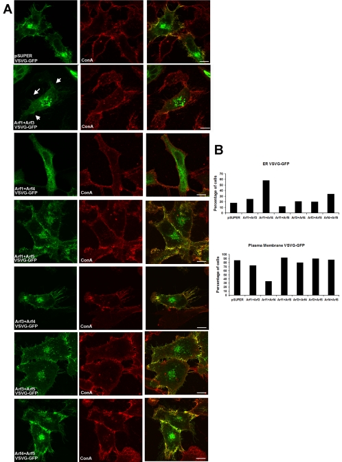
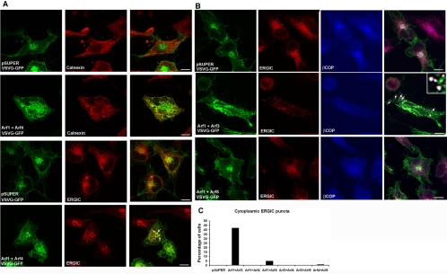
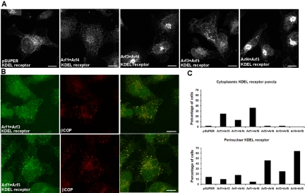
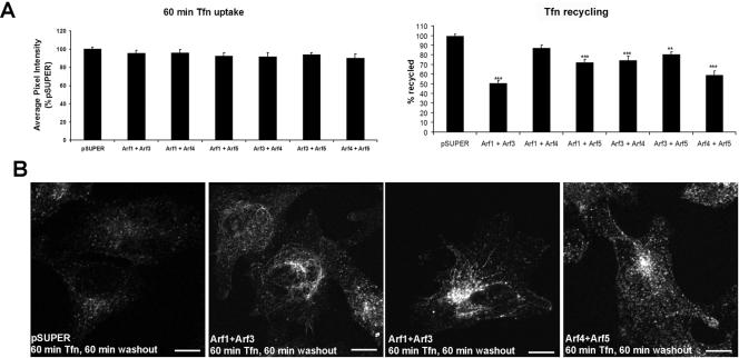
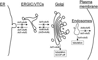
The ADP-ribosylation factors (Arfs) are six proteins within the larger Arf family and Ras superfamily that regulate membrane traffic. Arfs all share numerous biochemical activities and have very similar specific activities. The use of dominant mutants and brefeldin A has been important to the discovery of the cellular functions of Arfs but lack specificity between Arf isoforms. We developed small interference RNA constructs capable of specific depletion of each of the cytoplasmic human Arfs to examine the specificity of Arfs in live cells. No single Arf was required for any step of membrane traffic examined in HeLa cells. However, every combination of the double knockdowns of Arf1, Arf3, Arf4, and Arf5 yielded a distinct pattern of defects in secretory and endocytic traffic, demonstrating clear specificity for Arfs at multiple steps. These results suggest that the cooperation of two Arfs at the same site may be a general feature of Arf signaling and provide candidates at several cellular locations that when paired with data on the localization of the many different Arf guanine nucleotide exchange factors, Arf GTPase activating proteins, and effectors will aid in the description of the mechanisms of specificity in this highly conserved and primordial family of regulatory GTPases.


Figures

Figure 1.
Figure 1.
[Q71L]Arf3 expression caused an expansion of the Golgi and delayed the BFA-induced release of βCOP, whereas [N126I]Arf3 caused a dispersion of the Golgi and dissociation of βCOP. (A) NRK cells stably transfected with wild-type and mutant forms of Arf3 and Arf4 were incubated for varying times with interferon and the levels of Arf3 and Arf4 proteins were determined by immunoblotting. (B) Uninduced or induced cells were stained for mannosidase II to analyze the effect of Arf3, [Q71L]Arf3, and [N126I]Arf3 on Golgi morphology. (C) Uninduced or induced cells were stained for βCOP. Expression of [N126I]Arf3 caused a release of βCOP from the Golgi in untreated cells, whereas expression of [Q71L]Arf3 slowed the release of βCOP in cells treated with 10 μM BFA for 10 min.
Figure 2.
Figure 2.
Knockdown of each Arf isoform by siRNA is effective and specific. (A) HeLa cells were transfected with pSUPER-based plasmids directing expression of siRNAs targeted to human Arf1, Arf3, Arf4, or Arf5 or the empty pSUPERvector. Two different plasmids expressing siRNAs targeting different sequences within the open reading frame of each Arf isoform were used. The level of expression of each Arf was assessed 3 d after transfection, using antisera specific to each Arf isoform. Loading controls for each blot are presented beneath the blot for the respective Arf isoform, as described under Materials and Methods. (B) Immunoblots were scanned and quantified using the Odyssey infrared imaging system. The pixel intensity of each band was normalized to its respective loading control and quantified as described under Materials and Methods. The data represent the average knockdown (±SEM) from the following number of independent experiments: Arf1, n = 5; Arf3, n = 4; Arf4, n = 5; and Arf5, n = 4. (C) HeLa cells were cotransfected with the following combinations of pSUPER-based plasmids: pSUPER control, Arf1a+Arf3b, Arf1a+Arf4b, Arf1a+Arf5b, Arf3b+Arf4b, and Arf3b+Arf5b, Arf4b+Arf5b. Immunoblots show that the double transfectants result in knockdowns of each targeted Arf isoform that are comparable in magnitude to those seen with single knockdowns.
Figure 3.
Figure 3.
Decreased expression of both Arf1 and Arf4 caused tubulation and vesiculation of the Golgi. (A) HeLa cells were transfected with pSUPER control or siRNA constructs targeting dual combinations of Arfs and 3 d later were fixed, labeled with antibodies to GM130 to visualize the Golgi apparatus, and confocal images were collected. With the exception of the Arf1+Arf4 double knockdown, all combinations of Arf siRNAs yielded Golgi morphologies that were indistinguishable from controls. In cells cotransfected with Arf1+Arf4, GM130 staining seemed more punctate with elongated tubules extending from the Golgi. For comparison, GM130 staining in HeLa cells treated with BFA (5 μg/ml) for 30 min showed a punctate, dispersed pattern. Bar, 10 μm. (B) HeLa cells were labeled with antibodies to giantin, which showed a perinuclear ribbon-like appearance in pSUPER-transfected control cells. Decreased expression of Arf1+Arf4 caused giantin staining to seem dispersed throughout the cell, similar to the effects of BFA treatment on giantin localization. Bar, 10 μm. (C) The percentage of cells showing punctate and/or tubulated GM130 localization without the condensed perinuclear morphology visualized in control cells were counted (pSUPER, n = 150; Arf1+Arf3, n = 150; Arf1+Arf4, n = 150; Arf1+Arf5, n = 100; Arf3+Arf4, n = 100; Arf3+Arf5, n = 100; and Arf4+Arf5, n = 150).
Figure 4.
Figure 4.
Decreased expression of Arf1+Arf4 caused βCOP to disperse throughout the cytoplasm, whereas Arf1+Arf3 and Arf1+Arf5 double knockdowns caused βCOP to localize to large cytoplasmic puncta. (A) After 3 d, control pSUPER- and siRNA-transfected HeLa cells were labeled with antibodies to βCOP to visualize the localization of the COPI coat complex. In cells depleted of Arf1+Arf4, βCOP was dispersed throughout the cell, similar to what was observed in cells treated for 3 min with BFA (row 2, panel 4). In cells depleted of Arf1+Arf3 or Arf1+Arf5, the cytoplasmic βCOP puncta seemed larger. Bar, 10 μm. (B) Cells were counted as having a “dispersed” βCOP localization if staining was cytoplasmic with no perinuclear localization, and the percentage of such cells are shown in the bar graph to the left (pSUPER, n = 250; Arf1+Arf3, n = 250; Arf1+Arf4, n = 250; Arf1+Arf5, n = 150; Arf3+Arf4, n = 150; Arf3+Arf5, n = 150; Arf4+Arf5, n = 150). The percentage of cells showing enlarged cytoplasmic βCOP-positive puncta was counted in cells expressing ts045-VSVG-GFP after an overnight incubation at 40°C, and 1.5 h at 32°C, and are shown in the bar graph shown at the right (pSUPER, n = 100; Arf1+Arf3, n = 100; Arf1+Arf4, n = 100; Arf1+Arf5, n = 100; Arf3+Arf4, n = 100; Arf3+Arf5, n = 100; Arf4+Arf5, n = 100).
Figure 5.
Figure 5.
Decreased expression of Arf1+Arf4 and Arf1+Arf3 altered traffic of ts045-VSVG-GFP. HeLa cells were cotransfected with pSUPER-based plasmids on day 0. After 2 d, cells were transfected with the plasmid directing expression of ts045-VSVG-GFP. Cells were then incubated at the restrictive temperature (40°C) for 16 h before switching to the permissive temperate (32°C) for 90 min before fixing, before collection of confocal images. (A) Equal amounts of pSUPER control or the two plasmids directing knockdown of pairs of Arfs, as indicated in each panel, were used for transfections. Nonpermeabilized cells were incubated with ConA-Alexa594 to label the cell surface. Colocalization of ts045-VSVG-GFP (green) and ConA (red) was visualized as yellow in the merged images. In Arf1+Arf4 knockdown cells, ts045-VSVG-GFP displayed an ER-like appearance, did not localize to the cell surface and did not colocalize with ConA. In Arf1+Arf3 knockdown cells, ts045-VSVG-GFP localized to enlarged puncta. Although ts045-VSVG-GFP traveled to the cell surface in these cells (arrows), the amount of staining at the cell surface seemed diminished. Bar, 10 μm. (B) Percentage of cells in which ts045-VSVG-GFP showed a reticular/ER distribution throughout the entire cell (i.e., seemed similar to the distribution of the ER marker calnexin) was quantified (pSUPER, n = 100; Arf1+Arf3, n = 100; Arf1+Arf4, n = 150; Arf1+Arf5, n = 100; Arf3+Arf4, n = 100; Arf3+Arf5, n = 100; Arf4+Arf5, n = 150). The percentage of cells displaying fluorescence at the plasma membrane also was quantified (pSUPER, n = 150; Arf1+Arf3, n = 100; Arf1+Arf4, n = 150; Arf1+Arf5, n = 100; Arf3+Arf4, n = 100; Arf3+Arf5, n = 100; and Arf4+Arf5, n = 100). Cells were scored a 0 if they showed no cell surface localization and a 1 if they showed any localization to the plasma membrane regardless of the intensity. Therefore, these cell counts are a conservative estimate of whether ts045-VSVG-GFP travels to the plasma membrane under these conditions but do not reflect the amount of ts045-VSVG-GFP at the cell surface.
Figure 6.
Figure 6.
Reduced levels of Arf1+Arf3 and Arf1+Arf4 trap ts045-VSVG-GFP early in the secretory pathway. HeLa cells were cotransfected with pSUPER-based and ts045-VSVG-GFP plasmids and incubated as described in the legend to Figure 5. (A) Cells were then incubated with antibodies to calnexin or ERGIC53 to label the ER or VTCs, respectively. Control cells show no colocalization of ts045-VSVG-GFP (green) with calnexin (red) and a minimal amount of colocalization with ERGIC53 (red). In cells depleted of Arf1+Arf4 ts045-VSVG-GFP colocalized extensively with calnexin (visualized as yellow in the merged images). In some Arf1+Arf4 knockdown cells ts045-VSVG-GFP was also seen in large puncta that colocalized with ERGIC53 (arrows). (B) Cells were prepared as described in A before staining with antibodies to ERGIC53 (red) or βCOP (blue). Ts045-VSVG-GFP, ERGIC53 and βCOP colocalized in the perinuclear region of control cells (visualized as white in the merged images). In Arf1+Arf3 depleted cells, ts045-VSVG-GFP localized to large puncta that costained with ERGIC53 and βCOP (white). Arrows point out examples of colocalization. In Arf1+Arf5 knockdown cells, ERGIC53 localization seemed similar to control cells and ts045-VSVG-GFP and ERGIC53 did not colocalize. Bar, 10 μm.
Figure 7.
Figure 7.
Double knockdowns alter the localization of the retrograde transport KDEL receptor. Cells were transfected with pSUPER-based and ts045-VSVG-GFP plasmids and incubated as described in the figure legend to Figure 5. (A) Cells were fixed, incubated with antibodies to the KDEL receptor, and confocal images were collected, as described under Materials and Methods. In control cells and those depleted of Arf1+Arf4 or Arf3+Arf5, the KDEL receptor localized to small puncta distributed throughout the cytoplasm with a slight concentration near the nucleus. In Arf4+Arf5 knockdown cells, the KDEL receptor was substantially more concentrated in the perinuclear region. This effect was also seen in Arf3+Arf4 knockdown cells. All images were captured at the same gain. Bar, 10 μm. (B) In Arf1+Arf3 or Arf1+Arf5 knockdown cells, the KDEL receptor (green) in a proportion of cells localized to larger cytoplasmic puncta that colocalized with βCOP (red). (C) The percentage of cells showing cytoplasmic KDEL receptor puncta and perinuclear KDEL receptor were quantified (pSUPER, n = 100; Arf1+Arf3, n = 100; Arf1+Arf4, n = 100; Arf1+Arf5, n = 100; Arf3+Arf4, n = 100; Arf3+Arf5, n = 100; Arf4+Arf5, n = 100).
Figure 8.
Figure 8.
Double Arf knockdowns alter the recycling of Tfn. (A) Cells were incubated with Tfn-Alexa488 for 60 min and either fixed or rinsed several times and incubated in media for another 60 min to allow internalized Tfn to return to the cell surface and be released into the medium. Cells were scanned using confocal microscopy and the average pixel intensity (±SEM) corresponding to the amount of Alexa-488 Tfn in the cells was determined. All images were captured at the same gain. Tfn recycling was calculated as the average pixel intensity in the recycling experiment subtracted from the average pixel intensity of the 60-min time point. The data are expressed as a percentage of control cells. The double knockdowns produced no significant changes in the extent of Tfn uptake under these conditions. **p < 0.01, ***p < 0.001. (B) Confocal images (captured at the same gain) show the localization of Tfn-Alexa 488 after 60-min uptake and 60-min washout, as described under Materials and Methods. In control cells, Tfn-Alexa 488 localized to small puncta in the cytoplasm. Two examples of the Arf1+Arf3 knockdowns show that Tfn-positive endosomes are extensively tubulated. In Arf4+Arf5 knockdown cells, Tfn Alexa 488 is concentrated in the perinuclear compartment. (C) The percentage of cells showing tubulated endosomes after 60-min Tfn uptake was determined. The endosomes were counted as tubulated if the TfnR staining showed long tubules or finger-like projections extending from the perinuclear region (pSUPER, n = 150; Arf1+Arf3, n = 150; Arf1+Arf4, n = 150; Arf1+Arf5, n = 100; Arf3+Arf4, n = 100; Arf3+Arf5, n = 100; and Arf4+Arf5, n = 100).
Figure 9.
Figure 9.
Model showing the sites of action described for different combinations of Arfs in traffic between the ER and Golgi and in endosome recycling to the plasma membrane. Note that only paired combinations are shown because no single Arf knockdown yielded a discernible effect. An “X” through a line indicates a defect in that step was observed with depletion of that combination of Arfs. Pairs shown within parentheses yielded effects but less than the other pair shown.

Similar articles

Cited by

References

    1. Aridor, M., Bannykh, S. I., Rowe, T., and Balch, W. E. (1995). Sequential coupling between COPII and COPI vesicle coats in endoplasmic reticulum to Golgi transport. J. Cell Biol. 131, 875–893. - PMC - PubMed
    1. Balch, W. E., Kahn, R. A., and Schwaninger, R. (1992). ADP-ribosylation factor is required for vesicular trafficking between the endoplasmic reticulum and the cis-Golgi compartment. J. Biol. Chem. 267, 13053–13061. - PubMed
    1. Barlowe, C., Orci, L., Yeung, T., Hosobuchi, M., Hamamoto, S., Salama, N., Rexach, M. F., Ravazzola, M., Amherdt, M., and Schekman, R. (1994). COPII: a membrane coat formed by Sec proteins that drive vesicle budding from the endoplasmic reticulum. Cell 77, 895–907. - PubMed
    1. Brown, H. A., Gutowski, S., Kahn, R. A., and Sternweis, P. C. (1995). Partial purification and characterization of Arf-sensitive phospholipase D from porcine brain. J. Biol. Chem. 270, 14935–14943. - PubMed
    1. Brummelkamp, T. R., Bernards, R., and Agami, R. (2002). A system for stable expression of short interfering RNAs in mammalian cells. Science 296, 550–553. - PubMed

Publication types

LinkOut - more resources