. 2005 Jul 18;170(2):213-23.
doi: 10.1083/jcb.200501042.

Meiotic telomere clustering requires actin for its formation and cohesin for its resolution

Affiliations

Meiotic telomere clustering requires actin for its formation and cohesin for its resolution

Edgar Trelles-Sticken et al. J Cell Biol. .

Abstract

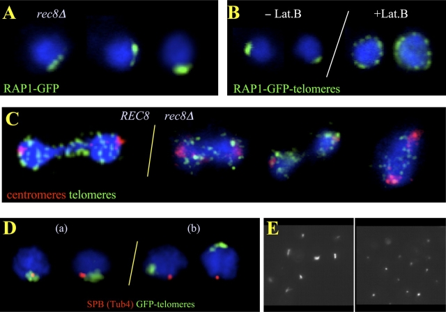
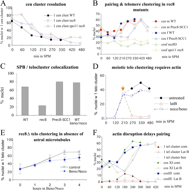
In diploid organisms, meiosis reduces the chromosome number by half during the formation of haploid gametes. During meiotic prophase, telomeres transiently cluster at a limited sector of the nuclear envelope (bouquet stage) near the spindle pole body (SPB). Cohesin is a multisubunit complex that contributes to chromosome segregation in meiosis I and II divisions. In yeast meiosis, deficiency for Rec8 cohesin subunit induces telomere clustering to persist, whereas telomere cluster-SPB colocalization is defective. These defects are rescued by expressing the mitotic cohesin Scc1 in rec8delta meiosis, whereas bouquet-stage exit is independent of Cdc5 pololike kinase. An analysis of living Saccharomyces cerevisiae meiocytes revealed highly mobile telomeres from leptotene up to pachytene, with telomeres experiencing an actin- but not microtubule-dependent constraint of mobility during the bouquet stage. Our results suggest that cohesin is required for exit from actin polymerization-dependent telomere clustering and for linking the SPB to the telomere cluster in synaptic meiosis.


Figures

Figure 1.
Figure 1.
Meiotic telomere dynamics and exit from the bouquet stage require functional cohesin. (A) Projections of trajectories (red threads) of tracked Rap1-GFP telomere spots from a time-lapse image series displayed over an image frame of the respective nucleus. Vegetative telomere clusters (veg.) displaying locally accumulated trajectory projections as a result of constrained motility range. Leptotene telomeres (L) move across the nuclear periphery, which leads to a web of trajectories around the nucleus. Bouquet nucleus (B) with trajectories confined to a limited sector of the nuclear periphery, indicative of telomere clustering. Pachytene nucleus (P) with trajectories around the nucleus, consistent with unconstrained, peripheral telomere movements around the nucleus. Bar, 1 μm. (B) Cumulative plot displaying the velocities of single telomere signals derived from movements of tracked GFP-Rap1 signals in an image series captured every 0.5 s during 7 min. Each trajectory represents a traceable telomere signal through an image series (focal plane fixed at nuclear equator). The trajectories are ranked according to speed. It is evident that in the wild type, vegetative telomere movements are slower than leptotene/bouquet movements, whereas pachytene telomere movements are the most rapid ones. In rec8Δ cells, vegetative telomere velocities match those of wild type, whereas telomere velocities in bouquet-like rec8Δ meiocytes are variable. (C) Analysis of bouquet frequencies by telomere FISH to meiotic time courses. The wild-type frequency of nuclei with a single telomere cluster reaches a maximum after 210 min in sporulation medium (SPM). Wild-type time courses were shorter because meiotic divisions appeared after 270 min and rendered FISH patterns difficult to interpret. rec8Δ and rec8Δ spo11Δ nuclei maintain a telomere cluster up to meiosis I division (compare C with E). Telomere cluster frequency drops after 420 min in rec8Δ spo11Δ meiosis when meiotic divisions become abundant (E). Meiotic expression of the mitotic cohesin SCC1 (Prec8-SCC1) restores bouquet-stage exit in a rec8Δ background, leading to low frequencies of nuclei with a single telomere cluster. Reduction of meiotic CDC5 expression by SCC1 promoter control in REC8 meiosis (Pscc1-CDC5) does not increase telomere clustering. A delay in bouquet formation is evident in red1Δ, Prec8-SCC1, and Pscc1-CDC5 meiosis. (D) Development of FACS profiles in wild-type (WT), rec8Δ, and rec8Δ spo11Δ sporulation indicates that all strains pass through S-phase. G1/2, cell-cycle stage gap1/2. (E) Timing of meiotic divisions in wild-type, rec8Δ, and rec8Δ spo11Δ strains derived by scoring bi- and tetranucleate cells (100–200 per time point). Divisions were significantly delayed in rec8Δ meiosis and occurred at a maximum of 20% (even in longer time courses; not depicted), whereas the spo11Δ mutation eliminated the DSB-dependent prophase I arrest.
Figure 2.
Figure 2.
Telomere clustering in rec8Δ meiosis is displaced from the SPB and requires actin polymerization. (A) Rap1-GFP–tagged telomeres (green) in rec8Δ meiocytes display one telomere cluster. (B) rec8Δ meiocyte nuclei before (−Lat B) and after (+Lat B) inhibition of actin polymerization by Lat B treatment, which leads to peripherally dispersed telomeres. (C) Anaphase I figures of wild-type (left) and rec8Δ meiocytes (right) show absence of a telomere cluster. Centromeres (red) lead the anaphase movements, whereas telomeres (green) are seen as numerous spots trailing behind. (D) Spatial relationships of the telomere cluster (green, Rap1-GFP) and SPB (red, Tub4) in rec8Δ meiocytes. Class a nuclei depict abundant patterns of telomere cluster–SPB association in wild-type meiosis. In rec8Δ meiosis, the telomere cluster is often separated from the SPB (class b; Table I). (E) Image field showing tubulin staining (FITC channel, gray scale) of cells from meiotic cultures without (left) and with (right) benomyl and nocodazole treatment. The treated cells display spots as a result of residual SPB staining that is resistant to MT drugs (Hasek et al., 1987; Lillie and Brown, 1998).
Figure 3.
Figure 3.
Cohesin and actin polymerization are required for normal telomere and chromosome dynamics in SK1 meiosis. (A) Centromere cluster resolution as measured by the frequency of nuclei with a single centromere FISH cluster (Trelles-Sticken et al. 1999). Centromere cluster resolution is similar in wild-type (WT), rec8Δ (rec8), and rec8Δ spo11Δ (spo11 rec8) time courses. (B) Cosmid FISH pairing (cos f, interstitial to XI; cos m, localizing to HML; Trelles-Sticken et al., 2000) as scored by the presence of one large FISH cos-signal cluster in rec8Δ strains. In rec8Δ and rec8Δ spo11Δ cells, this was only seen at a low level (cos f), and meiosis-specific pairing levels never installed with progression through sporulation. Similar values were noted for cos m (not depicted for clarity of display). It should be noted that up to four FISH signals were seen in rec8Δ meiosis, consistent with a lack of sister chromatid cohesion. A single FISH signal may relate to persistent premeiotic association and to a few rec8Δ cells not entering meiosis. Pairing is restored by meiotic expression of the mitotic cohesin SCC1 (Prec8-SCC1). The interstitial chromosome XI region shows a delay in homologue pairing, whereas the telomere of III does not (n > 100 in all time points). (C) SPB colocalization with the telomere cluster in wild-type (WT; n = 60) cells as measured by FISH and Tub4 costaining. In rec8Δ meiocytes (rec8; n = 100), this colocalization is significantly reduced, whereas it is restored in cells expressing the mitotic cohesin Scc1 (Prec8-SCC1; n = 100). Telomere–SPB colocalization is not affected by the presence of benomyl/nocodazole MT drugs (beno/noco; n = 60; anti–Rap1-GFP and Tub4 costaining). (D) Time course experiments with addition of MT and actin poisons 210 min after meiosis induction (orange arrow) in rec8Δ SK1 meiosis. Combined nocodazole/benomyl treatment induced only a slight reduction in the frequency of bouquet-like meiocytes, consistent with results in live cells (C). However, Lat B treatment (latB) dissociated the telomere cluster (n ≥ 100 nuclei were evaluated in all time points and strains). (E) Benomyl/nocodazole treatment of live rec8Δ Rap1-GFP meiocytes fails to eliminate telomere clustering. MT drugs were added 4 h after meiosis induction, and samples were taken before addition (0). After each hour, cells were evaluated for the presence of a single telomere cluster. Error bars represent SD of three experiments; n = 100. (F) Pairing of cos f and m (B) in MT-depleted (ben), and wild-type REC8 (closed symbols, cont.) time courses as measured by FISH in actin-depleted (Lat B) meiosis. The frequency of cells with one telomere cluster is marked by red, open triangles. Inhibition of actin polymerization throughout sporulation prevents telomere clustering and delays homologous pairing for 1–2 h.

Similar articles

Cited by

References

    1. Abe, T., K. Takano, A. Suzuki, Y. Shimada, M. Inagaki, N. Sato, T. Obinata, and T. Endo. 2004. Myocyte differentiation generates nuclear invaginations traversed by myofibrils associating with sarcomeric protein mRNAs. J. Cell Sci. 117:6523–6534. - PubMed
    1. Armstrong, S.J., A.P. Caryl, G.H. Jones, and F.C. Franklin. 2002. Asy1, a protein required for meiotic chromosome synapsis, localizes to axis-associated chromatin in Arabidopsis and Brassica. J. Cell Sci. 115:3645–3655. - PubMed
    1. Bannister, L.A., L.G. Reinholdt, R.J. Munroe, and J.C. Schimenti. 2004. Positional cloning and characterization of mouse mei8, a disrupted allelle of the meiotic cohesin Rec8. Genesis. 40:184–194. - PubMed
    1. Bass, H.W. 2003. Telomere dynamics unique to meiotic prophase: formation and significance of the bouquet. Cell. Mol. Life Sci. 60:2319–2324. - PMC - PubMed
    1. Bergerat, A., B. de Massy, D. Gadelle, P.C. Varoutas, A. Nicolas, and P. Forterre. 1997. An atypical topoisomerase II from Archaea with implications for meiotic recombination. Nature. 386:414–417. - PubMed

Publication types

MeSH terms