. 2005 Aug;25(15):6722-33.
doi: 10.1128/MCB.25.15.6722-6733.2005.

Individual palmitoyl residues serve distinct roles in H-ras trafficking, microlocalization, and signaling

Affiliations

Individual palmitoyl residues serve distinct roles in H-ras trafficking, microlocalization, and signaling

Sandrine Roy et al. Mol Cell Biol. 2005 Aug.

Abstract

H-ras is anchored to the plasma membrane by two palmitoylated cysteine residues, Cys181 and Cys184, operating in concert with a C-terminal S-farnesyl cysteine carboxymethylester. Here we demonstrate that the two palmitates serve distinct biological roles. Monopalmitoylation of Cys181 is required and sufficient for efficient trafficking of H-ras to the plasma membrane, whereas monopalmitoylation of Cys184 does not permit efficient trafficking beyond the Golgi apparatus. However, once at the plasma membrane, monopalmitoylation of Cys184 supports correct GTP-regulated lateral segregation of H-ras between cholesterol-dependent and cholesterol-independent microdomains. In contrast, monopalmitoylation of Cys181 dramatically reverses H-ras lateral segregation, driving GTP-loaded H-ras into cholesterol-dependent microdomains. Intriguingly, the Cys181 monopalmitoylated H-ras anchor emulates the GTP-regulated microdomain interactions of N-ras. These results identify N-ras as the Ras isoform that normally signals from lipid rafts but also reveal that spacing between palmitate and prenyl groups influences anchor interactions with the lipid bilayer. This concept is further supported by the different plasma membrane affinities of the monopalmitoylated anchors: Cys181-palmitate is equivalent to the dually palmitoylated wild-type anchor, whereas Cys184-palmitate is weaker. Thus, membrane affinity of a palmitoylated anchor is a function both of the hydrophobicity of the lipid moieties and their spatial organization. Finally we show that the plasma membrane affinity of monopalmitoylated anchors is absolutely dependent on cholesterol, identifying a new role for cholesterol in promoting interactions with the raft and nonraft plasma membrane.


Figures
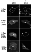
FIG. 1.
FIG. 1.
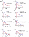
H-ras palmitate groups play distinct roles in H-ras trafficking. BHK cells transiently expressing GFP-H-rasG12V, GFP-N-rasG12V, GFP-H-rasG12V C181S, or GFP-H-rasG12V C184S were imaged by confocal microscopy before (−) or after (+) incubation for 5 h in cycloheximide (CHX) to inhibit de novo protein synthesis. The disappearance of ER staining that is particularly striking with GFP-H-rasG12V C184S and to a lesser extent with GFP-H-rasG12V C181S was evident in all cells imaged. Similar distributions were seen when the same set of Ras proteins were expressed in COS cells (data not shown).
FIG. 2.
FIG. 2.
Inhibiting palmitoylation causes redistribution of H-ras monopalmitoylated mutants to the ER. A. BHK cells stably transfected with GFP-ras proteins were pretreated for 2 h with cycloheximide to inhibit de novo protein synthesis. Cells were then incubated for 3 h in cycloheximide with or without 2-BP. The cells were visualized in a confocal microscope and scored for predominant plasma membrane (PM), Golgi, or endoplasmic reticulum (ER) localization. Examples of typical cells scored in these three categories are shown in panel B. At least 300 cells were evaluated from three independent experiments for each Ras protein. C. BHK cells transiently expressing GFP-H-rasG12V C181S or GFP-H-rasG12V C184S were labeled with [3H]palmitic acid in the presence of cycloheximide. Duplicate anti-GFP immunoprecipitates prepared from whole-cell lysates were immunoblotted for Ras input and fluorographed for [3H]palmitic acid. Blots and scans were quantified, and the ratio of [3H]palmitic acid units per Ras unit was calculated. Representative blots and 3H scans are shown. The ratios of [3H]palmitic acid/Ras (in arbitrary units) for H-rasG12V C181S and H-rasG12V C184S, respectively, were 0.68 ± 0.01 and 0.66 ± 0.04 (means ± standard errors of the means, n = 3). IP, immunoprecipitation. IB, immunoblot.
FIG. 3.
FIG. 3.
The plasma membrane microlocalization of monopalmitoylated H-rasG12V C184S emulates that of GFP-N-rasG12V. Apical plasma membrane sheets from BHK cells transiently expressing GFP-ras proteins were labeled with anti-GFP antibodies coupled directly to 4-nm gold. Where indicated, cells were incubated with 1% β-methyl-cyclodextrin for 30 min to deplete cell surface cholesterol. The immunogold point patterns were analyzed using Ripley's K-function. The graphs show weighted mean K-functions (n = 8 to 17 sheets) standardized on the 99% confidence interval (CI) for complete spatial randomness. The average number of gold particles per plasma membrane sheet evaluated was 939 μm−2. In this analysis a pattern is significantly clustered if the L(r) − r curve leaves the CI (i.e., is >1), whereas in a random pattern the L(r) − r approximates to 0 for all values of r (for examples see references and 42). Differences between control and cholesterol-depleted membranes were evaluated for each construct using bootstrap tests. A statistically significant effect of cholesterol depletion is seen on the clustering of GFP-H-ras (P = 0.017), GFP-N-rasG12V (P = 0.039), and GFP-H-rasG12V C184S (P = 0.001).
FIG. 4.
FIG. 4.
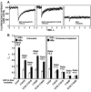
Monopalmitoylated Ras proteins exhibit different dynamic interactions with the plasma membrane than dual-palmitoylated H-ras. A. FRAP experiments (with a 63× objective) were conducted on COS-7 cells transiently expressing the depicted GFP constructs. The dots represent the fluorescence intensity. Solid lines show the best fit of a nonlinear regression analysis. GFP exhibits free diffusion in the cytoplasm, resulting in extremely fast fluorescence recovery. Thus, free diffusion in the cytoplasm occurs on a faster time scale and does not contribute significantly to the measurements depicted for GFP-H-rasG12V and GFP-H-rasG12V C181S. B. FRAP measurements were also performed in untreated and in cholesterol-depleted cells using two beam sizes that were generated by 63× and 40× objectives. The fluorescence recovery time (τ) determined with each objective and the τ(40×)/τ(63×) ratios derived (means ± standard errors of the means of 15 to 60 measurements) are shown. The mobile fractions were high for all proteins (>90%). We used t tests to determine whether the ratios differed significantly from that expected for pure lateral diffusion, an experimentally determined ratio between the areas illuminated by the laser beam using the two objectives (means ± standard errors of the means value of 2.56 ± 0.30, n = 39). ARB., arbitrary.
FIG. 5.
FIG. 5.
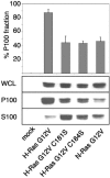
Monopalmitoylated Ras proteins have a reduced affinity for the plasma membrane. BHK cells expressing Ras proteins were fractionated into S100 and P100 fractions and immunoblotted to determine the relative extent of membrane association. The graph shows the percentage of Ras protein associated with the P100 fraction (means ± standard errors of the means, n = 3). WCL, whole-cell lysates.
FIG. 6.
FIG. 6.
Monopalmitoylated RasG12V proteins recruit Raf to membranes more efficiently than H-rasG12V. A. BHK cells coexpressing GFP-ras proteins and mRFP-RBD were incubated for 5 h in cycloheximide and visualized by confocal microscopy. Representative cells are shown. B. BHK cells transiently expressing GFP-ras proteins and mRFP-RBD were fractionated into membrane (P100) and cytosolic (S100) fractions by high-speed centrifugation. GFP-ras and mRFP-RBD content of the P100 fraction was estimated using quantitative immunoblotting. The graphs show mRFP-RBD recruitment per unit of Ras expressed in arbitrary phosphorimager units (means ± standard errors of the means, n = 3). Significant differences from control (H-rasG12V), evaluated in t tests, are shown on the graph (an asterisk indicates P value of <0.05).
FIG. 7.
FIG. 7.
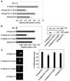
Monopalmitoylated RasG12V proteins activate the Raf/MEK/MAPK cascade with varying efficiencies. A. P100 fractions from BHK cells transiently expressing equivalent amounts of H-rasG12V, N-rasG12V, H-rasG12V C181S, or H-rasG12V C184S were assayed for Raf activity in a coupled mitogen-activated protein kinase assay with myelin basic protein phosphorylation as a read out (49). P100 fractions were immunoblotted for Raf-1, and the kinase assay results were standardized on the Raf content of the P100 fraction to estimate Raf-1-specific activity. The graph shows Raf specific activity (means ± standard errors of the means, n = 3) expressed relative to the activity of Raf-1 recruited and activated by H-rasG12V. B. The same lysates assayed in panel A for Raf activity were analyzed for Erk activation by quantitative immunoblotting for phosphorylated Erk (ppErk). A representative blot is shown, and the graph shows results (means ± standard errors of the means, n = 3) for three independent experiments. Significant differences from control (H-rasG12V), evaluated in t tests, are shown on the graph (*, P < 0.05). C. PC12 cells transiently expressing GFP-ras proteins were visualized 72 h after transfection by fluorescence microscopy. The percentage of GFP-positive, differentiated cells (with neurite outgrowth of at least twice the diameter of the cell body) was estimated by counting >300 cells per construct per experiment. Images of differentiated cells are shown. The graph shows results obtained from three independent experiments (means ± standard errors of the means, n = 3). Significant differences from control (H-rasG12V), evaluated in t tests, are shown on the graph (**, P < 0.01).

Similar articles

Cited by

References

    1. Apolloni, A., I. A. Prior, M. Lindsay, R. G. Parton, and J. F. Hancock. 2000. H-ras but not K-ras traffics to the plasma membrane through the exocytic pathway. Mol. Cell. Biol. 20:2475-2487. - PMC - PubMed
    1. Axelrod, D., D. E. Koppel, J. Schlessinger, E. Elson, and W. W. Webb. 1976. Mobility measurement by analysis of fluorescence photobleaching recovery kinetics. Biophys. J. 16:1055-1069. - PMC - PubMed
    1. Baker, T. L., H. Zheng, J. Walker, J. L. Coloff, and J. E. Buss. 2003. Distinct rates of palmitate turnover on membrane-bound cellular and oncogenic H-ras. J. Biol. Chem. 278:19292-19300. - PubMed
    1. Besag, J. E. 1977. Contribution to the discussion of Dr. Ripley's paper. J. R. Statist. Soc. B. 39:193-195.
    1. Bivona, T. G., I. Perez De Castro, I. M. Ahearn, T. M. Grana, V. K. Chiu, P. J. Lockyer, P. J. Cullen, A. Pellicer, A. D. Cox, and M. R. Philips. 2003. Phospholipase Cgamma activates Ras on the Golgi apparatus apparatus by means of RasGRP1. Nature 424:694-698. - PubMed

Publication types