. 2005 Jul 18;170(2):237-48.
doi: 10.1083/jcb.200503148. Epub 2005 Jul 11.

The WD40 protein Caf4p is a component of the mitochondrial fission machinery and recruits Dnm1p to mitochondria

Affiliations

The WD40 protein Caf4p is a component of the mitochondrial fission machinery and recruits Dnm1p to mitochondria

Erik E Griffin et al. J Cell Biol. .

Abstract

The mitochondrial division machinery regulates mitochondrial dynamics and consists of Fis1p, Mdv1p, and Dnm1p. Mitochondrial division relies on the recruitment of the dynamin-related protein Dnm1p to mitochondria. Dnm1p recruitment depends on the mitochondrial outer membrane protein Fis1p. Mdv1p interacts with Fis1p and Dnm1p, but is thought to act at a late step during fission because Mdv1p is dispensable for Dnm1p localization. We identify the WD40 repeat protein Caf4p as a Fis1p-associated protein that localizes to mitochondria in a Fis1p-dependent manner. Caf4p interacts with each component of the fission apparatus: with Fis1p and Mdv1p through its NH2-terminal half and with Dnm1p through its COOH-terminal WD40 domain. We demonstrate that mdv1delta yeast contain residual mitochondrial fission due to the redundant activity of Caf4p. Moreover, recruitment of Dnm1p to mitochondria is disrupted in mdv1delta caf4delta yeast, demonstrating that Mdv1p and Caf4p are molecular adaptors that recruit Dnm1p to mitochondrial fission sites. Our studies support a revised model for assembly of the mitochondrial fission apparatus.


Figures

Figure 1.
Figure 1.
Construction of M 9 TH-FIS1 and Caf4p/Mdv1p alignment. (A) A 9xMyc-TEV-URA3-TEV-His8 cassette was PCR amplified with FIS1-targeting primers and integrated in-frame into the NH2 terminus of FIS1. Pop-out of the URA3 cassette by recombination between flanking TEV sites yielded M9TH-FIS1 under the control of the endogenous FIS1 promoter. UTR: untranslated region. (B) Schematic of Mdv1p and Caf4p. The NH2-terminal extension (NTE), coiled-coil, and WD40 regions are shown with percent identity. Overall identity is 37% and overall similarity is 57%.
Figure 2.
Figure 2.
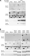
Caf4p and Mdv1p coimmunoprecipitation experiments. (A) Yeast carrying the indicated HA- and Myc-tagged constructs were lysed and immunoprecipitated with an anti-Myc antibody. Total lysates (labeled “Lysate”) and immunoprecipitated samples (labeled “Myc IP”) were analyzed by immunoblotting with anti-Myc (9E10) and anti-HA (12CA5) antibodies as indicated. The expression constructs were: Caf4p wt (residues 1–659), Caf4p N (residues 1–274), Caf4p C (residues 275–659), Mdv1 wt (residues 1–714), Mdv1p N (residues 1–300), and Mdv1p C (residues 301–714). The yeast backgrounds were: (A) wild-type, lanes 1–6; caf4Δ M3TH-FIS1, lanes 7–9; mdv1Δ M3TH-FIS1, lanes 10–12; (B) wild-type, lanes 1–6; caf4Δ MDV1-HTM, lanes 7–9; mdv1Δ CAF4-HTM, lanes 10–12; CAF4-HTM, lanes 13–15; MDV1-HTM, lanes 16–18. (B) Yeast carrying the indicated HA- and Myc-tagged constructs were immunoprecipitated and analyzed as in A. Immunoprecipitated samples were loaded at 10 (A) and 20 (B) equivalents of the lysate samples. HA-tagged proteins in the lysate are marked with an asterisk. The HA-tagged Caf4p C polypeptide co-migrates with a background band in the total lysate blot probed with HA antibody.
Figure 3.
Figure 3.
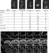
CAF4 regulates mitochondrial morphology. Strains expressing mitochondrially targeted GFP were grown in YP dextrose to mid-log phase and fixed. The percentage of cells (n = 400) with mitochondria having wild-type (A), collapsed net (B), or spread net morphology (C and D) is tabulated. The spread net phenotype encompasses a distribution of morphologies ranging from simple structures containing one or two loops (C) to complexly fenestrated mitochondria with dozens of loops (D). For both wild-type and caf4Δ strains, the wild-type category includes 1% fragmented cells. Bar, 1 μm.
Figure 4.
Figure 4.
CAF4 mediates residual fission in mdv1 Δ cells. Top: mid-log cultures grown in YP dextrose were treated for 60 min with 200 μM latrunculin A (+) or vehicle (−). For each strain, 200 cells were scored into the following phenotypic categories: wild-type (A), fragments and short tubules (B), collapsed net (C), spread net (D), or partial net (E). Numbers shown are percentages. The fragments and short tubules category encompasses a range of morphologies from completely fragmented (as shown in B) to a mixture of fragments and short tubules. (F–H) Still images from time-lapse movies showing fission events in mdv1Δ yeast treated with 200 μM latrunculin A. The boxed area in the first frame is magnified in the subsequent sequence of five images. Arrows indicate fission events. Mitochondria were visualized with the outer membrane marker OM45-GFP. Bars, 1 μm.
Figure 5.
Figure 5.
Caf4p overexpression blocks mitochondrial fission. Wild-type yeast (DCY1979) carrying the pRS416 GalL vector with no insert, full-length CAF4-HA, CAF4-HA N, or CAF4-HA C were grown in rich dextrose or galactose media for 180 min and fixed. Cells were scored into the following phenotypic categories: wild-type (A), connected tubules (B), or spread nets with tubules (C). Numbers shown are percentages (n = 200). Overexpression in galactose cultures was estimated to result in 20-fold greater expression than endogenous levels by Western blots of serially diluted lysates (not depicted). Bar, 1 μm.
Figure 6.
Figure 6.
Suppression of the glycerol growth defect of fzo1 Δ cells. Individual spores of the indicated genotypes were assayed by serial dilution on YP glycerol and YP dextrose plates to determine the percent survival on glycerol-containing medium. Each point represents the viability of an individual spore. For clarity, spores showing 1% or less survival were plotted as 1%.
Figure 7.
Figure 7.
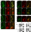
Mitochondrial localization of Caf4p and Mdv1p requires Fis1p. Immunofluorescence (red, middle panels) was used to localize Myc-tagged Caf4p (Caf4p-HTM; A–-F and M–U) and Mdv1p (Mdv1p-HTM; G–L and P–R) in wild-type (A–C, G–I, and M–U) and fis1Δ cells (D–F and J–L). Caf4p-HTM and Mdv1p-HTM are expressed from the endogenous loci and are functional. Mitochondria were labeled with mitochondrially targeted GFP (A–R, left, green). The majority of Dnm1p-GFP puncta colocalize with Caf4p-HTM (S–U). Overlays of the two signals are shown in the merged images (right). Note that both Caf4p and Mdv1p localize to mitochondria in wild-type cells, but are diffusely cytosolic in fis1Δ cells. Cells were grown in YP dextrose (A–L) or YP galactose (M–U). Representative maximum intensity projections of deconvolved z-stacks are shown. Bar, 1 μm. (V) Caf4p-HTM and Mdv1p-HTM were analyzed by subcellular fractionation. The total cell lysate (Total), high-speed supernatant (Cyto), and mitochondrial pellet (Mito) were analyzed by Western blot with an anti-Myc antibody in wild-type (left) and fis1Δ (right) yeast. PGK (3-phosphoglycerate kinase) is a cytosolic marker, and porin is a mitochondrial outer membrane marker.
Figure 8.
Figure 8.
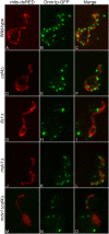
Fis1p mediates Dnm1p-GFP localization through either Mdv1p or Caf4p. The localization of Dnm1p-GFP (middle, green) was compared to mito-DsRed (left, red) in yeast of the indicated genotype. Merged images are shown on the right. Representative maximum intensity projections of deconvolved z-stacks are shown.

Similar articles

Cited by

References

    1. Bleazard, W., J.M. McCaffery, E.J. King, S. Bale, A. Mozdy, Q. Tieu, J. Nunnari, and J.M. Shaw. 1999. The dynamin-related GTPase Dnm1 regulates mitochondrial fission in yeast. Nat. Cell Biol. 1:298–304. - PMC - PubMed
    1. Boldogh, I., N. Vojtov, S. Karmon, and L.A. Pon. 1998. Interaction between mitochondria and the actin cytoskeleton in budding yeast requires two integral mitochondrial outer membrane proteins, Mmm1p and Mdm10p. J. Cell Biol. 141:1371–1381. - PMC - PubMed
    1. Cerveny, K.L., and R.E. Jensen. 2003. The WD-repeats of Net2p interact with Dnm1p and Fis1p to regulate division of mitochondria. Mol. Biol. Cell. 14:4126–4139. - PMC - PubMed
    1. Cerveny, K.L., J.M. McCaffery, and R.E. Jensen. 2001. Division of mitochondria requires a novel DMN1-interacting protein, Net2p. Mol. Biol. Cell. 12:309–321. - PMC - PubMed
    1. Denis, C.L., and J. Chen. 2003. The CCR4-NOT complex plays diverse roles in mRNA metabolism. Prog. Nucleic Acid Res. Mol. Biol. 73:221–250. - PubMed

Publication types

MeSH terms

Substances