

Opsin activation of transduction in the rods of dark-reared Rpe65 knockout mice
- PMID: 15994181
- PMCID: PMC1474752
- DOI: 10.1113/jphysiol.2005.091942
Opsin activation of transduction in the rods of dark-reared Rpe65 knockout mice
Abstract
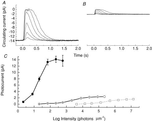
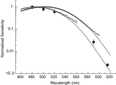
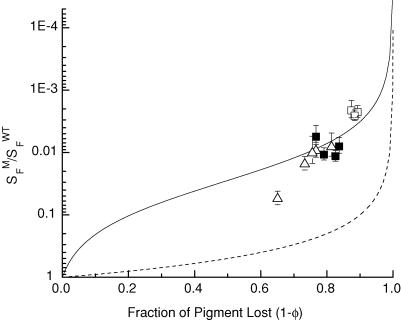
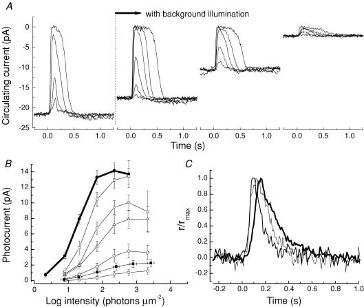
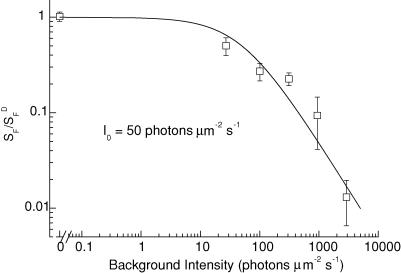
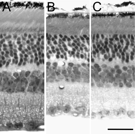
Rpe65 knockout mice (Rpe65-/-) are unable to synthesize the visual pigment chromophore 11-cis retinal; however, if these animals are reared in complete darkness, the rod photoreceptors accumulate a small amount of 9-cis retinal and its corresponding visual pigment isorhodopsin. Suction-electrode recording of single rods from dark-reared Rpe65-/- mice showed that the rods were about 400 times less sensitive than wild-type control rods and that the maximum responses were much smaller in amplitude. Spectral sensitivity measurements indicated that Rpe65-/- rod responses were generated by isorhodopsin rather than rhodopsin. Sensitivity and pigment concentration were compared in the same mice by measuring light responses from rods of one eye and pigment concentration from the retina of the other eye. Retinas had 11-35% of the normal pigment level, but the rods were of the order of 20-30 times less sensitive than could be accounted for by the loss in quantum catch. This extra desensitization must be caused by opsin-dependent activation of the visual cascade, which leads to a state equivalent to light adaptation in the dark-adapted rod. By comparing the sensitivity of dark-reared Rpe65-/- rods to that produced in normal rods by background light, we estimate that Rpe65-/- opsin is of the order of 2.5x10(-5) as efficient in activating transduction as photoactivated rhodopsin (Rh*) in WT mice. Dark-reared Rpe65-/- rods are less desensitized than rods from cyclic light-reared Rpe65-/- mice, have about 50% more photocurrent and degenerate at a slower rate. Retinas sectioned after 9 months in darkness show a larger number of photoreceptor nuclei in dark-reared animals than in cyclic light-reared animals, though both have fewer nuclei than in cyclic light-reared wild-type retinas. Both also have shorter outer segments and a lower free-Ca2+ concentration. These experiments provide the first quantitative measurement of opsin activation in physiologically responding mammalian rods.
Figures
Similar articles
-
Correlation of regenerable opsin with rod ERG signal in Rpe65-/- mice during development and aging.Invest Ophthalmol Vis Sci. 2003 Jan;44(1):310-5. doi: 10.1167/iovs.02-0567. Invest Ophthalmol Vis Sci. 2003. PMID: 12506090
-
Isorhodopsin rather than rhodopsin mediates rod function in RPE65 knock-out mice.Proc Natl Acad Sci U S A. 2003 Nov 11;100(23):13662-7. doi: 10.1073/pnas.2234461100. Epub 2003 Oct 24. Proc Natl Acad Sci U S A. 2003. PMID: 14578454 Free PMC article.
-
9-cis Retinal increased in retina of RPE65 knockout mice with decrease in coat pigmentation.Photochem Photobiol. 2006 Nov-Dec;82(6):1461-7. doi: 10.1562/2006-02-02-RA-793. Photochem Photobiol. 2006. PMID: 16553465
-
Vitamin A and Vision.Subcell Biochem. 2016;81:231-259. doi: 10.1007/978-94-024-0945-1_9. Subcell Biochem. 2016. PMID: 27830507 Review.
-
The 11-cis Retinal Origins of Lipofuscin in the Retina.Prog Mol Biol Transl Sci. 2015;134:e1-12. doi: 10.1016/bs.pmbts.2015.07.022. Prog Mol Biol Transl Sci. 2015. PMID: 26310175 Review.
Cited by
-
Dark adaptation of human rod bipolar cells measured from the b-wave of the scotopic electroretinogram.J Physiol. 2006 Sep 1;575(Pt 2):507-26. doi: 10.1113/jphysiol.2006.108027. Epub 2006 Jun 15. J Physiol. 2006. PMID: 16777945 Free PMC article.
-
Leber congenital amaurosis due to RPE65 mutations and its treatment with gene therapy.Prog Retin Eye Res. 2010 Sep;29(5):398-427. doi: 10.1016/j.preteyeres.2010.04.002. Epub 2010 Apr 24. Prog Retin Eye Res. 2010. PMID: 20399883 Free PMC article. Review.
-
Flavonoids enhance rod opsin stability, folding, and self-association by directly binding to ligand-free opsin and modulating its conformation.J Biol Chem. 2019 May 17;294(20):8101-8122. doi: 10.1074/jbc.RA119.007808. Epub 2019 Apr 3. J Biol Chem. 2019. PMID: 30944172 Free PMC article.
-
The mammalian cone visual cycle promotes rapid M/L-cone pigment regeneration independently of the interphotoreceptor retinoid-binding protein.J Neurosci. 2011 May 25;31(21):7900-9. doi: 10.1523/JNEUROSCI.0438-11.2011. J Neurosci. 2011. PMID: 21613504 Free PMC article.
-
Rod and cone visual pigments and phototransduction through pharmacological, genetic, and physiological approaches.J Biol Chem. 2012 Jan 13;287(3):1635-41. doi: 10.1074/jbc.R111.303008. Epub 2011 Nov 10. J Biol Chem. 2012. PMID: 22074928 Free PMC article. Review.
References
-
- Ablonczy Z, Crouch RK, Goletz PW, Redmond TM, Knapp DR, Ma JX, Rohrer B. 11-cis-retinal reduces constitutive opsin phosphorylation and improves quantum catch in retinoid-deficient mouse rod photoreceptors. J Biol Chem. 2002;277:40491–40498. - PubMed
Publication types
MeSH terms
Substances
Grants and funding
LinkOut - more resources
Full Text Sources
Miscellaneous