Comparative Study
. 2005 Jul;11(7):1017-29.
doi: 10.1261/rna.7218905.

The positional, structural, and sequence requirements of the Drosophila TLS RNA localization element

Affiliations
Comparative Study

The positional, structural, and sequence requirements of the Drosophila TLS RNA localization element

Robert S Cohen et al. RNA. 2005 Jul.

Abstract

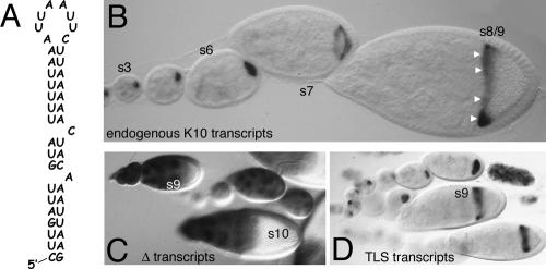
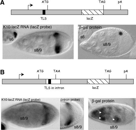
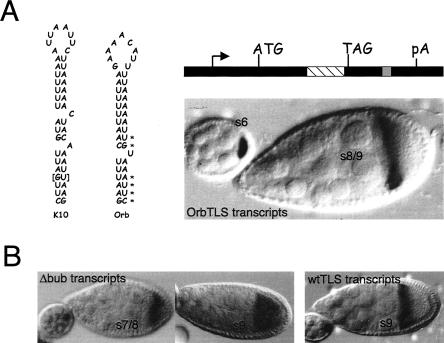
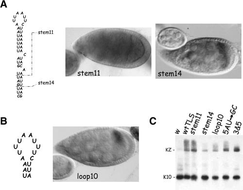
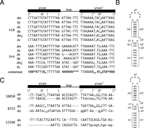
The subcellular localization of mRNAs is a key step in the polarization of cells in organisms from yeast to man. Here, we use a transgenic fly/in situ hybridization assay system to define the positional, structural, and sequence requirements of the TLS, a stem loop RNA sequence element that mediates the subcellular localization of K10 and Orb transcripts in Drosophila oocytes. We find that the TLS is a highly robust and modular element. It mediates efficient RNA localization regardless of sequence context or position within the transcript. Site-specific mutagenesis experiments indicate that the size and shape of the stem and loop regions are critical determinants of TLS activity. Such experiments also identify specific base residues that are important for TLS activity. All such residues map to the stem portion of the structure. Significantly, mutations at these residues interfere with TLS activity only when they alter the stereochemistry of the stem's minor groove. For example, mutation of the A:U base pair at position 3 of the TLS stem to G:C severely reduces TLS activity, while mutation of the same base pair to U:A has no effect. Extensive searches for TLS-like elements in other Drosophila mRNAs using sequence and structural parameters defined by our experiments indicate that the TLS is unique to K10 and Orb mRNAs. This unexpected finding raises important questions as to how the many hundreds of other mRNAs that are known or thought to exhibit K10 and Orb-like localization are localized.


Figures

FIGURE 1.
FIGURE 1.
The TLS is required and sufficient for the transport and anterior localization of K10-LacZ fusion transcripts. (A) Sequence and putative secondary structure of the 44 nucleotide TLS. (B) Whole-mount in situ hybridization to endogenous K10 transcripts in wild-type egg chambers, arranged Left to Right in order of increasing stage of development. Stages (s) of select egg chambers are indicated. The oocyte lies to the Right (posterior) of the nurse cell cluster in each of these and in all other displayed egg chambers. In young (s1–6) egg chambers, K10 mRNA fills the entire oocyte. Localization to the oocyte’s anterior cortex (arrowheads) is evident in the stage 8/9 egg chamber. In the stage 7 egg chamber, K10 mRNA is moving toward, but is not yet completely localized to, the oocyte’s anterior cortex. (C, D) Whole-mount in situ hybridization to K10-lacZ transcripts in egg chambers containing the Δ transgene (C), which lacks the TLS, or the wtTLS transgene (D), which contains the TLS. The probe in C and D and in all other experiments designed to detect K10-lacZ transcripts is complementary to the lacZ portion of the RNA and thus does not detect endogenous K10 transcripts.
FIGURE 2.
FIGURE 2.
The TLS directs transport and anterior localization when placed in the 5′UTR or intron portion of the K10-lacZ reporter transcript. (A) Structure and expression pattern of the 5TLS transgene. (Top) Diagram to approximate scale of the 5TLS transgene. The TLS is located in the 5′UTR of the gene, 49 nucleotides upstream of the ATG translation start codon. (Bottom Left) In situ hybridization for K10-lacZ transcripts in representative egg chambers containing the 5TLS transgene. Wild-type transport and anterior localization is observed. Note, for example, the intense labeling of the stage 6 oocyte and the sharp localization of the K10-lacZ transcripts to the anterior cortex of the stage 8/9 oocyte. (Bottom Right) Immunodetection of β-galactosidase (β-gal) fusion protein. The single dot of staining in the stage 8/9 egg chamber corresponds to the oocyte nucleus. (B) Structure and expression pattern of the intronTLS transgene. (Top) Diagram to approximate scale of the intronTLS transgene. The TLS (black box) is located in the intron (white box), 15 nucleotides downstream of the intron/exon junction. The intron contains an in-frame TAA stop codon, such that translation of the lacZ portion of the transcript is dependent on removal of the intron by splicing. (Bottom Left) In situ hybridization for intronTLS transcripts in transgenic ovaries using the lacZ probe. The nurse cells and oocyte both stain, consistent with the presence of unspliced and spliced transcripts (see Results). (Bottom middle) In situ hybridization for intronTLS transcripts in transgenic ovaries using the intron-specific probe (see text). (Bottom Right) Immunodetection of β-gal fusion protein. Protein is detected in nurse cell nuclei (arrowheads) only, consistent with the idea that only the nontransported (nurse cell) transcripts are spliced (see Results).
FIGURE 3.
FIGURE 3.
A TLS-like sequence in Orb mediates RNA transport and anterior localization and highlights essential features of the TLS. (A) The nucleotide sequences and putative secondary structures of the TLS elements of the D. melanogaster K10 and Orb genes are shown at the Left. The overall structure of the two elements is very similar, except that the Orb TLS contains only one bulge, while the K10 TLS contains two. The asterisks highlight base pairs in the Orb stem whose orientation is reversed compared to the corresponding base pairs in the K10 stem. As described in the Results, differences in base pair orientation (e.g., 5′A:U vs. 5′U:A) alter the stereochemistry of H-bond donor and acceptor groups in the major groove, but not in the minor groove, of the double helix. The brackets highlight a base pair that is exclusive to the K10 TLS. The structure of the OrbTLS transgene is shown at the top Right. It is identical to the TLS construct of Figure 1, except that it contains a copy of the Orb TLS (gray box) in place of the K10 TLS. The lacZ portion of this construct is denoted with the striped box. In situ hybridization for K10-lacZ transcripts (bottom Right) shows that the Orb TLS mediates wild-type transport and anterior localization. (B) In situ hybridization experiments showing that maximal TLS localization activity requires at least one bulge. (Left) In situ hybridization for Δbub transcripts, which contain a TLS that lack both bulges. Transport into the oocyte is normal; however, localization to the anterior cortex is transient. Thus, while some localization to the anterior cortex is apparent in the stage 7/8 oocyte (s7/8), no localization is apparent in the stage 9 (s9) oocyte. (Right) Control in situ hybridization to a stage 9 oocyte showing persistent localization of transcripts that contain a wild-type copy of the TLS (wtTLS transcripts) to the anterior cortex.
FIGURE 4.
FIGURE 4.
The lengths of the TLS stem and loop are critical for transport and anterior localization activity. (A) Structure and expression patterns of TLS stem truncation mutants. The wild-type TLS with its 17 base pair stem is shown at the Left. The breakpoints of the stem14 and stem11 mutants, which reduce the stem length by 3 and 6 base pairs, respectively, are indicated by the dashed lines. In situ hybridization for K10-lacZ transcripts in representative egg chambers of stem11 and stem14 transgenic lines are shown to the Right of the diagram. The stem11 TLS has no detectable activity; stem11 transcripts never become enriched in oocytes and no localization to the anterior cortex is ever observed. The stem14 TLS possesses only weak activity; stem14 transcripts do not become enriched in the oocyte until ~stage 6 and exhibit only transient and diffuse localization to the oocyte’s anterior cortex. (B) Structure and expression patterns of the loop10 mutant. The sequence of the mutated loop (and only a portion of the stem) is shown on the Left. The Right panel shows that this TLS possesses only weak transport and anterior localization activity; no accumulation is evident in the oocyte until stage 6 and localization to the anterior cortex is diffuse and transient. (C) Northern blot analysis of K10-lacZ (KZ) and endogenous K10 transcripts from transgenic ovaries. The KZ transcripts encoded by the stem11, stem14 and loop10 transcripts all migrate as a single major band of the expected size, ruling out the possibility that their poor transport/anterior localization is due to message instability. The Northern blot also rules out message instability as the reason for the poor transport/anterior localization of 5AUGC and 3&5 transcripts (see text and Fig. 6 for further details). The w and wtTLS lanes are controls containing RNA from nontransgenic flies and flies harboring the wtTLS transgene, respectively.
FIGURE 5.
FIGURE 5.
Alterations of the major groove of the TLS stem do not significantly interfere with TLS activity. The wild-type K10 TLS is shown at the far Left for reference. The altered regions of the seven reversal (rev) mutants are shown to the Right of the wild-type TLS. Five of the seven reversal mutants exhibit wild-type transport and anterior localization, including rev8 −17 (see figure for representative in situ hybridization experiment) in which each of the last 10 base pairs are reversed. The other two reversal mutants, rev5 (top Right) and rev3 −7 (Table 1) exhibit moderate activity, distinguishable from wild-type only by their inability to mediate complete localization to the anterior cortex.
FIGURE 6.
FIGURE 6.
Alterations of the minor groove of the TLS can greatly reduce or eliminate TLS activity. The wild-type K10 TLS is shown at the far Left , with the arrows and numbers indicating those base pairs that were mutated individually or in pairs to alter the minor groove. For example, 3UACG denotes mutation of the third base pair of the stem from UA to CG. The middle panels show representative in situs to reporter transcripts containing TLS elements with minor groove mutations at the indicated base pairs. The far Right panels show representative in situs of the 3&5 TLS in which the 3rd and 5th base pairs were simultaneously mutated and of the 8&10 TLS in which the 8th and 10th base pairs were simultaneously mutated. The 3&5 TLS possesses no activity, while the 8&10 TLS possess weak activity, indistinguishable from that of the 10th base pair single mutation.
FIGURE 7.
FIGURE 7.
The TLS element is highly conserved, but not found in other genes. (A) Sequence alignments of the D. melanogaster K10 and Orb TLS elements with their counterparts in other Drosophila species. A linear diagram of the TLS is shown above the alignments for reference, where the numbers indicate the positions of the nucleotides that comprise the 1st, 7th, 10th, and 17th base pairs. Abbreviations are as follows: dm, D. melanogaster; dy, D. yakuba; da, D. ananassae; dp, D. pseudoobscura; dmj, D. mojavensis; dv, D. virilis. Gaps in the sequence are denoted with dashes. Unpaired bases (bulges and bubbles) that lie outside of the loop are indicated in subscripts. Abbreviations for the deduced consensus sequences are as follows: V, not T; W, A or T; Y, C or T; S, C or G; N, any nucleotide; *, optional nucleotide of type N; R, A or G; B, not A. (B) Schematic representation of the TLS primary sequence and secondary structure as used for database searches with RNABOB. For these searches, the TLS is conceptually divided into three helical (h1/h1′, h2/h2′, and h3/h3′) and four single-stranded (s1–4) motifs as shown. The acceptable sequences for each motif is indicated, using the same code described in A. The top structure fits most closely with our data (see text), but identifies no TLS-like elements apart from those in the K10 and Orb genes. The bottom structure depicts the more relaxed search conditions (see text), that led to the identification of three candidate elements. (C) Attempted sequence alignments of the candidate TLS elements, which are found in the 3′UTRs of the CG10850 and CG8233 genes and in the 5′UTR of the CG111598 gene, respectively. Species abbreviations are as in A. Residues that are unable to make their normal base pairs (as defined by the K10 and Orb elements) are denoted with lowercase letters. The deletion at the beginning of the D. ananassae CG15598 sequence is based on more extensive sequence alignments (data not shown). The deletion is flanked on its 5′side by the sequence AAAGACACAG. When substituted for the deleted segment, this sequence only regenerates one base pair, i.e., less than the number expected for a random sequence.

Similar articles

Cited by

References

    1. Arn, E.A., Cha, B.J., Theurkauf, W.E., and Macdonald, P.M. 2003. Recognition of a bicoid mRNA localization signal by a protein complex containing Swallow, Nod, and RNA binding proteins. Dev. Cell 4: 41–51. - PubMed
    1. Bashirullah, A., Cooperstock, R.L., and Lipshitz, H.D. 1998. RNA localization in development. Annu. Rev. Biochem. 67: 335–394. - PubMed
    1. Bullock, S.L. and Ish-Horowicz, D. 2001. Conserved signals and machinery for RNA transport in Drosophila oogenesis and embryogenesis. Nature 414: 611–616. - PubMed
    1. Cheung, H.-K., Serano, T.L., and Cohen, R.S. 1992. Evidence for a highly selective RNA transport system and its role in establishing the dorsoventral axis of the Drosophila egg. Development 114: 653–661. - PubMed
    1. Cohen, R.S. and Meselson, M. 1985. Separate regulatory elements for the heat-inducible and ovarian expression of the Drosophila hsp26 gene. Cell 42: 737–746. - PubMed

Publication types

MeSH terms

LinkOut - more resources