. 2005 Jul;79(13):8493-505.
doi: 10.1128/JVI.79.13.8493-8505.2005.

Activation of the Kaposi's sarcoma-associated herpesvirus major latency locus by the lytic switch protein RTA (ORF50)

Affiliations

Activation of the Kaposi's sarcoma-associated herpesvirus major latency locus by the lytic switch protein RTA (ORF50)

Satoko Matsumura et al. J Virol. 2005 Jul.

Abstract

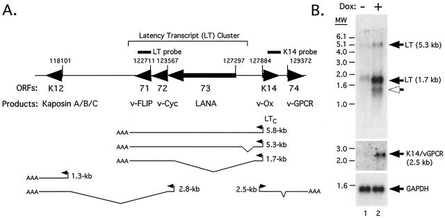
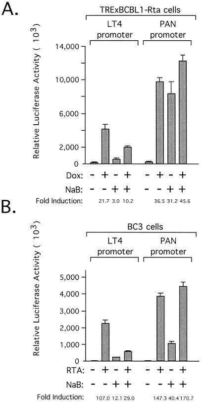
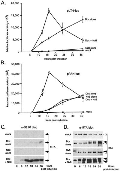
Kaposi's sarcoma-associated herpesvirus (KSHV) maintains a latent infection in primary effusion lymphoma cells but can be induced to enter full lytic replication by exposure to a variety of chemical inducing agents or by expression of the KSHV-encoded replication and transcription activator (RTA) protein. During latency, only a few viral genes are expressed, and these include the three genes of the so-called latency transcript (LT) cluster: v-FLIP (open reading frame 71 [ORF71]), v-cyclin (ORF72), and latency-associated nuclear antigen (ORF73). During latency, all three open reading frames are transcribed from a common promoter as part of a multicistronic mRNA. Subsequent alternative mRNA splicing and internal ribosome entry allows for the expression of each protein. Here, we show that transcription of LT cassette mRNA can be induced by RTA through the activation of a second promoter (LT(i)) immediately downstream of the constitutively active promoter (LT(c)). We identified a minimal cis-regulatory region, which overlaps with the promoter for the bicistronic K14/v-GPCR delayed early gene that is transcribed in the opposite direction. In addition to a TATA box at -30 relative to the LT(i) mRNA start sites, we identified three separate RTA response elements that are also utilized by the K14/v-GPCR promoter. Interestingly, LT(i) is unresponsive to sodium butyrate, a potent inducer of lytic replication. This suggests there is a previously unrecognized class of RTA-responsive promoters that respond to direct, but not indirect, induction of RTA. These studies highlight the fact that induction method can influence the precise program of viral gene expression during early events in reactivation and also suggest a mechanism by which RTA contributes to establishment of latency during de novo infections.


Figures

FIG. 1.
FIG. 1.
The LT promoter can be induced by RTA. (A) A total of 107 TRExBCBL1-Rta cells were transiently transfected using Lipofectamine 2000 with 2 μg of the following luciferase reporter constructs: promoterless vector, pGL3basic-luc; LT promoter, pLT1-luc; PAN promoter, pPAN(−102/+15); ORF50 promoter, pORF50-luc; and human bFGF-2 promoter, pFGF-2-luc. After 24 h, the transfected cells were split into two flasks, one of which was mock treated with sterile water and the other treated with Dox (1 μg/ml). After a further 18 h, cell extracts were prepared and assayed for luciferase activity. Bars represent the mean and standard deviation of three independent transfections. (B) TRExBCBL1 cells were cotransfected with 0.5 μg pPAN(−102/+15) or pLT4-luc (see Fig. 3 for details) and either mock or Dox treated. In addition, parallel cultures we cotransfected with 1.5 μg of an expression vector encoding full-length KSHV RTA (pCGFlag-RTA). (C) The reporter constructs (0.5 μg) used in the results shown in panel A were electroporated into 106 HeLa cells, together with 1 μg of empty expression vector or pCGFlag-RTA. Luciferase activity was measured 24 h after transfection. (D) Comparison of RTA expression in mock-treated (lane 1) or Dox-treated (lane 2) TRExBCBL1-Rta cells and HeLa cells transfected with 2 μg empty expression vector (lane 3) or pCGFlag-RTA (lane 4). Total cell lysates (5 × 10⁴ cell equivalents per lane) were resolved on 15% SDS-PAGE, transferred to a nitrocellulose membrane, and probed with a polyclonal antibody against RTA (71).
FIG. 2.
FIG. 2.
LT transcripts are induced by RTA expression. (A) Arrangement of known transcripts encoding the three genes of the LT cluster and neighboring open reading frames. In latently infected PEL cells, transcription from a constitutively active promoter (LTc) gives rise to a ∼5.8-kb precursor RNA that undergoes alternative splicing to produce ∼5.3-kb tricistronic (ORF71, -72, and -73) and ∼1.7-kb dicistronic (ORF71 and -72) transcripts (9, 56, 65). A less abundant 1.1-kb monocistronic (ORF71) transcript (not shown) has also been detected in PEL lines (19). Open reading frame K12 (T0.7) encoding the kaposins is actively transcribed during latency from both proximal and upstream promoters and is induced further during lytic replication (31, 54). The bicistronic mRNA encoding K14 (v-Ox) and v-GPCR (ORF74) is essentially silent during latency but strongly induced by RTA (28, 65). (B) Autoradiograms from Northern blot analyses of poly(A)+-selected RNA isolated from TRExBCBL1-Rta cells either mock treated (lane 1) or Dox treated (lane 2) for 36 h. The probes used (indicated by solid bars in panel A) correspond to the coding region of ORF71 (top) and K14 (middle). As a loading control, the blot was reprobed with a GAPDH-specific probe (bottom). Positions of size markers (in kilobases) are shown on the left. Filled arrowheads indicate the major LT mRNAs at 5.3 and 1.7 kb and major K14/v-GPCR mRNA at 2.5 kb. An induced transcript at ∼1.4 kb is indicated by an open arrowhead.
FIG. 3.
FIG. 3.
Separation of the constitutive (LTc) and inducible (LTi) promoters. (A) Schematic showing the organization of the three open reading frames (ORF71, -72, and -73) encoded by the LT mRNAs and two open reading frames (K14 and ORF74) transcribed in the opposite direction as a bicistronic mRNA (9, 56, 57, 65). Reporter construct pLT1-luc contains a 1.8-kb fragment of KSHV genomic DNA spanning nucleotides 127609 to 129375 (nucleotide positions refer to sequences for GenBank accession number KSU75698) (53), orientated such that the luciferase gene is placed downstream of the LT promoter. Note that this fragment contains the entire K14 ORF (open arrow). Boundaries of further truncations within this fragment are shown: LT3 (nucleotides 127609 to 128282), LT4 (nucleotides 127609 to 127807), LT5 (nucleotides 127609 to 127992), LT6 (nucleotides 127816 to 129375), LT7 (nucleotides 127816 to 127992), LT8 (nucleotides 127764 to 127992), and LT9 (nucleotides 127609 to 127948). Note: LT2 of this series is only slightly shorter than LT1 and was omitted from the analysis. (B) HeLa cells were transiently transfected with a promoterless reporter (pGL3basic-luc) or plasmids pLT1-luc through pLT9-luc together with an empty expression vector (open bars) or a vector encoding full-length RTA (pCGFlag-RTA) (shaded bars). Luciferase activity was measured after 24 h and displayed as a histogram showing the mean and standard deviation of three independent assays. (C) As in panel B, except that each reporter was cotransfected with either empty vector (open bars) or a vector encoding LANANC (pCGT-LANANC) (shaded bars).
FIG. 4.
FIG. 4.
Structure of the LTi promoter. (A) Mapping of the 5′ end of the RTA-inducible transcript using primer extension with 32P-labeled LUCPE1 oligonucleotide annealed to poly(A)+ RNA isolated from transiently transfected HeLa cells. HeLa cells were transfected with promoterless reporter pGL3basic-luc (lanes 5 to 6), pLT1-luc (lanes 7 to 8), and pLT4-luc (lanes 9 to 10) and pCGFlag-RTA where indicated (lanes 6, 8, and 10). To accurately size the extension products, a sequencing ladder generated with the same primer and pLT4-luc plasmid DNA was run along side (lanes 1 to 4). Locations of primers used for primer extension (LUCPE1) or 5′RLM-RACE (RACE3 and RACE4) are shown schematically. Sizes of the principal extension products (dotted line) are given. (B) DNA sequences surrounding the transcriptional start sites of pLT1-luc/pLT4-luc (top) or KHSV genome (bottom). Transcriptional start sites detected by primer extension in transfected HeLa cells or by 5′ RLM-RACE from Dox-treated TRExBCBL1-Rta cells are shown, and a TATA box 30 to 35 bp upstream of the initiation sites is highlighted.
FIG. 5.
FIG. 5.
Truncation analysis of the LTi promoter. (A) Sequence of the LTi promoter highlighting potential binding sites for the cellular transcription factors Sp1, C/EBP, CSL (CBF1/RBP-Jκ), interferon regulatory factor 8 (IRF/ICSBP), and nuclear factor 1 (NF1). A candidate TATA is located at −30 relative to the transcriptional start sites (+1) of the newly mapped LTi promoter. Three RTA-responsive elements (A, B, and C) used by the K14 promoter are indicated, and binding of CSL to RRE-A and C has been demonstrated (33). Sequences present in pLT4-luc are shown in boldface uppercase letters. Upstream end points for additional promoter truncations (LT4.1 to LT4.5) are indicated by arrows below the sequence. (B) Luciferase assays of the promoter truncations tested in TRExBCBL1-Rta cells in presence (+) or absence (−) of Dox. (C) Luciferase assays of the truncations in HeLa cells in the presence (+) or absence (−) of transfected RTA expression plasmid (pCGFlag-RTA). (D) Schematic showing the distribution of candidate binding sites and known RTA-response elements relative to truncations (LT4 to LT4.5).
FIG. 6.
FIG. 6.
The LTi and K14 promoters use common elements to respond to RTA. (A) Schematic showing candidate transcription factor binding sites targeted for mutagenesis (m1 to m9). (B) Wild-type and mutant versions of pLT4-luc were transfected into TRExBCBL1-Rta cells and induced with Dox. (C) As in panel B, except that each reporter was cotransfected with the pCGFlag-RTA expression plasmid.
FIG. 7.
FIG. 7.
LTi promoter is not induced by sodium butyrate. (A) TRExBCBL1-Rta cells were transfected with pLT4-luc or pPAN-luc reporters, cultured for 24 h, and then treated with Dox, sodium butyrate (NaB), or a combination of Dox and NaB. After a further 36 h, extracts were prepared and assayed for luciferase activity. (B) BC3 cells transfected with 0.5 μg pLT4-luc or pPAN-luc reporters, together with 1.5 μg of pCGFlag-RTA (+RTA) or empty expression vector (−RTA). After 24 h, NaB was added to the medium, and extracts were prepared 36 h later.
FIG. 8.
FIG. 8.
Delayed suppression of the LTi promoter by sodium butyrate. (A and B) Time course analysis of TRExBCBL1-Rta cells transfected with pLT4-luc (A) and pPAN-luc (B). Time 0 h corresponds to addition of inducing agents: mock (filled squares), Dox (filled circles), NaB (filled triangles), or a combination of Dox and NaB (open squares). (C) Dox-dependent expression of myc-tagged RTA in TRExBCBL1-Rta cells detected by immunoblotting using mouse monoclonal antibody 9E10. Cells were lysed in SDS-sample buffer, and proteins were resolved by 10% SDS-PAGE. The time shown indicates hours after addition of inducing agents. (D) As in panel C, except that protein extracts were separated by 5% SDS-PAGE and immunoblotted with an α-RTA polyclonal antibody. A band that is present in uninduced cells and might represent a cross-reacting cellular protein or an inactive form of RTA is shown, indicated by open arrowheads.

Similar articles

Cited by

References

    1. Arvanitakis, L., E. A. Mesri, R. G. Nador, J. W. Said, A. S. Asch, D. M. Knowles, and E. Cesarman. 1996. Establishment and characterization of a primary effusion (body cavity-based) lymphoma cell line (BC-3) harboring Kaposi's sarcoma-associated herpesvirus (KSHV/HHV-8) in the absence of Epstein-Barr virus. Blood 88:2648-2654. - PubMed
    1. AuCoin, D. P., K. S. Colletti, S. A. Cei, I. Papouskova, M. Tarrant, and G. S. Pari. 2004. Amplification of the Kaposi's sarcoma-associated herpesvirus/human herpesvirus 8 lytic origin of DNA replication is dependent upon a cis-acting AT-rich region and an ORF50 response element and the trans-acting factors ORF50 (K-Rta) and K8 (K-bZIP). Virology 318:542-555. - PubMed
    1. Bannister, A. J., and E. A. Miska. 2000. Regulation of gene expression by transcription factor acetylation. Cell. Mol. Life Sci. 57:1184-1192. - PMC - PubMed
    1. Byun, H., Y. Gwack, S. Hwang, and J. Choe. 2002. Kaposi's sarcoma-associated herpesvirus open reading frame (ORF) 50 transactivates K8 and ORF57 promoters via heterogeneous response elements. Mol. Cells 14:185-191. - PubMed
    1. Cesarman, E. 2003. Kaposi's sarcoma-associated herpesvirus-the high cost of viral survival. N. Engl. J. Med. 349:1107-1109. - PubMed

Publication types

MeSH terms