. 2005 Jul;79(13):8046-56.
doi: 10.1128/JVI.79.13.8046-8056.2005.

Effects of a temperature sensitivity mutation in the J1R protein component of a complex required for vaccinia virus assembly

Affiliations

Effects of a temperature sensitivity mutation in the J1R protein component of a complex required for vaccinia virus assembly

Wen-Ling Chiu et al. J Virol. 2005 Jul.

Abstract

Vaccinia virus J1R protein is required for virion morphogenesis (W. L. Chiu and W. Chang, J. Virol. 76:9575-9587, 2002). In this work, we further characterized the J1R protein of wild-type vaccinia virus and compared it with the protein encoded by the temperature-sensitive mutant virus Cts45. The mutant Cts45 was found to contain a Pro-to-Ser substitution at residue 132 of the J1R open reading frame, which is responsible for a loss-of-function phenotype. The half-life of the J1R-P132S mutant protein was comparable at both 31 and 39 degrees C, indicating that the P132S mutation did not affect the stability of the J1R protein. We also showed that the J1R protein interacts with itself in the virus-infected cells. The N-terminal region of the J1R protein, amino acids (aa) 1 to 77, interacted with the C-terminal region, aa 84 to 153, and the P132 mutation did not abolish this interaction, as determined by two-hybrid analysis. Furthermore, we demonstrated that J1R protein is part of a viral complex containing the A30L, G7L, and F10L proteins in virus-infected cells. In immunofluorescence analyses, wild-type J1R protein colocalized with the A30L, G7L, and F10L proteins in virus-infected cells but the loss-of-function P132 mutant did not. Furthermore, without a functional J1R protein, rapid degradation of A30L and the 15-kDa forms of the G7L and F10L proteins was observed in cells infected with Cts45 at 39 degrees C. This study thus demonstrated the importance of the J1R protein in the formation of a viral assembly complex required for morphogenesis.


Figures
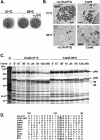
FIG. 1.
FIG. 1.
(A) Marker rescue of Cts45. CV-1 cells were transfected with either the plasmid vector (−) or the plasmid containing the WT J1R ORF (+J1R) and infected with Cts45 as described in Materials and Methods (34, 36). These cells, incubated at 31 or 39°C, were fixed, stained with crystal violet, and photographed. (B) Electron microscopy of BSC40 cells infected with viJ1R or Cts45 at 31 or 39°C. BSC40 cells were infected with viJ1R, with or without 50 μM IPTG, or with Cts45. The cells were cultured at 31°C or 39°C for 24 h and processed for EM as described elsewhere (6). G, granular materials. Arrowheads represent double-layer viral membranes that are accumulated at 39°C. (C) Pulse-chase experiment for precursor p4a/p4b protein processing. BSC40 cells were either mock infected (m) or infected with Cts45 at an MOI of 10 PFU per cell and incubated at 31°C or 39°C. At 8 h p.i., the cells were pulse-labeled with [35S]methionine for 30 min and chased with normal medium for 0 min, 15 min, 30 min, 1 h, 2 h, 4 h, 12 h, or 24 h. Proteins were denatured and analyzed by SDS-12% PAGE followed by autoradiography. The mobilities of p4a and p4b and their mature processed forms 4a and 4b are shown on the right. (D) Amino acid mutation of J1R protein in Cts45. DNA sequences of the J1R gene in Cts45 revealed a single nucleotide change from C to T, resulting in a P-to-S change at residue 132. P132 is boxed, indicating that it is invariable in all the J1R orthologues. The alignment shown here contains J1R protein sequences only from amino acid residue120 to 140. VACV-WR, vaccinia virus strain WR; VARV, variola virus (INDIA-1967/isolate IND3); SWPV, swinepox virus; SPPV, sheep poxvirus; LSDV, lumpy skin disease virus; MYXV, myxoma virus; SFV, Shope fibroma virus; YLDV, Yaba-like disease virus; MCV, molluscum contagiosum virus subtype 1; FPV, fowlpox virus.
FIG. 2.
FIG. 2.
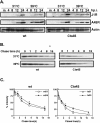
Stability of J1R protein in cells infected with the WT or Cts45 virus under permissive and nonpermissive conditions. (A) Expression of J1R protein in cells infected with WT VV or Cts45 at 31 or 39°C. BSC40 cells were either mock infected (m) or infected with WT VV or Cts45, incubated at 31 or 39°C, and harvested at different times as indicated for immunoblot analyses with an anti-J1R, anti-A45R, or anti-actin Ab. (B) Pulse-chase experiments with J1R protein. BSC40 cells were infected with WT VV or Cts45 at an MOI of 10 PFU per cell and incubated at 31°C or 39°C. At 8 h p.i., the cells were pulse-labeled with [35S]methionine (50 μCi/ml) for 30 min and then chased with normal medium for 0, 1, 2, 4, 8, or 16 h. Cell lysates were immunoprecipitated with anti-J1R (1:100), analyzed by SDS-15% PAGE, and autoradiographed. (C) Quantification of J1R protein from scanning of autoradiograms shown in panel B. The y axis represents the percentage of J1R protein present during the chase period, calculated as (intensity of J1R with chase)/(intensity of J1R without chase) × 100%.
FIG. 3.
FIG. 3.
Self-interaction of J1R protein. (A) Recombinant T7-J1R protein was expressed and purified from E. coli as described in Materials and Methods. (B) Gel filitration analysis of the recombinant J1R protein. Purified J1R (50 μg) in PBS was loaded onto a HiPrep 26/60 Sephacryl S-100 High Resolution column (Amersham Pharmacia), and individual fractions were collected. Every three fractions were pooled, separated on an SDS-12% PAGE gel, and analyzed by immunoblotting with an anti-T7 Ab (1:5,000). Markers used for gel filtration analyses included blue dextran (2 MDa), RNase A (15.7 kDa), and ovalbumin (48.6 kDa). (C) Coimmunoprecipitation of WT J1R protein with T7-J1R protein in virus-infected cells. BSC40 cells were coinfected with T7-J1R virus and viJ1R at an MOI of 5 PFU per cell and cultured in a medium containing IPTG. Cell lysates were prepared at 24 h p.i. for immunoprecipitation (IP) using an anti-T7 MAb (1:1,000). The immunoprecipitated products were separated on an SDS-12% PAGE gel, and both T7-J1R (21 kDa) and WT J1R protein (16 kDa) were detected by an anti-J1R Ab (1:1,000). WB, Western blotting.
FIG. 4.
FIG. 4.
Deletion constructs of J1R protein used in the yeast two-hybrid analyses. A hydrophathy plot of J1R protein is shown at the top, and all the N- or C-terminal J1R deletion constructs are shown below. The white boxes indicate the two hydrophobic regions previously described (6). Inclusive positions of amino acids present in each J1R deletion construct are given on the right.
FIG. 5.
FIG. 5.
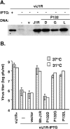
Transient complementation of viJ1R with various P132 mutant J1R proteins. (A) Immunoblot analysis of J1R mutant proteins. 293T cells at 31 or 37°C were infected with viJ1R in the presence (+) or absence (−) of IPTG and transfected with either vector DNA (v), WT J1R (J1R), or a P132 mutant J1R construct (P132D, P132G, or P132L). Extracts were harvested at 24 h p.i., resolved by SDS-15% PAGE, and analyzed by immunoblotting using an anti-J1R Ab (1:1,000). Only the J1R protein expression at 37°C is shown here. (B) Virus titer determinations by transient complementation assays. Aliquots of cell extracts harvested from 293T cells as described for panel A were used for plaque assays on BSC40 cells at either 31 or 37°C in the presence of 50 μM IPTG as described in Materials and Methods.
FIG. 6.
FIG. 6.
Coimmunoprecipitation of J1R protein with F10L, G7L, and A30L proteins from VV-infected cells. (A) Coimmunoprecipitation of J1R protein with an anti-HA Ab recognizing A30L protein in virus-infected cells. BSC-1 cells were infected with vT7LacOI or vA30iHA virus in the presence (+) of 50 μM IPTG. At 24 h p.i., cell extracts were prepared and incubated with the anti-HA antibody conjugated to agarose beads. The immunoprecipitated (IP) products were analyzed by electrophoresis on an SDS-10 to 20% Tricine gel, followed by immunoblotting using an anti-G7L Ab, an anti-J1R Ab, or an anti-HA MAb conjugated to HRP. (B) Coimmunoprecipitation of J1R protein with an anti-V5 Ab recognizing F10L in virus-infected cells. BSC-1 cells were infected with wild-type VV (WR) or vWT-F10V5 (WR containing a V5-tagged copy of the F10 protein) as described previously (33). At 24 h p.i., cell extracts were prepared for immunoprecipitation with the anti-V5 MAb conjugated to agarose beads and were analyzed as described for panel A by using either an anti-V5 Ab conjugated to HRP or an anti-G7L, anti-J1R, or anti-A30L Ab, as indicated. Numbers on the left correspond to molecular masses (in kilodaltons) of the marker proteins.
FIG. 7.
FIG. 7.
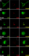
Intracellular localization of J1R, A30L, G7L, and F10L proteins in virus-infected cells by confocal microscopy. BSC40 cells were infected with viJ1R and transfected with either T7-tagged WT J1R (A, C, and E) or P132D mutant (B, D, and F) constructs. Cells were cultured in medium without IPTG for 24 h, fixed, permeabilized, and stained for viral proteins as shown in each panel. Insets in panel E show immunofluorescence of J1R and F10L proteins in virus-infected cells harvested at 8 h p.i.
FIG. 8.
FIG. 8.
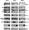
Immunoblots of J1R-associated viral proteins in virus-infected cells at different temperatures. BSC40 cells were infected with WT VV (wt), Cts45 (ts), or viJ1R in the presence (+J1) or absence (−J1) of 50 μM IPTG at an MOI of 5 PFU per cell and were cultured at 31, 37, or 39°C. At 24 h p.i., cells were harvested for immunoblot analysis with an anti-A30L, anti-G7L, anti-F10L, anti-A17L-N or -C, anti-J1R,or anti-A45R Ab. The anti-G7L Ab recognized the 42-kDa precursor protein and the 15-kDa C-terminal cleavage product, as described previously (31). A background band (bg) that was already present in mock-infected cell lysates was marked with an asterisk.

Similar articles

Cited by

References

    1. Almazan, F., D. C. Tscharke, and G. L. Smith. 2001. The vaccinia virus superoxide dismutase-like protein (A45R) is a virion component that is nonessential for virus replication. J. Virol. 75:7018-7029. - PMC - PubMed
    1. Bartel, P. L., and S. Fields. 1995. Analyzing protein-protein interactions using two-hybrid system. Methods Enzymol. 254:241-263. - PubMed
    1. Betakova, T., E. J. Wolffe, and B. Moss. 1999. Regulation of vaccinia virus morphogenesis: phosphorylation of the A14L and A17L membrane proteins and C-terminal truncation of the A17L protein are dependent on the F10L kinase. J. Virol. 73:3534-3543. - PMC - PubMed
    1. Blasco, R., and B. Moss. 1991. Extracellular vaccinia virus formation and cell-to-cell virus transmission are prevented by deletion of the gene encoding the 37,000-dalton outer envelope protein. J. Virol. 65:5910-5920. - PMC - PubMed
    1. Blasco, R., and B. Moss. 1992. Role of cell-associated enveloped vaccinia virus in cell-to-cell spread. J. Virol. 66:4170-4179. - PMC - PubMed

Publication types

LinkOut - more resources