. 2005 Jul 6;24(13):2265-83.
doi: 10.1038/sj.emboj.7600688. Epub 2005 Jun 9.

Essential role of Hrs in a recycling mechanism mediating functional resensitization of cell signaling

Affiliations

Essential role of Hrs in a recycling mechanism mediating functional resensitization of cell signaling

Aylin C Hanyaloglu et al. EMBO J. .

Abstract

Hepatocyte growth factor-regulated tyrosine kinase substrate (Hrs) is well known to terminate cell signaling by sorting activated receptors to the MVB/lysosomal pathway. Here we identify a distinct role of Hrs in promoting rapid recycling of endocytosed signaling receptors to the plasma membrane. This function of Hrs is specific for receptors that recycle in a sequence-directed manner, in contrast to default recycling by bulk membrane flow, and is distinguishable in several ways from previously identified membrane-trafficking functions of Hrs/Vps27p. In particular, Hrs function in sequence-directed recycling does not require other mammalian Class E gene products involved in MVB/lysosomal sorting, nor is receptor ubiquitination required. Mutational studies suggest that the VHS domain of Hrs plays an important role in sequence-directed recycling. Disrupting Hrs-dependent recycling prevented functional resensitization of the beta(2)-adrenergic receptor, converting the temporal profile of cell signaling by this prototypic G protein-coupled receptor from sustained to transient. These studies identify a novel function of Hrs in a cargo-specific recycling mechanism, which is critical to controlling functional activity of the largest known family of signaling receptors.


Figures
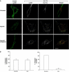
Figure 1ab
Figure 1ab
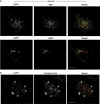
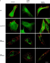
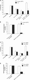
Hrs overexpression inhibits recycling of the β2ADR. (A) HeLa cells expressing Flag-β2ADR and either pcDNA3 (control) or myc-Hrs were fed with rabbit anti-Flag antibody to label the surface receptor prior to treatment with agonist isoproterenol (30 min, 10 μM). Cells were washed and treated with antagonist alprenolol (1 h, 10 μM) to allow for recycling and prevent further receptor internalization. Cells were fixed and stained for confocal microscopy with anti-myc antibodies as described in Materials and methods. Merged and unmerged images are shown for cells co-expressing Flag-β2ADR (green) and myc-Hrs (red). Yellow indicates colocalization. White bar=10 μm. (B) Cell surface receptor was measured by flow cytometry in HeLa cells co-expressing Flag-β2ADR and GFP together with pcDNA3 (control) or myc-Hrs. Cells were treated as for (A). Percentage of internalization refers to the fractional reduction of surface receptor in response to 30-min agonist exposure. Percentage of receptor recycled refers to the fractional recovery of surface receptor following agonist washout for 1 h. Data represent the mean±s.e.m. from three independent experiments. ***P<0.001.
Figure 1cd
Figure 1cd
(C) HeLa cells expressing Flag-β2ADR and myc-Hrs were fed with mouse anti-Flag antibody to label the surface receptor prior to treatment with agonist isoproterenol (30 min, 10 μM). Cells were washed and treated with antagonist alprenolol (1 h, 10 μM) to allow for recycling and prevent further receptor internalization. Cells were fixed and stained for confocal microscopy with anti-EEA-1-FITC or anti-LAMP1 antibodies as described in Materials and methods. Only cells treated with antagonist, following agonist washout, are shown. Merged and unmerged images are shown for cells co-expressing Flag-β2ADR (red), EEA-1 (green) or LAMP1 (green). Yellow indicates colocalization. White bar=10 μm. (D) HeLa cells expressing Flag-β2ADR were fed with mouse anti-Flag antibody to label the surface receptor prior to treatment with agonist isoproterenol (30 min, 10 μM). Cells were fixed and stained for confocal microscopy with rabbit anti-Hrs antibody. Merged and unmerged images are shown for cells co-expressing Flag-β2ADR (green) and Hrs (red). Yellow indicates colocalization. Arrows indicate examples of colocalized vesicles. White bar=10 μm.
Figure 2ab
Figure 2ab
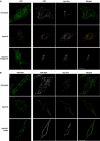
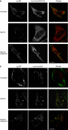
Hrs-dependent recycling is cargo specific. (A, B) HeLa cells expressing Flag-μOR or Flag-V2R 362T with either pcDNA3 (control) or myc-Hrs were fed with anti-Flag antibody to label surface receptor prior to treatment with agonist etorphine for μOR and arginine vasopressin for V2R 362T (30 min, 10 μM). Cells were washed and treated with antagonist naloxone (1 h, 10 μM) for μOR to allow for recycling and prevent further receptor internalization. V2R 362T-expressing cells were incubated in media only following agonist washout.
Figure 2cd
Figure 2cd
(C) Cell surface receptor was measured by flow cytometry in HeLa cells co-expressing Flag-μOR or Flag-V2R 362T, together with pcDNA3 (control) or myc-Hrs. Cells were treated as for (A). (D) HeLa cells expressing Flag-V2R 362/β2ADR with either pcDNA3 (control) or myc-Hrs were fed with anti-Flag antibody to label surface receptor prior to treatment with agonist arginine vasopressin (30 min, 10 μM). Cells were washed and incubated in media only for 1 h. Cell surface receptor was measured in HeLa cells co-expressing Flag-V2R 362/β2ADR together with pcDNA3 (control) or myc-Hrs. Cells were treated and measured as in (C). For all images, merged and unmerged images are shown for cells co-expressing receptor (green) and myc-Hrs (red). Yellow indicates colocalization. White bar=10 μm. For all flow cytometry experiments, a minimum of 10 000 cells were counted. Data represent the mean±s.e.m. from three independent experiments. **P<0.01.
Figure 3
Figure 3
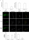
Hrs-dependent recycling does not require receptor ubiquitination. (A) HeLa cells expressing Flag-β2ADR 0K with either pcDNA3 (control) or myc-Hrs were treated and stained for confocal microscopy as described in Figure 1A. White bar=10 μm. (B) Cell surface receptor was measured by flow cytometry in HeLa cells co-expressing either Flag-β2ADR or Flag-β2ADR 0 K and either pcDNA3 (control) or myc-Hrs. A minimum of 10 000 cells were counted. Data represent the mean±s.e.m. of four independent experiments. **P<0.01.
Figure 4abc
Figure 4abc
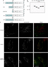
The VHS domain of Hrs is required for its effect on β2ADR recycling. (A) Schematic of the domain organization of full-length Hrs and the truncated mutants used as indicated. (B) Western blot to verify similar expression levels of all proteins with an anti-myc antibody in transfected HeLa lysates. (C–F) HeLa cells were transfected with Flag-β2ADR and myc-Hrs truncated mutants as indicated. Cells were treated and stained for receptor and myc-Hrs as for Figure 1A. White bar=10 μm.
Figure 4de
Figure 4de
(C–F) HeLa cells were transfected with Flag-β2ADR and myc-Hrs truncated mutants as indicated. Cells were treated and stained for receptor and myc-Hrs as for Figure 1A. White bar=10 μm.
Figure 4fg
Figure 4fg
(C–F) HeLa cells were transfected with Flag-β2ADR and myc-Hrs truncated mutants as indicated. Cells were treated and stained for receptor and myc-Hrs as for Figure 1A. White bar=10 μm. (G) Quantification of β2ADR internalization and recycling by flow cytometry in the absence and presence of WT and mutant myc-Hrs constructs. Data shown are the mean±s.e.m. of five separate experiments.
Figure 5acd
Figure 5acd
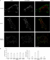
Hrs, but not Tsg101 or Vps4, is essential for the sequence-directed recycling mechanism. (A) Western blot of total cellular levels of Hrs in HeLa cells following transfection with either control (nonsilencing) siRNA or Hrs siRNA. Lysates were also blotted for GapDH to test the specificity of knockdown. (C) Surface receptor was measured by flow cytometry in HeLa cells co-expressing Flag-β2ADR, Flag-μOR, or Flag-V2R 362T, and either control (non-silencing) siRNA or Hrs siRNA. Cells were treated as in Figures 1B and 2C. A minimum of 10 000 cells were counted. Data represent the mean±s.e.m. from three independent experiments. *P<0.05. (D) Tsg101 levels in cells transfected with either control (nonsilencing) siRNA or Tsg101 siRNA were assessed by Western blotting. Lysates were also blotted for GapDH to test the specificity of knockdown. HeLa cells expressing Flag-β2ADR with control (nonsilencing) siRNA or Tsg101 siRNA were assessed for recycling (as in (B)) by flow cytometry. All flow cytometry results shown are the mean±s.e.m. of three independent experiments.
Figure 5b
Figure 5b
(B) HeLa cells expressing Flag-β2ADR and either control (nonsilencing) siRNA or Hrs siRNA were fed with anti-Flag antibody to label surface receptor prior to treatment with agonist isoproterenol (30 min, 10 μM). Cells were washed and treated with antagonist alprenolol (1 h, 10 μM) to allow for recycling and prevent further receptor internalization. Cells were fixed and stained for EEA-1 as described in Materials and methods. Merged and unmerged images are shown. Receptor is in red, EEA-1 is in green, and yellow indicates colocalization. White bar=10 μm.
Figure 5e
Figure 5e
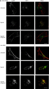
(E) Cells transfected with Flag-β2ADR and either GFP, WT Vps4/GFP, Vps4 E228Q/GFP or Vps4 K172Q/GFP were treated as in (B) and assessed by confocal microscopy. White bar=10 μm.
Figure 6
Figure 6
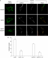
Manipulation of Hrs prevents functional resensitization of β2ADR signaling. (A, B) HeLa cells co-expressing Flag-β2ADR and either pcDNA3 or myc-Hrs, or (C, D) HeLa cells transfected with Flag-β2ADR and either control (nonsilencing) siRNA or Hrs siRNA were incubated with 0.5 mM IBMX prior to agonist treatment (isoproterenol (Iso), 10 μM) as indicated. Cells were measured for whole-cell cAMP production as described in Materials and methods. For (A) and (C), the concentration of cAMP was normalized to 90 s Iso treatment. (B) and (D) show the agonist-dependent increase from the first and second challenges of agonist. All results shown are the mean±s.e.m. of four independent experiments. *P<0.05.

Similar articles

Cited by

References

    1. Babst M, Odorizzi G, Estepa EJ, Emr SD (2000) Mammalian tumor susceptibility gene 101 (TSG101) and the yeast homologue, Vps23p, both function in late endosomal trafficking. Traffic 1: 248–258 - PubMed
    1. Babst M, Sato TK, Banta LM, Emr SD (1997) Endosomal transport function in yeast requires a novel AAA-type ATPase, Vps4p. EMBO J 16: 1820–1831 - PMC - PubMed
    1. Bache KG, Raiborg C, Mehlum A, Stenmark H (2003) STAM and Hrs are subunits of a multivalent ubiquitin-binding complex on early endosomes. J Biol Chem 278: 12513–12521 - PubMed
    1. Bilodeau PS, Urbanowski JL, Winistorfer SC, Piper RC (2002) The Vps27p Hse1p complex binds ubiquitin and mediates endosomal protein sorting. Nat Cell Biol 4: 534–539 - PubMed
    1. Bishop N, Horman A, Woodman P (2002) Mammalian class E vps proteins recognize ubiquitin and act in the removal of endosomal protein–ubiquitin conjugates. J Cell Biol 157: 91–101 - PMC - PubMed

Publication types

MeSH terms

Substances

LinkOut - more resources