. 2005 Jul 6;24(13):2379-90.
doi: 10.1038/sj.emboj.7600711. Epub 2005 Jun 9.

Altered nucleosome occupancy and histone H3K4 methylation in response to 'transcriptional stress'

Affiliations

Altered nucleosome occupancy and histone H3K4 methylation in response to 'transcriptional stress'

Lian Zhang et al. EMBO J. .

Abstract

We report that under 'transcriptional stress' in budding yeast, when most pol II activity is acutely inhibited, rapid deposition of nucleosomes occurs within genes, particularly at 3' positions. Whereas histone H3K4 trimethylation normally marks 5' ends of highly transcribed genes, under 'transcriptional stress' induced by 6-azauracil (6-AU) and inactivation of pol II, TFIIE or CTD kinases Kin28 and Ctk1, this mark shifted to the 3' end of the TEF1 gene. H3K4Me3 at 3' positions was dynamic and could be rapidly removed when transcription recovered. Set1 and Chd1 are required for H3K4 trimethylation at 3' positions when transcription is inhibited by 6-AU. Furthermore, Deltachd1 suppressed the growth defect of Deltaset1. We suggest that a 'transcriptional stress' signal sensed through Set1, Chd1, and possibly other factors, causes H3K4 hypermethylation of newly deposited nucleosomes at downstream positions within a gene. This response identifies a new role for H3K4 trimethylation at the 3' end of the gene, as a chromatin mark associated with impaired pol II transcription.


Figures

Figure 1
Figure 1
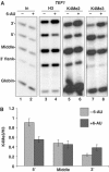
Inhibition of transcription by 6-AU alters the 5′-3′ distribution of histones within a gene. (A) Treatment with 6-AU results in an increase in histone occupancy at the 3′ end and a modest reduction at the 5′ end of TEF1. WT cells (BY4741) were treated without (−) or with (+) 6-AU. ChIP analysis was carried out using anti-pol II, -H2B, -H3 and -H4, and 32P-labeled PCR products of 5′, middle and 3′ region on TEF1 gene are shown. Mitochondrial COXIII, telomere VIR (TEL) and globin are controls. Note that the COXIII background signal is low relative to input chromatin (in). Ratios of ChIP signal +6-AU/−6-AU normalized to input are graphed in the bottom panel with mean and standard deviations from three different WT strains: BY4741, DY103 and HTB1. (B) 6-AU specifically affects histone occupancy on transcribed sequences. 6-AU had little effect on H3 occupancy in the nontranscribed gene GAL10 ORF (lanes 5 and 6) or telomere. 3′/5′ ratios of normalized ChIP signals are shown. (C) The 5′-3′ redistribution of pol II and histones caused by 6-AU is partially reversed by guanine. DY108 (rpb2-10Δdst1) was treated with 6-AU or 6-AU+guanine. Note that guanine restored pol II density (lanes 5 and 6) and reversed the increase in H3 occupancy at the TEF1 3′ end (lanes 8 and 9).
Figure 2
Figure 2
6-AU-induced transcription inhibition increases histone occupancy within the PMA1 and ASC1 genes. DY103 cells were treated without (−) or with (+) 6-AU. ChIP analysis was carried out using anti-pol II, -H2B, -H3 or -H4, and PCR products of 5′, middle and 3′ region on PMA1(A) or ASC1(B) are shown. TEL and globin are controls. Ratios of ChIP signals normalized to input and the globin control + and −6-AU were determined. Means and standard deviation from three PCR determinations are shown.
Figure 3
Figure 3
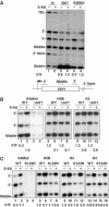
Inhibition of transcription by 6-AU increases H3K4Me3 at the 3′ end of TEF1 gene. (A) DY103 cells were treated without (−) and with (+) 6-AU for 1 h and ChIP analysis was carried out using anti-H3, -H3K4Me2 and -H3K4Me3. 32P-labeled PCR products of 5′, middle and 3′ region in TEF1 gene are shown. Note that H3K4Me3 crosslinking decreased at the 5′ end and increased at the 3′ end (lanes 7 and 8) whereas H3K4Me2 did not decrease at the 5′ end with 6-AU treatment (lanes 5 and 6). (B) 6-AU reduces K4 trimethylation of H3 at the 5′ end and increases it at the 3′ end of TEF1. ChIP signals for H3K4Me3 relative to total H3 were graphed from seven independent experiments: three in DY103 and one each in BY4741, W303mycSet1, W1588-4C and HTB1.
Figure 4
Figure 4
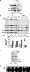
5′-3′ histone redistribution in 6-AU is independent of H3K4 methylation. (A) Set1 is localized throughout the TEF1 gene − and +6-AU. Set1myc W303 cells were treated without (−) or with (+) 6-AU and analyzed by ChIP using anti-myc and -H3K4Me3. A small enrichment of Set1 within TEF1 relative to the 3′ flank and telomere was observed. 3′/5′ ratios of normalized ChIP signals are shown. Note that stronger globin signals that appear in lanes 3 and 4 are due to larger amount of samples analyzed. (B) Set1 is not required for the 5′-3′ repositioning of histones on TEF1 in 6-AU. Isogenic WT (DY103) and Δset1 (DBY531) strains were treated without (−) or with (+) 6-AU, and analyzed by ChIP with the indicated antibodies. H2B was immunoprecipitated with antibody against N-terminal Flag epitope. Although there is no methylated H3K4 in Δset1 (lanes 3 and 4), 5′-3′ redistribution of H2B and H3 still occurred as shown by the increased 3′/5′ ratios of ChIP signals with 6-AU treatment (lanes 7, 8, 11 and 12). (C) H2B K123 ubiquitylation is not required for the 5′-3′ repositioning of histones on TEF1 with 6-AU treatment. Isogenic HTB1 and htb1K123R cells were treated without (−) or with (+) 6-AU and analyzed by ChIP with antibodies as indicated. 3′/5′ ratios of ChIP signals show that the 5′-3′ redistribution of histones H2B, H3 and H4 with 6-AU treatment is approximately equivalent in WT and htb1K123R although the mutation abolished methylation of H3K4 (lanes 3 and 4).
Figure 5
Figure 5
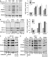
Chd1 is required for 6-AU-induced H3K4 trimethylation at the 3′ end of TEF1 gene. (A) Increased H3K4 trimethylation at the TEF1 3′ end with 6-AU treatment is abolished in Δisw1Δisw2Δchd1 triple mutant (YTT227). WT (W1588-4C) and mutant (YTT227) cells were treated without (−) or with (+) 6-AU and ChIP analysis was carried out with anti-H3 and -H3K4Me3. 3′/5′ ratio of normalized ChIP signals is shown. The loss of H3 at the 5′ end and the increase of H3K4Me3 at the 3′ end in 6-AU are both inhibited in the mutant (lanes 3, 4, 7 and 8). (B) Δchd1 but not Δisw1 or Δisw2 disrupts the chromatin response to 6-AU. ChIP analysis was carried out as in panel A with cells with single deletion of ISW1, ISW2 or CHD1. The enhanced H3K4 trimethylation normalized to total H3 at the 3′ end in 6-AU is abolished only in Δchd1 (lanes 16 and 17 and graph). Results from three determinations are summarized in the graph. (C) Set1 localization is independent of Chd1. Set1 localization in BY4741 (WT) and Δchd1 cells was determined by ChIP analysis with monoclonal anti-Set1. 3′/5′ ratios of normalized ChIP signals are shown. (D) Δchd1 suppresses the growth defect of Δset1. Dilution series of isogenic strains WT (BY4741), Δchd1, Δset1 (DBY804) and Δset1Δchd1 (DBY805) were plated on YPD, −Ura, and −Ura+100 μg/ml 6-AU at 25°C.
Figure 6
Figure 6
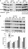
‘Transcriptional stress' causes chromatin reconfiguration of transcribed sequences. (A) Glucose starvation reduced pol II and increased histone occupancy in the TEF1 ORF but not in the nontranscribed 5′ or 3′ flank regions. DY103 cells were shifted from YPD (2% glucose (+)) to YP lacking glucose (−) for 30 min, and analyzed by ChIP. Mean ratios of normalized ChIP signals from − and +glucose were quantified and are indicated in the graph (n=3). (B) Heat shock rapidly stimulates histone deposition on TEF1 but eviction from HSP104. WT cells (DBY175) in YPD were shifted from 25 to 37°C for 2 and 4 min and then analyzed by ChIP. Mean ChIP signals for the TEF1 3′ end relative to values at 25°C are indicated in the graph (n=3). (C) TFIIE inactivation stimulates H3 deposition and H3K4 trimethylation on TEF1. tfa1-21 cells in YPD were shifted from 25 to 37°C for 1 h and analyzed by ChIP. 3′/5′ ratios of normalized ChIP signals for H3 before and after Tfa1 inactivation are 1.4 and 2.9 (lanes 5 and 6) indicating a 5′:3′ shift in histone occupancy. The ratios of H3K4Me3/H3 ChIP signals for the middle and 3′ end of TEF1 gene are shown. (D) Histone deposition and H3K4 trimethylation in response to inactivation of Rpb1 are reversible. rpb1-1 (DBY120) cells were shifted from 25 to 37°C for 1 h and then recovered at 25°C for 1 h. The ratios of H3K4Me3/H3 ChIP signals for the middle and 3′ end of TEF1 gene are shown. 3′/5′ ratios of normalized H3 ChIP signals for lanes 10–12 are 1.5, 2.4 and 1.0.
Figure 7
Figure 7
Inactivation of CTD kinases causes chromatin reconfiguration of transcribed sequences. (A) Ctk1 deletion causes a shift of H3K4Me3 toward the 3′ end of TEF1. Isogenic WT (BY4741) and Δctk1 cells were treated with 6-AU and analyzed by ChIP. Ser-2P is an antibody against Ser2 phosphorylated CTD. The ratios of H3K4Me3/H3 ChIP signals for the middle and 3′ end are shown. Note the elevated H3K4Me3 level at the 3′ end of TEF1 in untreated Δctk1 cells (lane 15) mimicking the effect of 6-AU on WT cells (lane 14). (B) Histone deposition and H3K4 trimethylation on TEF1 are elevated with Kin28 inactivation. kin28 ts3 cells in YPD were shifted from 25 to 37°C for 1 h. Loss of pol II (lanes 3 and 4) was associated with increased histone occupancy (lanes 5–10) particularly in the middle and 3′ regions of the TEF1 ORF relative to the 3′ flank. Note the elevated H3K4Me3 relative to total H3, particularly at the middle and 3′ end at 37°C (lanes 7, 8, 11 and 12 and graph). The graph shows mean ratios of normalized ChIP signals at 37°C relative to 25°C (n=3).
Figure 8
Figure 8
Model for chromatin reconfiguration in response to ‘transcriptional stress'. Impaired pol II transcription causes preferential recruitment of nucleosomes to promoter distal regions of the transcription unit, which can be accompanied by increased H3K4 trimethylation indicated with stars. This chromatin reconfiguration could be mediated by Chd1 and yet unidentified assembly factor(s). Inhibition of transcription by 6-AU also caused net loss of H3 and H3K4 trimethylation at the 5′ end.

Similar articles

Cited by

References

    1. Adkins MW, Howar SR, Tyler JK (2004) Chromatin disassembly mediated by the histone chaperone Asf1 is essential for transcriptional activation of the yeast PHO5 and PHO8 genes. Mol Cell 14: 657–666 - PubMed
    1. Ahmad K, Henikoff S (2002) The histone variant H3.3 marks active chromatin by replication-independent nucleosome assembly. Mol Cell 9: 1191–1200 - PubMed
    1. Alen C, Kent NA, Jones HS, O'Sullivan J, Aranda A, Proudfoot NJ (2002) A role for chromatin remodeling in transcriptional termination by RNA polymerase II. Mol Cell 10: 1441–1452 - PubMed
    1. Almer A, Rudolph H, Hinnen A, Horz W (1986) Removal of positioned nucleosomes from the yeast PHO5 promoter upon PHO5 induction releases additional upstream activating DNA elements. EMBO J 5: 2689–2696 - PMC - PubMed
    1. Boeger H, Griesenbeck J, Strattan JS, Kornberg RD (2003) Nucleosomes unfold completely at a transcriptionally active promoter. Mol Cell 11: 1587–1598 - PubMed

Publication types

MeSH terms