. 2005 Aug;16(8):3692-704.
doi: 10.1091/mbc.e05-01-0020. Epub 2005 Jun 8.

Phosphatidylinositol-4-kinase type II alpha is a component of adaptor protein-3-derived vesicles

Affiliations

Phosphatidylinositol-4-kinase type II alpha is a component of adaptor protein-3-derived vesicles

Gloria Salazar et al. Mol Biol Cell. 2005 Aug.

Abstract

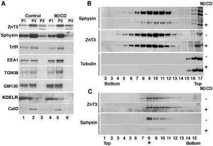
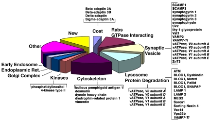
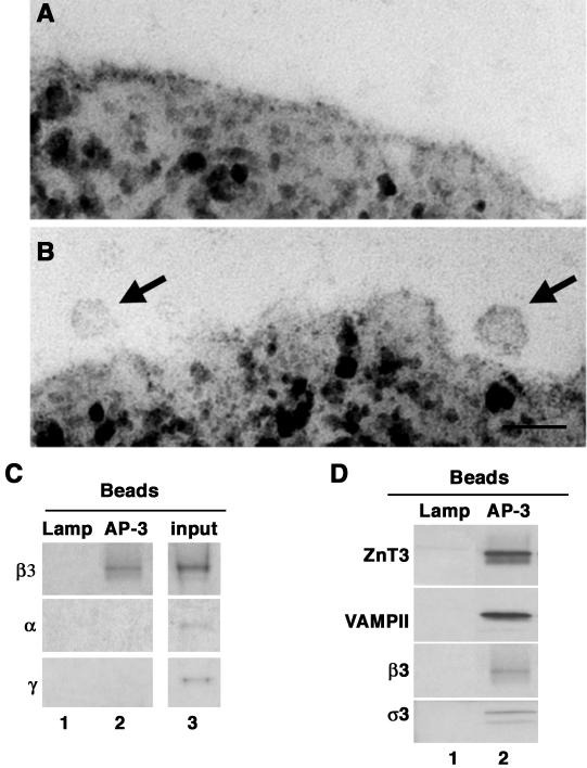
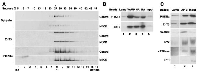
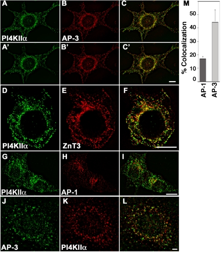
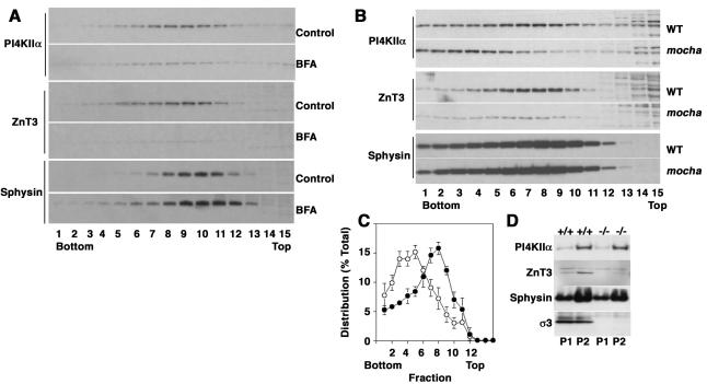
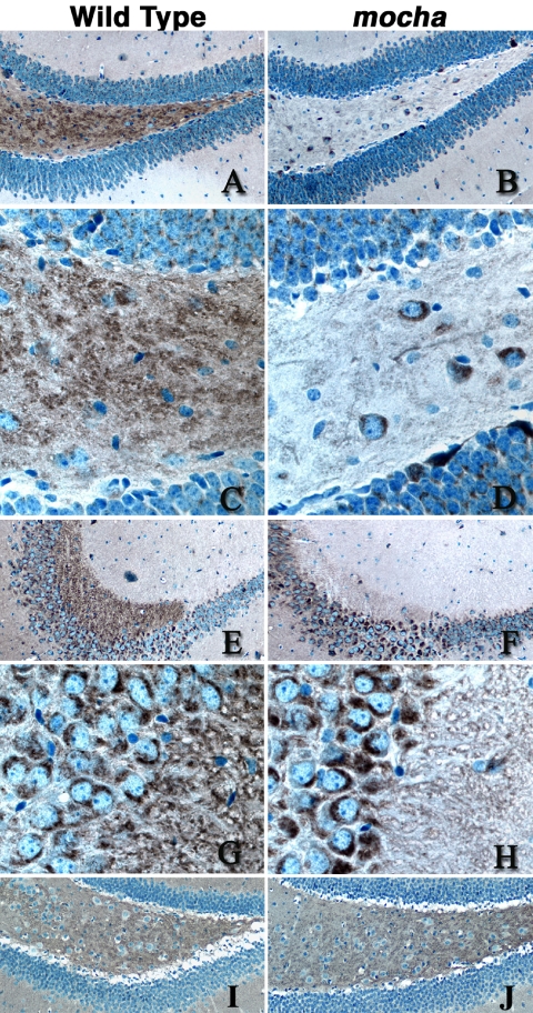
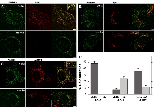
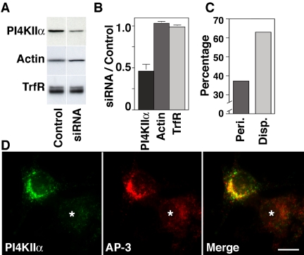
A membrane fraction enriched in vesicles containing the adaptor protein (AP) -3 cargo zinc transporter 3 was generated from PC12 cells and was used to identify new components of these organelles by mass spectrometry. Proteins prominently represented in the fraction included AP-3 subunits, synaptic vesicle proteins, and lysosomal proteins known to be sorted in an AP-3-dependent way or to interact genetically with AP-3. A protein enriched in this fraction was phosphatidylinositol-4-kinase type IIalpha (PI4KIIalpha). Biochemical, pharmacological, and morphological analyses supported the presence of PI4KIIalpha in AP-3-positive organelles. Furthermore, the subcellular localization of PI4KIIalpha was altered in cells from AP-3-deficient mocha mutant mice. The PI4KIIalpha normally present both in perinuclear and peripheral organelles was substantially decreased in the peripheral membranes of AP-3-deficient mocha fibroblasts. In addition, as is the case for other proteins sorted in an AP-3-dependent way, PI4KIIalpha content was strongly reduced in nerve terminals of mocha hippocampal mossy fibers. The functional relationship between AP-3 and PI4KIIalpha was further explored by PI4KIIalpha knockdown experiments. Reduction of the cellular content of PI4KIIalpha strongly decreased the punctate distribution of AP-3 observed in PC12 cells. These results indicate that PI4KIIalpha is present on AP-3 organelles where it regulates AP-3 function.


Figures

Figure 1.
Figure 1.
Isolation of a membrane fraction enriched in AP-3-derived vesicles. PC12 cells expressing ZnT3-HA were treated in the absence or presence of MβCD to interfere with SLMV biogenesis from plasma membrane. Plasma membrane-derived SLMV were monitored with synaptophysin and AP-3-derived vesicles with ZnT3. Cell homogenates were fractionated by differential centrifugation (A) to generate P1 (lanes 1 and 4), P2 (lanes 2 and 5), and P3 (lanes 3 and 6) membranes. P3 membranes contained the synaptic vesicle markers synaptophysin (Sphysin) and ZnT3, yet they were greatly depleted for other contaminating membranes. Cholesterol depletion did not affect the overall fractionation of Golgi, endosome, and lysosomes; however, it selectively decreased the synaptophysin content in P3 membranes (compare lanes 3 and 6). (B) Glycerol velocity gradient fractions probed with synaptophysin, ZnT3, and tubulin antibodies. The ZnT3-enriched peak is found in fractions 8–11, whereas the bulk of the cytosol, assessed with tubulin antibodies, remains in the top fractions (15–17). MβCD treatment decreases the synaptophysin content in SLMV fractions while sparing ZnT3. (C) Vesicles from glycerol fractions 8–11 were floated to equilibrium in sucrose gradients. ZnT3 and synaptophysin equilibrate at 26.4 ± 0.76% sucrose. MβCD treatment selectively decreases the synaptophysin content in SLMV without affecting ZnT3 (n = 3).
Figure 2.
Figure 2.
Classification of the ZnT3-enriched vesicle proteome. Relevant proteins for the categories described in Supplemental Table 1 are depicted in brackets. Entries in italics correspond to proteins described in both synaptic vesicles and lysosomes. For a complete list of all the proteins identified in the different categories refer to Supplemental Table 1.
Figure 3.
Figure 3.
Identification of AP-3-derived vesicles. Vesicle fractions isolated by sequential glycerol and sucrose sedimentation were immunomagnetically isolated with beads coated with either a luminal human-specific Lamp1 antibody or AP-1 γ as controls, or coated with antibodies against AP-3 δ adaptin. Beads were analyzed by transmission electron microscopy (A and B) or immunoblot (C and D). AP-1 γ beads did not bind membranes (A). AP-3 δ beads bind 40-nm vesicles (see arrows) (B). Bar, 55 nm. (C) AP-3 vesicles are free of AP-1 or AP-2 adaptins. β3 Adaptin blots were exposed for 3 min. α and γ Adaptin blots were exposed overnight. (D) AP-3-coated vesicles contain synaptic vesicle markers. Membranes bound to beads coated with antibodies against the indicated antigens were resolved in SDS-PAGE and analyzed by immunoblot with antibodies against synaptic vesicle proteins (ZnT3 and VAMP II) and the AP-3 subunits β3 and σ3. Control immunoisolations in C and D, lane 1, were performed with LAMP antibodies. Input corresponds to 10%.
Figure 4.
Figure 4.
PI4KIIα is present in AP-3-derived vesicles. (A) PC12 cells expressing ZnT3-HA were treated in the absence or presence of MβCD, and ZnT3-enriched fractions were isolated by sequential velocity and isopycnic sedimentation. Fractions were analyzed by immunoblot with antibodies against synaptophysin, ZnT3, and PI4KIIα. The kinase comigrated with ZnT3. The abundance of this kinase was not affected by MβCD treatment. (B) PI4KIIα is present in vesicles containing synaptic vesicle markers. Vesicles were isolated with magnetic beads decorated with control (luminal domain of LAMP, lane 1), VAMP II (lane 2), or HA (lanes 3 and 4) antibodies to recognize the tag in ZnT3. Bound vesicle content was analyzed by immunoblot with kinase and ZnT3 antibodies. (C) PI4KIIα is present in AP-3-coated vesicles. Vesicles were bound to beads coated with either a luminal human-specific Lamp I antibody as negative control, or coated with antibodies against AP-3 δ adaptin. PI4KIIα is present in AP-3-coated vesicles together with synaptic vesicle markers (ZnT3, VAMPII, SV2, and vacuolar ATPase). No contamination with transferrin receptor was detected in the AP-3-coated vesicles. Transferrin receptor blot was exposed 20 times longer that the other blots. Vesicles used in B and C were isolated by velocity sedimentation from untreated cells. Inputs represent 10%.
Figure 5.
Figure 5.
PI4KIIα colocalizes predominantly with AP-3 and its cargo ZnT3. PC12 cells expressing HA-tagged ZnT3 were costained with antibodies against PI4KIIα (A and A′, D, and G) and either AP-3 δ adaptin (B and B′), HA antibodies to detect ZnT3 (E), or AP-1 γ adaptin (H). Similarly, primary mouse skin fibroblasts were stained with AP-3 δ and PI4KIIα antibodies (J–L). Cells were imaged by wide field deconvolution microscopy. (M) Colocalization of PI4KIIα and the adaptor complexes was determined by MetaMorph. The kinase extensively colocalizes with AP-3 and ZnT3 and to a lower extent with AP-1. A–C to A′–C′ series represent two optical planes taken every 0.75 μm. Bar, 6 μm.
Figure 6.
Figure 6.
PI4KIIα is targeted by AP-3-dependent mechanisms. (A) PC12 cells were treated either in the absence or presence of brefeldin A to deplete AP-3-generated vesicles and were subsequently fractionated in glycerol velocity gradients. Synaptophysin levels were not affected by brefeldin A; in contrast, ZnT3 and PI4KIIα levels were dramatically reduced (n = 2). (B) High-speed supernatants (S2) from wild-type and mocha brain homogenates were fractionated in glycerol gradients to resolve synaptic vesicles and AP-3 vesicles. Synaptic vesicle antigen levels across gradients were determined by immunoblot using antibodies against PI4KIIα, ZnT3, and synaptophysin (Sphysin). PI4KIIα and ZnT3 sedimentation pattern and the antigen content in membranes were specifically altered in mocha brain vesicles. (C) Depicts the normalized content distribution of PI4KIIα (n = 4). Closed circles represent wild-type membranes, and open circles correspond to mocha vesicles. (D) Distribution of synaptic vesicle antigens in P1 and P2 membranes from wild-type (+/+) and mocha brain (–/–).
Figure 7.
Figure 7.
PI4KIIα content decreases in mocha hippocampal mossy fiber nerve terminals. Wild-type (A, C, E, G, and I) and mocha (B, D, F, H, and J) hippocampal brains sections were stained with antibodies against PI4KIIα (A–H), and VAMP II (I–J). Immunocomplexes were detected with the ABC peroxidase reagent and DAB deposition. (A–D) Representative images of the dentate gyrus and hilus at low (A and B, 20×) and high magnification (C and D, 63×), whereas E–H correspond to the CA3 region of the hippocampus acquired at low (E–F) or high power (G–H). Note the reduction of PI4KIIα immunoreactivity in the hilus mossy fibers and CA3 mossy fiber synaptic terminals. Images are representative of sections obtained from four wild-type and mocha brains simultaneously stained in three independent experiments using either of two PI4KIIα antibodies.
Figure 8.
Figure 8.
PI4KIIα redistributes to perinuclear compartments in the absence of AP-3. Mocha mouse fibroblasts transduced with retroviruses carrying the δ subunit (delta) or without insert (mocha, mh) were double labeled with antibodies against PI4KIIα (A–D) and one of the following antigens: AP-3 δ adaptin (A), AP-1 γ adaptin (B), and the AP-3 cargo LAMP1 (C). Cells were imaged by wide field deconvolution microscopy, and the extent of colocalization was determined in at least 10 randomly selected cells collected from two independent experiments for each condition (D). In the absence of AP-3 (mocha, mh), PI4KIIα redistributes to perinuclear compartments increasing it colocalization with AP-1 and decreasing its localization with the AP-3 cargo LAMP-1. Bars, 5 μm.
Figure 9.
Figure 9.
PI4KIIα siRNA alters the subcellular distribution of AP-3 complexes. (A) Rat-specific PI4KIIα siRNA and control oligonucleotide-transfected PC12 cells were analyzed by immunoblot with antibodies against PI4KIIα, actin, and transferrin receptor (TrfR). (B) PI4KIIα siRNA selectively reduced kinase content by 50% of control values (n = 8). (C and D) Rat-specific PI4KIIα siRNA-treated PC12 cells were stained with antibodies against PI4KIIα and AP-3 delta subunit and examined by immunomicroscopy. Kinase down-regulation was observed only in PI4KIIα siRNA-transfected cells (asterisk). Reduction of the cell PI4KIIα levels resulted in disappearance of AP-3 from perinuclear (peri.) membranous compartments adopting a disperse appearance (disp.). (C) Phenotypes were scored in 113 cells collected in three independent experiments. Bar, 5 μm.
Figure 10.
Figure 10.
Overexpression of PI4KIIα increases the recovery of AP-3-derived vesicles. PC12 clone 4 cells expressing ZnT3 were transfected with GFP-tagged PI4KIIα. (A) Double transfected clonal cells expressing PI4KIIα-GFP and similar amounts of ZnT3 and endogenous PI4KIIα were selected and further analyzed by subcellular fractionation. (B) Cells were fractionated by differential sedimentation to obtain a P3 fraction. Expression of PI4KIIα shifts ZnT3, VAMPII, and β3 adaptin from endosome-containing P2 fractions to P3 membranes, which are enriched in small vesicles, without obvious changes in the sedimentation pattern of the transferrin receptor (TrfR). (C) P3 membranes contained in S2 fractions were fractionated by glycerol velocity sedimentation. Fractions were probed with antibodies against PI4KIIα, ZnT3, and AP-3 β3 adaptin. Kinase expression increases the content of synaptic vesicle markers and AP-3 in microvesicle fractions (n = 3). All gradients were probed with antibodies against tubulin to confirm equal loading.

Similar articles

Cited by

References

    1. Advani, R. J., Yang, B., Prekeris, R., Lee, K. C., Klumperman, J., and Scheller, R. H. (1999). VAMP-7 mediates vesicular transport from endosomes to lysosomes. J. Cell Biol. 146, 765–776. - PMC - PubMed
    1. Balla, A., Tuymetova, G., Barshishat, M., Geiszt, M., and Balla, T. (2002). Characterization of type II phosphatidylinositol 4-kinase isoforms reveals association of the enzymes with endosomal vesicular compartments. J. Biol. Chem. 277, 20041–20050. - PubMed
    1. Barlow, C., et al. (2000). ATM is a cytoplasmic protein in mouse brain required to prevent lysosomal accumulation. Proc. Natl. Acad. Sci. USA 97, 871–876. - PMC - PubMed
    1. Barylko, B., Gerber, S. H., Binns, D. D., Grichine, N., Khvotchev, M., Sudhof, T. C., and Albanesi, J. P. (2001). A novel family of phosphatidylinositol 4-kinases conserved from yeast to humans. J. Biol. Chem. 276, 7705–7708. - PubMed
    1. Benson, K. F., et al. (2003). Mutations associated with neutropenia in dogs and humans disrupt intracellular transport of neutrophil elastase. Nat. Genet. 35, 90–96. - PubMed

Publication types

MeSH terms

Substances

LinkOut - more resources