

doi: 10.1073/pnas.0503596102.
Epub 2005 Jun 6.
Robust hepatitis C virus infection in vitro
Affiliations
- PMID: 15939869
- PMCID: PMC1166622
- DOI: 10.1073/pnas.0503596102
Item in Clipboard
Robust hepatitis C virus infection in vitro
Proc Natl Acad Sci U S A.
.
Abstract
The absence of a robust cell culture model of hepatitis C virus (HCV) infection has severely limited analysis of the HCV life cycle and the development of effective antivirals and vaccines. Here we report the establishment of a simple yet robust HCV cell culture infection system based on the HCV JFH-1 molecular clone and Huh-7-derived cell lines that allows the production of virus that can be efficiently propagated in tissue culture. This system provides a powerful tool for the analysis of host-virus interactions that should facilitate the discovery of antiviral drugs and vaccines for this important human pathogen.
Figures
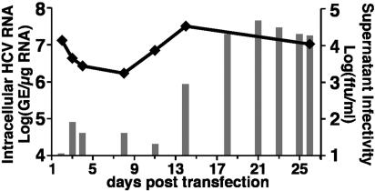
Production of infectious HCV after transfection of genomic JFH-1 RNA. Ten micrograms of in vitro transcribed JFH-1 RNA was electroporated into 4 × 10⁶ Huh-7.5.1 cells. Transfected cells and supernatant were harvested at the indicated time points posttransfection. Intracellular HCV RNA was analyzed by RT-QPCR and displayed as genome equivalents (GE)/μg total RNA (line). Supernatant infectivity titers were determined in naïve Huh-7.5.1 cells and are expressed as ffu/ml (bars).
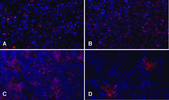
Detection of infected cells by NS5A immunofluorescence. (Upper) Immunofluorescent detection of NS5A in transfected cells: (A) day 5 and (B) day 24 posttransfection. (Lower) Infectivity titration of transfected cell supernatant on naïve Huh-7.5.1 cells; (C) undiluted supernatant; (D) 10-fold diluted supernatant. NS5A staining in red; nuclei stained with Hoechst (blue) (×50).
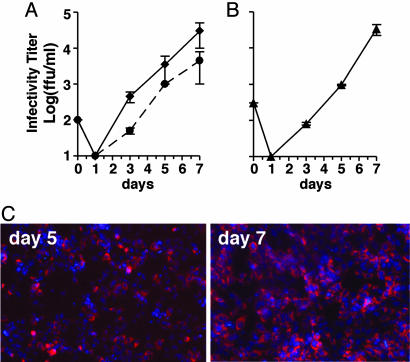
HCV infection kinetics and passage in tissue culture cells. Naïve Huh 7.5.1 cells were inoculated with culture supernatants at an moi of 0.01. Supernatants from the inoculated cells were collected at the indicated times p.i. and evaluated for infectivity (ffu/ml). Data represent the average of two or more experiments with error bars. (A) Huh-7.5.1 cells inoculated with supernatant harvested at day 19 after transfection of Huh-7.5.1 cells with JFH-1 genomic RNA by electroporation (dashed line) or day 24 after lipofection (solid line). (B) Huh-7.5.1 cells inoculated with supernatant collected at day 5 from the infection shown as a solid line in A; (C) Increasing NS5A immunostaining in Huh-7.5.1 cells between days 5 and 7 p.i. in the experiment shown in B.
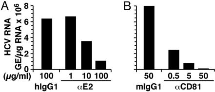
Inhibition of HCV infection by anti-E2 and anti-CD81 antibodies. (A) JFH-1 virus was preincubated with the indicated concentrations of anti-E2 antibody or irrelevant human IgG1 antibody for 1 h at 37°C before inoculating Huh-7.5.1 cells. Total cellular RNA was analyzed by RT-QPCR at day 3 p.i. (B) Huh-7.5.1 cells were preincubated with the indicated concentrations of anti-human CD81 or control mouse IgG1 antibody for 1 h at 37°C before inoculation with JFH-1 virus at an moi of 0.3. Total cellular RNA was analyzed by RT-QPCR at day 3 p.i.
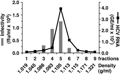
Sucrose gradient sedimentation of infectious HCV. Supernatant from infected Huh-7.5.1 cells was fractionated as described in Materials and Methods. Fractions (1-9) were collected from the top of the gradient and analyzed by RT-QPCR for HCV RNA (line). The infectivity of each fraction was determined (bars) by titration. Fraction densities are expressed as g/ml.
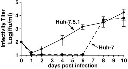
Kinetics of JFH-1 HCV infection in Huh-7.5.1 and Huh-7 cells. A virus stock generated in Huh-7.5.1 was diluted to infect Huh-7.5.1 and Huh-7 cells at an moi of 0.01. Culture supernatant was collected at the indicated times and titrated. Infectious titers in Huh-7.5.1 (solid lines) and Huh-7 cells (dashed lines) are expressed as ffu/ml. Average values of two independent infection experiments are shown.
Comment in
-
Efficient hepatitis C virus cell culture system: what a difference the host cell makes.Proc Natl Acad Sci U S A. 2005 Jul 12;102(28):9739-40. doi: 10.1073/pnas.0504296102. Epub 2005 Jul 5. Proc Natl Acad Sci U S A. 2005. PMID: 15998731 Free PMC article. No abstract available.
Similar articles
-
Regulation of Hepatitis C Virus Infection by Cellular Retinoic Acid Binding Proteins through the Modulation of Lipid Droplet Abundance.J Virol. 2019 Apr 3;93(8):e02302-18. doi: 10.1128/JVI.02302-18. Print 2019 Apr 15. J Virol. 2019. PMID: 30728260 Free PMC article.
-
Production of infectious hepatitis C virus in tissue culture from a cloned viral genome.Nat Med. 2005 Jul;11(7):791-6. doi: 10.1038/nm1268. Epub 2005 Jun 12. Nat Med. 2005. PMID: 15951748 Free PMC article.
-
Tupaia CD81, SR-BI, claudin-1, and occludin support hepatitis C virus infection.J Virol. 2011 Mar;85(6):2793-802. doi: 10.1128/JVI.01818-10. Epub 2010 Dec 22. J Virol. 2011. PMID: 21177818 Free PMC article.
-
Depressing time: Waiting, melancholia, and the psychoanalytic practice of care.In: Kirtsoglou E, Simpson B, editors. The Time of Anthropology: Studies of Contemporary Chronopolitics. Abingdon: Routledge; 2020. Chapter 5. In: Kirtsoglou E, Simpson B, editors. The Time of Anthropology: Studies of Contemporary Chronopolitics. Abingdon: Routledge; 2020. Chapter 5. PMID: 36137063 Free Books & Documents. Review.
-
Mapping the scientific knowledge and approaches to defining and measuring hate crime, hate speech, and hate incidents: A systematic review.Campbell Syst Rev. 2024 Apr 28;20(2):e1397. doi: 10.1002/cl2.1397. eCollection 2024 Jun. Campbell Syst Rev. 2024. PMID: 38686101 Free PMC article. Review.
Cited by
-
Hepatitis C virus and natural compounds: a new antiviral approach?Viruses. 2012 Oct 17;4(10):2197-217. doi: 10.3390/v4102197. Viruses. 2012. PMID: 23202460 Free PMC article. Review.
-
Expression of microRNA miR-122 facilitates an efficient replication in nonhepatic cells upon infection with hepatitis C virus.J Virol. 2012 Aug;86(15):7918-33. doi: 10.1128/JVI.00567-12. Epub 2012 May 16. J Virol. 2012. PMID: 22593164 Free PMC article.
-
New hepatitis C virus drug discovery strategies and model systems.Expert Opin Drug Discov. 2012 Sep;7(9):849-59. doi: 10.1517/17460441.2012.711312. Epub 2012 Aug 4. Expert Opin Drug Discov. 2012. PMID: 22861052 Free PMC article. Review.
-
Hepatitis C virus and antiviral innate immunity: who wins at tug-of-war?World J Gastroenterol. 2015 Apr 7;21(13):3786-800. doi: 10.3748/wjg.v21.i13.3786. World J Gastroenterol. 2015. PMID: 25852264 Free PMC article. Review.
-
Temporal analysis of hepatitis C virus cell entry with occludin directed blocking antibodies.PLoS Pathog. 2013 Mar;9(3):e1003244. doi: 10.1371/journal.ppat.1003244. Epub 2013 Mar 21. PLoS Pathog. 2013. PMID: 23555257 Free PMC article.
References
-
- Hoofnagle, J. H. (2002) Hepatology 36, S21-S29. - PubMed
-
- Kanto, T., Hayashi, N., Takehara, T., Tatsumi, T., Kuzushita, N., Ito, A., Sasaki, Y., Kasahara, A. & Hori, M. (1999) J. Immunol. 162, 5584-5591. - PubMed
-
- Auffermann-Gretzinger, S., Keeffe, E. B. & Levy, S. (2001) Blood 97, 3171-3176. - PubMed
-
- Hiasa, Y., Horiike, N., Akbar, S. M., Saito, I., Miyamura, T., Matsuura, Y. & Onji, M. (1998) Biochem. Biophys. Res. Commun. 249, 90-95. - PubMed
-
- Alter, H. J. & Seeff, L. B. (2000) Semin. Liver Dis. 20, 17-35. - PubMed
Publication types
MeSH terms
Substances
Grants and funding
LinkOut - more resources
Full Text Sources
Other Literature Sources
Medical
Research Materials
