

Regulatory T cells suppress in vitro proliferation of virus-specific CD8+ T cells during persistent hepatitis C virus infection
- PMID: 15919939
- PMCID: PMC1143649
- DOI: 10.1128/JVI.79.12.7852-7859.2005
Regulatory T cells suppress in vitro proliferation of virus-specific CD8+ T cells during persistent hepatitis C virus infection
Abstract
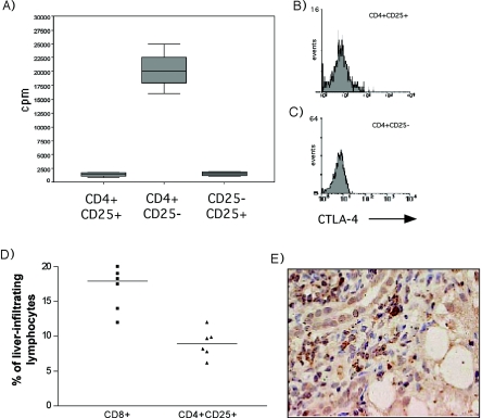
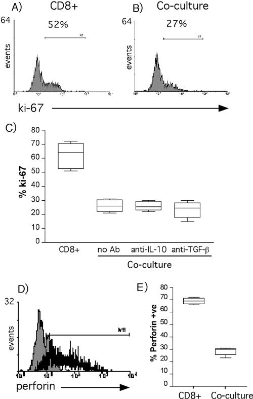
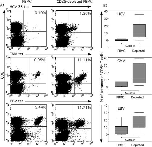
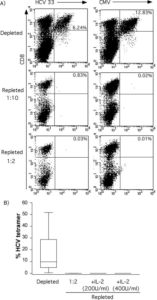
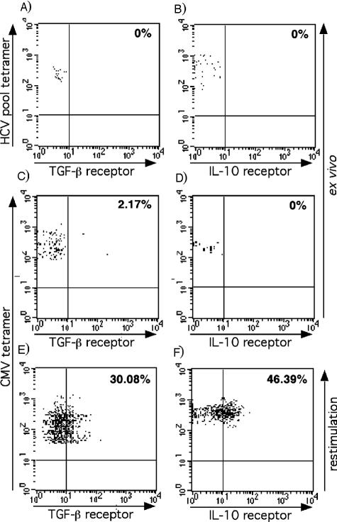
The basis of chronic infection following exposure to hepatitis C virus (HCV) infection is unexplained. One factor may be the low frequency and immature phenotype of virus-specific CD8(+) T cells. The role of CD4(+)CD25(+) T regulatory (T(reg)) cells in priming and expanding virus-specific CD8(+) T cells was investigated. Twenty HLA-A2-positive patients with persistent HCV infection and 46 healthy controls were studied. Virus-specific CD8(+) T-cell proliferation and gamma interferon (IFN-gamma) frequency were analyzed with/without depletion of T(reg) cells, using peptides derived from HCV, Epstein-Barr virus (EBV), and cytomegalovirus (CMV). CD4(+)CD25(+) T(reg) cells inhibited anti-CD3/CD28 CD8(+) T-cell proliferation and perforin expression. Depletion of CD4(+)CD25(+) T(reg) cells from chronic HCV patients in vitro increased HCV and EBV peptide-driven expansion (P = 0.0005 and P = 0.002, respectively) and also the number of HCV- and EBV-specific IFN-gamma-expressing CD8(+) T cells. Although stimulated CD8(+) T cells expressed receptors for transforming growth factor beta and interleukin-10, the presence of antibody to transforming growth factor beta and interleukin-10 had no effect on the suppressive effect of CD4(+)CD25(+) regulatory T cells on CD8(+) T-cell proliferation. In conclusion, marked CD4(+)CD25(+) regulatory T-cell activity is present in patients with chronic HCV infection, which may contribute to weak HCV-specific CD8(+) T-cell responses and viral persistence.
Figures

Similar articles
-
T cells with a CD4+CD25+ regulatory phenotype suppress in vitro proliferation of virus-specific CD8+ T cells during chronic hepatitis C virus infection.J Virol. 2005 Jun;79(12):7860-7. doi: 10.1128/JVI.79.12.7860-7867.2005. J Virol. 2005. PMID: 15919940 Free PMC article.
-
An immunomodulatory role for CD4(+)CD25(+) regulatory T lymphocytes in hepatitis C virus infection.Hepatology. 2004 Nov;40(5):1062-71. doi: 10.1002/hep.20454. Hepatology. 2004. PMID: 15486925
-
Hepatitis C virus core protein triggers expansion and activation of CD4(+)CD25(+) regulatory T cells in chronic hepatitis C patients.Cell Mol Immunol. 2015 Nov;12(6):743-9. doi: 10.1038/cmi.2014.119. Epub 2014 Dec 22. Cell Mol Immunol. 2015. PMID: 25531392 Free PMC article.
-
Viral and host immune regulatory mechanisms in hepatitis C virus infection.Eur J Gastroenterol Hepatol. 2006 Apr;18(4):327-31. doi: 10.1097/00042737-200604000-00004. Eur J Gastroenterol Hepatol. 2006. PMID: 16538102 Review.
-
Virus-Specific Cellular Response in Hepatitis C Virus Infection.Arch Immunol Ther Exp (Warsz). 2016 Apr;64(2):101-10. doi: 10.1007/s00005-015-0364-8. Epub 2015 Oct 1. Arch Immunol Ther Exp (Warsz). 2016. PMID: 26429740 Review.
Cited by
-
Human CD4+ CD25 high cells suppress proliferative memory lymphocyte responses to herpes simplex virus type 2.J Virol. 2006 Aug;80(16):8271-3. doi: 10.1128/JVI.00656-06. J Virol. 2006. PMID: 16873284 Free PMC article.
-
Transient regulatory T-cells: a state attained by all activated human T-cells.Clin Immunol. 2007 Apr;123(1):18-29. doi: 10.1016/j.clim.2006.10.014. Epub 2006 Dec 19. Clin Immunol. 2007. PMID: 17185041 Free PMC article.
-
Therapeutic vaccination in chronic hepatitis B: preclinical studies in the woodchuck.Hepat Res Treat. 2010;2010:817580. doi: 10.1155/2010/817580. Epub 2010 Sep 7. Hepat Res Treat. 2010. PMID: 21188201 Free PMC article.
-
Hepatocellular carcinoma in viral and autoimmune liver diseases: Role of CD4+ CD25+ Foxp3+ regulatory T cells in the immune microenvironment.World J Gastroenterol. 2021 Jun 14;27(22):2994-3009. doi: 10.3748/wjg.v27.i22.2994. World J Gastroenterol. 2021. PMID: 34168403 Free PMC article. Review.
-
Immunopathogenesis of Hepatitis C Virus Infection.Gastroenterol Clin North Am. 2015 Dec;44(4):735-60. doi: 10.1016/j.gtc.2015.07.004. Epub 2015 Aug 13. Gastroenterol Clin North Am. 2015. PMID: 26600217 Free PMC article. Review.
References
-
- Alter, H. J., and L. B. Seeff. 2000. Recovery, persistence, and sequelae in hepatitis C virus infection: a perspective on long-term outcome. Semin. Liver Dis. 20:17-35. - PubMed
-
- Altman, J. D., P. A. Moss, P. J. Goulder, D. H. Barouch, M. G. McHeyzer-Williams, J. I. Bell, A. J. McMichael, and M. M. Davis. 1996. Phenotypic analysis of antigen-specific T lymphocytes. Science 274:94-96. - PubMed
-
- Appay, V., P. R. Dunbar, M. Callan, P. Klenerman, G. M. Gillespie, L. Papagno, G. S. Ogg, A. King, F. Lechner, C. A. Spina, S. Little, D. V. Havlir, D. D. Richman, N. Gruener, G. Pape, A. Waters, P. Easterbrook, M. Salio, V. Cerundolo, A. J. McMichael, and S. L. Rowland-Jones. 2002. Memory CD8+ T cells vary in differentiation phenotype in different persistent virus infections. Nat. Med. 8:379-385. - PubMed
Publication types
MeSH terms
Substances
Grants and funding
LinkOut - more resources
Full Text Sources
Other Literature Sources
Molecular Biology Databases
Research Materials
