. 2005 May 15;19(10):1156-61.
doi: 10.1101/gad.1291205.

Zebrafish hairy/enhancer of split protein links FGF signaling to cyclic gene expression in the periodic segmentation of somites

Affiliations

Zebrafish hairy/enhancer of split protein links FGF signaling to cyclic gene expression in the periodic segmentation of somites

Akinori Kawamura et al. Genes Dev. .

Abstract

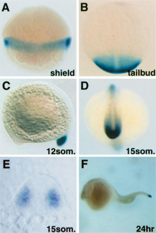
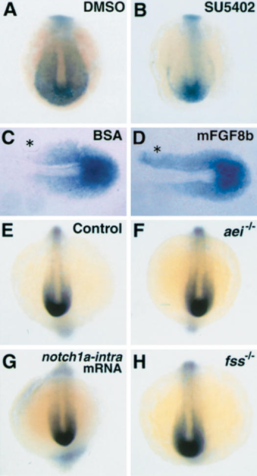
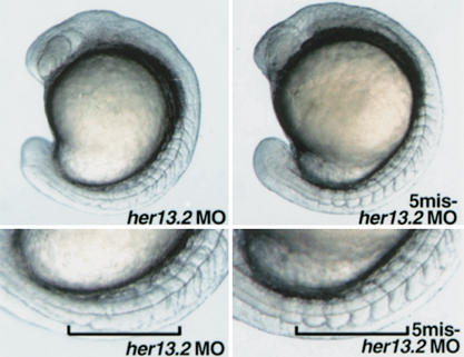
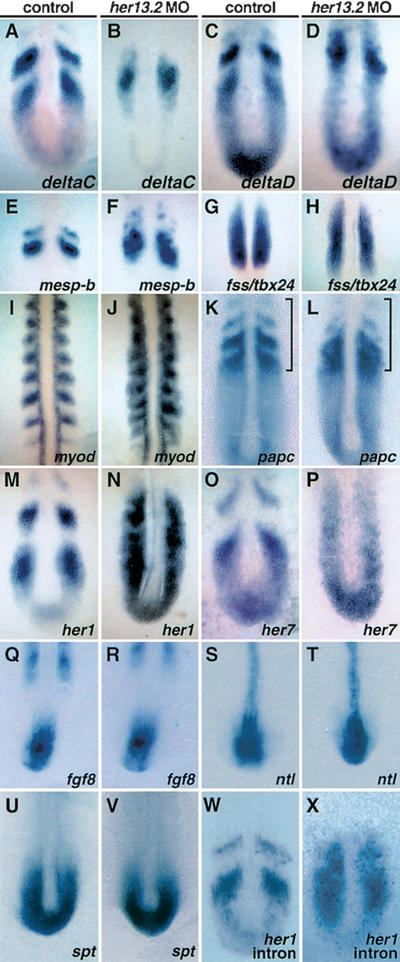
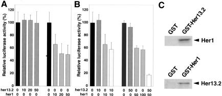
Notch and fibroblast growth factor (FGF) signaling pathways have been implicated in the establishment of proper periodicity of vertebrate somites. Here, we show evidence that a Hes6-related hairy/Enhancer of split-related gene, her13.2, links FGF signaling to the Notch-regulated oscillation machinery in zebrafish. Expression of her13.2 is induced by FGF-soaked beads and decreased by an FGF signaling inhibitor. her13.2 is required for periodic repression of the Notch-regulated genes her1 and her7, and for proper somite segmentation. Furthermore, Her13.2 augments autorepression of her1 in association with Her1 protein. Therefore, FGF signaling appears to maintain the oscillation machinery by supplying a binding partner, Her13.2, for Her1.


Figures

Figure 1.
Figure 1.
Zebrafish her13.2 mRNA is expressed in the posterior PSM and tailbud. (A,C,F) Lateral view. (B,D) Dorsal view. Whole-mount in situ hybridization was carried out by using her13.2 antisense probe and embryos at the developmental stages shown at the bottom right of each panel. her13.2 expression is first seen at the blastoderm margin at the shield stage. During the segmentation period, the signal remains confined to the posterior PSM and tailbud. (E) Transverse section of a stained embryo at the level of the posterior PSM. A signal is observed in the paraxial mesoderm.
Figure 2.
Figure 2.
Expression of her13.2 is regulated by FGF signaling. Each panel is shown from the dorsal aspect. (A,B) Transient inhibition of FGF signaling reduces the expression of her13.2 in the posterior PSM. her13.2 expression in embryos treated with DMSO (n =6; 0% affected) and SU5402 at the concentration of 660 μM (n = 6; 100% affected). Reduced expression of her13.2 was observed in embryos treated with SU5402 at any concentration that caused expansion of somites (150–660 μM; data not shown). (C,D) Implantation of mouse recombinant FGF8b-soaked bead leads to the ectopic expression of her13.2. her13.2 expression in flat-mounted embryos with BSA-soaked (n = 6; 0% affected) or mFGF8b-soaked (n = 9; 100% affected) bead. To minimize possible artifacts, beads were implanted lateral to the paraxial mesoderm. The anterior is left, and asterisks show the position of implanted beads. (EH) Independence of her13.2 expression from Notch signaling and fss/tbx24. Expression pattern of her13.2 was comparable between sibling (n = 16; 0% affected) and aei/deltaD–/– (n = 11; 0% affected) embryos. her13.2 expression was not altered in embryos injected with mRNA encoding a constitutively active form of notch1a (n = 20; 0% affected), although ectopic expression of her1 was observed in the entire PSM of these embryos (n = 23; 87% affected). her13.2 expression was also not altered in fss/tbx24–/– mutants (n = 7; 0% affected).
Figure 3.
Figure 3.
Altered somite patterning in her13.2 MO-injected embryos. Lateral views of her13.2 MO-injected and 5mis-her13.2 MO-injected embryos at the 12-somite stage. In her13.2 MO-injected embryos, the boundaries of somites are disrupted after the proper segmentation of the first eight somites, whereas no apparent defect is seen in 5mis-her13.2 MO-injected embryos. Injected embryos were incubated at 23.5°C after the dome stage (4.5 hpf), because this segmental defect was more prominent after this stage.
Figure 4.
Figure 4.
Ectopic expression of her1 and her7 in the PSM of her13.2 MO-injected embryos. Expression analysis of her13.2 MO-injected embryos by using probes indicated at the bottom right of each panel. Embryos were fixed at the 10–12-somite stages. Stained embryos were flat-mounted. (AF) The segmental expression of deltaC (n = 14; 86% affected), deltaD (n = 14; 93% affected), and mesp-b (n = 14; 86% affected) has vanished. Instead, scattered expression is observed in the anterior PSM of her13.2 MO-injected embryos. (G,H) fss/tbx24, normally expressed in the anterior and intermediate PSM, is not altered (n = 15; 0% affected). (IL) The polarity of somites is posteriorized in her13.2 morphants. The distribution of myod, expressed in the posterior region of somites, is expanded (n = 14; 93% affected). That of papc, expressed in the PSM and anterior part of somites, is scattered (n = 15; 87% affected) and emphasized by the bracket. (MP) Cyclic expression patterns of her1 and her7, categorized into three phases (Jiang et al. 2000; Oates and Ho 2002), are observed in the control embryos (n = 16; phase I, 5; phase II, 5; phase III, 6; and n = 10; phase I, 3; phase II, 3; phase III, 4, respectively). Expression pattern of her1 in M represents phase I, and that of her7 in O represents phase II. In her13.2 morphants, however, expression of her1 (n = 23; 87% affected) and her7 (n = 13; 85% affected) is seen in the entire PSM, distinct from the three phases observed in the control embryos. (QV) fgf8 (n = 13; 0% affected) and FGF-downstream genes such as ntl (n = 11; 0% affected) and spt (n = 13; 0% affected) are normally expressed in the posterior region of her13.2 MO-injected embryos. (W,X) Transcriptionally active state of her1 in the PSM of her13.2 MO-injected embryos. Cyclic expression of her1 primary transcripts is detected in control embryos by use of a her1 intron probe (n = 21; phase I, 6; phase II, 7; phase III, 8). Expression pattern of nascent her1 in W represents phase II. However, in her13.2 MO-injected embryos, her1 nascent mRNA is observed in the entire PSM (n = 21; 81% affected), as found with the her1 exon probe (Fig. 4N).
Figure 5.
Figure 5.
Her13.2 interacts with Her1 and synergistically represses the transcriptional activity of the her1 promoter. (A,B) Her13.2 alone has little transcriptional effect on the her1 promoter, but synergistically represses her1 activity in the presence of Her1. Luciferase assay using 293T cells was conducted to examine the effects of introduction of her13.2 and her1 on the 8.6-kb fragment upstream of the translational start site of her1 fused to the firefly luciferase gene. The amounts of pCS2+her13.2 and pCS2+her1 plasmids (in nanograms) used for transfection are shown under each bar. Total amount of DNA was adjusted with pCS2+ vector to be equal. All experiments were normalized for transfection efficiency by cotransfection with an expression vector for Renilla luciferase. The average normalized firefly luciferase without pCS2+her13.2 and pCS2+her1 was set at 100%, and the error bar represents the standardd. deviation. All transfections were performed in triplicate, and similar results were obtained in at least duplicate experiments. (C) Interaction between Her13.2 and Her1 in vitro. A pulldown assay was performed with in vitro labeled Her1 or Her13.2 proteins and purified Her13.2-GST or Her1-GST fusion proteins on glutathione Sepharose 4B, respectively. GST-Her13.2, not GST alone, associates with Her1 specifically, and vice versa. These specific interactions were confirmed in duplicate experiments.

Similar articles

Cited by

References

    1. Aulehla A., Wehrle, C., Brand-Saberi, B., Kemler, R., Gossler, A., Kanzler, B., and Herrmann, B.G. 2003. Wnt3a plays a major role in the segmentation clock controlling somitogenesis. Dev. Cell 4: 395–406. - PubMed
    1. Bae S., Bessho, Y., Hojo, M., and Kageyama, R. 2000. The bHLH gene Hes6, an inhibitor of Hes1, promotes neuronal differentiation. Development 127: 2933–2943. - PubMed
    1. Bessho Y. and Kageyama, R. 2003. Oscillations, clocks and segmentation. Curr. Opin. Genet. Dev. 13: 379–384. - PubMed
    1. Bessho Y., Sakata, R., Komatsu, S., Shiota, K., Yamada, S., and Kageyama, R. 2001. Dynamic expression and essential functions of Hes7 in somite segmentation. Genes & Dev. 15: 2642–2647. - PMC - PubMed
    1. Bessho Y., Hirata, H., Masamizu, Y., and Kageyama, R. 2003. Periodic repression by the bHLH factor Hes7 is an essential mechanism for the somite segmentation clock. Genes & Dev. 17: 1451–1456. - PMC - PubMed

Publication types

MeSH terms

LinkOut - more resources