Review
. 2005 Mar 29;360(1455):569-79.
doi: 10.1098/rstb.2004.1611.

The role of heterochromatin in centromere function

Affiliations
Review

The role of heterochromatin in centromere function

Alison L Pidoux et al. Philos Trans R Soc Lond B Biol Sci. .

Abstract

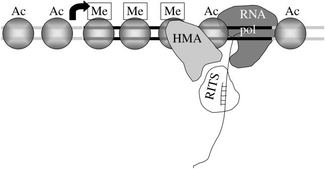
Chromatin at centromeres is distinct from the chromatin in which the remainder of the genome is assembled. Two features consistently distinguish centromeres: the presence of the histone H3 variant CENP-A and, in most organisms, the presence of heterochromatin. In fission yeast, domains of silent "heterochromatin" flank the CENP-A chromatin domain that forms a platform upon which the kinetochore is assembled. Thus, fission yeast centromeres resemble their metazoan counterparts where the kinetochore is embedded in centromeric heterochromatin. The centromeric outer repeat chromatin is underacetylated on histones H3 and H4, and methylated on lysine 9 of histone H3, which provides a binding site for the chromodomain protein Swi6 (orthologue of Heterochromatin Protein 1, HP1). The remarkable demonstration that the assembly of repressive heterochromatin is dependent on the RNA interference machinery provokes many questions about the mechanisms of this process that may be tractable in fission yeast. Heterochromatin ensures that a high density of cohesin is recruited to centromeric regions, but it could have additional roles in centromere architecture and the prevention of merotely, and it might also act as a trigger for kinetochore assembly. In addition, we discuss an epigenetic model for ensuring that CENP-A is targeted and replenished at the kinetochore domain.


Figures

Figure 1
Figure 1
Schematic of centromere 1. Centromere 1 spans approximately 35 kb and consists of a central core (cnt) of non-repetitive sequence flanked by innermost repeats (imr) and outer repeats (otr: made up of dg and dh elements, black and white arrows), which together form an almost perfect inverted repeat around the central core. Insertion of marker genes anywhere in the centromere results in their transcriptional silencing. The quality of silencing varies, depending on the insertion site: strong repression occurs at the outer repeat regions, while silencing in the central core is less robust. The centromere is divided into two domains: the central core domain (cnt and imr) and the outer repeat domain. Different classes of mutants affect silencing in each domain, and each is associated with a distinct set of proteins. Short vertical lines represent tRNA genes (Kuhn et al. 1991; Takahashi et al. 1991) which, intriguingly, occur at the transition between the domains. The central core region has a unique chromatin structure as indicated by the smear pattern which results upon partial digestion with micrococcal nuclease. CENP-Acnp1 replaces histone H3 in the central region, and upon this chromatin platform the kinetochore is assembled. Proteins such as Mis6, Mis12, Mal2 and Sim4 are specifically associated with the central core region. Mutants in these genes disrupt the unusual chromatin structure, and (where tested) cause alleviation of central core silencing. The outer repeats are packaged in nucleosomes, which are underacetylated on the N-terminal tails, owing to the action of the histone deacetylases Clr3, Clr6 and Sir2. This allows di-methylation of lysine 9 of histone H3 by the histone methyltransferase Clr4, providing a binding site for the chromodomain proteins Swi6 (HP1) and Chp1. This Swi6-containing heterochromatin is responsible for the recruitment of a high density of cohesin to the outer repeat region, which is important for proper biorientation of centromeres at mitosis, and may have other roles (see figure 5). The assembly of heterochromatin is dependent on the RNAi machinery and siRNAs derived from centromeric transcripts. This involves the RNAse III-like endonuclease Dicer, the RITS complex (Chp1, Tas3, Ago1 and siRNAs), the RNA dependent-RNA polymerase Rdp1, and the UVDDB protein Rik1. For clarity, not all components are shown in the diagram. See the text for further details and, for a more comprehensive review of the genes and proteins involved in the assembly of the two domains, see Pidoux & Allshire (2004) and Ekwall (2004).
Figure 2
Figure 2
Targeting and incorporation of CENP-A at functional centromeres. (a) At functional centromeres, CENP-A nucleosomes (white circles) form a platform for kinetochore assembly (grey lozenges) which allows proper interaction with spindle microtubules at mitosis. Tension is generated across correctly bi-oriented centromeres at metaphase. We propose that this tension induces an epigenetic ‘mark’ (*) to direct the incorporation of new CENP-A at the active centromere. CENP-A could conceivably be incorporated in a direct response to tension at metaphase, or, more probably, the mark would be read in the next cell cycle, at S phase. (b) Random partitioning would mean that some marked CENP-A nucleosomes (or other kinetochore component) remained on each chromatid as the replication fork passed. These would signal the incorporation of fresh CENP-A nucleosomes in the gaps, rather than H3-containing nucleosomes. Alternatively, H3-nucleosomes would be incorporated at replication, and the mark read post-replication to signal exchange of H3-nucleosomes for CENP-A nucleosomes by specialized chromatin remodelling activities. (c) At defective centromeres, the assembly of imperfect kinetochores (white lozenges) would cause reduced/absent MT interaction and tension at mitosis. The centromeres would fail to receive the epigenetic mark (and segregation would be impaired). (d) At unmarked centromeres and at euchromatin (e), H3-containing nucleosomes would be incorporated at S phase by default. Subsequent failure to properly biorient would lead to further CENP-A reduction and eventual loss of centromere function. This epigenetic mechanism would ensure that CENP-A is replenished at active centromeres but prevent ectopic incorporation at euchromatic sites. It guarantees long-term propagation of centromeres but also permits plasticity.
Figure 3
Figure 3
Silencing by RNA interference. Observations in several organisms suggest that the RNA interference machinery causes silencing by three different routes. This is illustrated for silencing by a plasmid-expressed hairpin RNA homologous to a 280 bp region of the ura4 gene in fission yeast. Box: dsRNA is cleaved by Dicer to produce siRNAs. Middle: silencing by mRNA degradation or post-transcriptional gene silencing (PTGS). siRNAs are incorporated into the RNA-induced silencing complex (RISC, which includes Argonaute—Ago1 in fission yeast), which causes degradation of mRNA homologous to the siRNAs. Left: chromatin-based silencing. In plants this involves DNA methylation to silence genes. In fission yeast, siRNAs incorporated into the RITS complex (Ago1, Chp1 and Tas3) lead to methylation of lysine 9 of histone H3, binding of Swi6 and recruitment of cohesin at homologous regions. The assembly of heterochromatin requires the histone deacetylases Clr3, Clr6 and Sir2, and the histone methyltransferase Clr4, Rik1 and Rdp1. In wild-type cells, spreading of heterochromatin occurs beyond the region of homology. In swi6Δ cells, lysine 9 methylation occurs only at the region of homology, indicating that Swi6 is required for spreading. Right: siRNA-dependent translational inhibition contributes to silencing in some organisms, but it is not known whether this occurs in fission yeast.
Figure 4
Figure 4
Chromatin-based silencing could involve siRNAs targeting nascent RNA transcripts. In this model, siRNAs in the RITS complex would home in on their target by hybridization with the nascent transcript produced by RNA polymerase II. Histone modifying activities (HMA), including histone deacetylases (Clr3, Clr6, Sir2) and histone methyltransferase (Clr4), would piggyback on the RITS complex. As the polymerase traverses the region, deaceylation and methylation of nucleosomes would occur, leading to the assembly of repressive heterochromatin. Ac is acetylation; Me is methylation. In an alternative model (not shown), targeting involves the hybridization of siRNAs to DNA.
Figure 5
Figure 5
Speculative models for the role of outer repeat heterochromatin in centromere specification and architecture. (a) The de novo assembly of kinetochores might be facilitated by heterochromatic ‘signposts’ which instruct the cell to incorporate CENP-A. (b) The Swi6-containing heterochromatin of the outer repeats is known to recruit a high density of cohesin for sister-chromatid cohesion. It might also have a role in intramolecular synapsis of the two sides of a single centromere to form a hypothetical loop structure. This specialised architecture might be required to present the central core in a favourable configuration for kinetochore assembly, and in ensuring a rigid structure in which multiple MT-binding sites are clamped together so they are all oriented towards the same spindle pole, thus reduce the likelihood of merotelic attachment. The diagram represents a 3-dimensional structure. The ends of each sister chromatid are indicated (* and *, § and §). Chromosome arms are shown in light grey; outer repeats, dark grey; central core, black; kinetochore, white boxes; MTs, thin black lines. Intermolecular heterochromatin/cohesin between sister chromatids is indicated by the horizontal stippled lines. Intramolecular heterochromatin/cohesin, holding the two sides of each centromere together is indicated by vertical stippled lines.

Similar articles

Cited by

References

    1. Aagaard L, et al. Functional mammalian homologues of the Drosophila PEV-modifier Su(var)3-9 encode centromere-associated proteins which complex with the heterochromatin component M31. EMBO J. 1999;18:1923–1938. - PMC - PubMed
    1. Allshire R.C, Javerzat J.P, Redhead N.J, Cranston G. Position effect variegation at fission yeast centromeres. Cell. 1994;76:157–169. - PubMed
    1. Allshire R.C, Nimmo E.R, Ekwall K, Javerzat J.P, Cranston G. Mutations derepressing silent centromeric domains in fission yeast disrupt chromosome segregation. Genes Dev. 1995;9:218–233. - PubMed
    1. Amor D.J, Choo K.H. Neocentromeres: role in human disease, evolution, and centromere study. Am. J. Hum. Genet. 2002;71:695–714. - PMC - PubMed
    1. Ayoub N, Goldshmidt I, Lyakhovetsky R, Cohen A. A fission yeast repression element cooperates with centromere-like sequences and defines a mat silent domain boundary. Genetics. 2000;156:983–994. - PMC - PubMed