. 2005 Jun;79(11):7146-61.
doi: 10.1128/JVI.79.11.7146-7161.2005.

Genetic and cell biological characterization of the vaccinia virus A30 and G7 phosphoproteins

Affiliations
Free PMC article

Genetic and cell biological characterization of the vaccinia virus A30 and G7 phosphoproteins

Jason Mercer et al. J Virol. 2005 Jun.
Free PMC article

Abstract

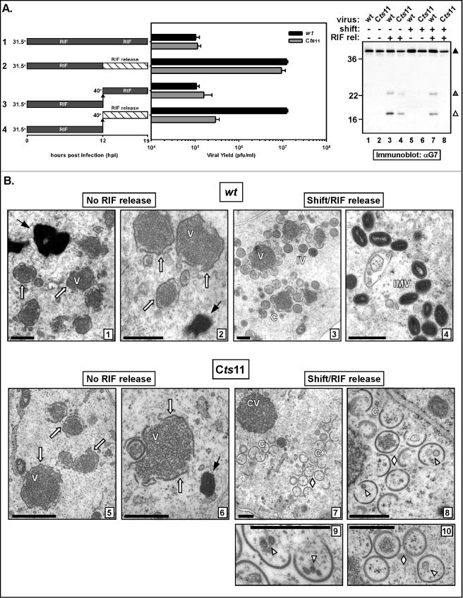
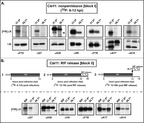
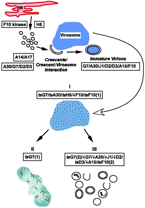
The vaccinia virus proteins A30 and G7 are known to play essential roles in early morphogenesis, acting prior to the formation of immature virions. Their repression or inactivation results in the accumulation of large virosomes, detached membrane crescents, and empty immature virions. We have undertaken further study of these proteins to place them within the context of the F10 kinase, the A14 membrane protein, and the H5 phosphoprotein, which have been the focus of previous studies within our laboratory. Here we confirm that both A30 and G7 undergo F10 kinase-dependent phosphorylation in vivo and recapitulate that modification of A30 in vitro. Although the detached crescents observed upon loss of A30 or G7 echo those seen upon repression of A14, no interaction between A30/G7 and A14 could be detected. We did, however, determine that the A30 and G7 proteins are unstable during nonpermissive tsH5 infections, suggesting that the loss of A30/G7 is the underlying cause for the formation of lacy or curdled virosomes. We also determined that the temperature-sensitive phenotype of the Cts11 virus is due to mutations in two codons of the G7L gene. Phenotypic analysis of nonpermissive Cts11 infections indicated that these amino acid substitutions compromise G7 function without impairing the stability of either G7 or A30. Utilizing Cts11 in conjunction with a rifampin release assay, we determined that G7 acts at multiple stages of virion morphogenesis that can be distinguished both by ultrastructural analysis and by monitoring the phosphorylation status of several viral proteins that undergo F10-mediated phosphorylation.


Figures

FIG. 1.
FIG. 1.
Immunoprecipitation analysis of G7 and A30 expression; A30 and G7 do not coprecipitate, but A30 associates with the F18 protein. (A) Immunoprecipitation analysis of nascent A30, G7, and A14. BSC40 cells infected with wt virus at an MOI of 5 were labeled with [35S]methionine from 6 to 9 hpi. Lysates were subjected to immunoprecipitation analysis with α-A30, α-G7, and α-A14 sera. The precipitates were resolved by SDS-PAGE and visualized by autoradiography. Each serum retrieved the cognate protein; in addition, the α-A30 precipitate contained a strongly labeled species of ∼11 kDa (formula image
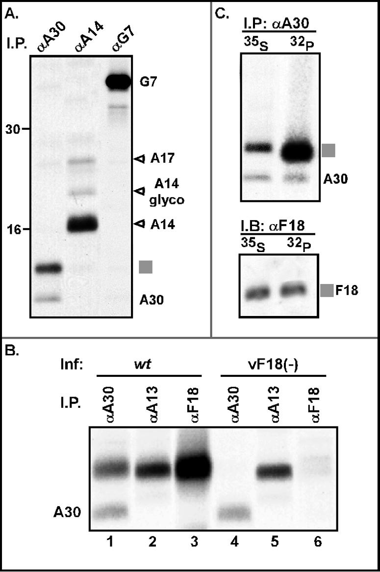
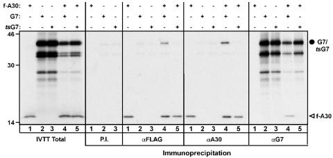
), and the α-A14 precipitate contained the ∼21-kDa A17 protein, as well as A14 and a small amount of glycosylated A14 (A14 glyco). No interaction between A30 and G7 was detected. The precipitated proteins are identified at the right; molecular markers are shown at the left, with their masses given in kilodaltons. (B) The 11-kDa protein coprecipitating with A30 migrates with A13 and F18 and is not retrieved when F18 expression is repressed. Cells were infected with either wt virus (lanes 1 to 3) or the inducible F18 recombinant in the absence of IPTG (vF18[−], lanes 4 to 6). Cells were metabolically labeled and subjected to immunoprecipitation as described for panel A. (C) The 11-kDa protein that coprecipitates with A30 is the viral F18 protein. Cells infected with wt virus (as described in panel A) were pulse-labeled with either [35S]methionine or [32P]Pi and subjected to immunoprecipitation with α-A30. The immunoprecipitates were resolved in duplicate by SDS-PAGE; one set was visualized directly by autoradiography (I.P.; top), and the second was subjected to immunoblot analysis with α-F18 serum (I.B.; bottom).
FIG. 2.
FIG. 2.
A30 and G7 are phosphorylated on serine residues in vivo in an F10-dependent manner; A30 is a substrate for the F10 and B1 kinases in vitro (A) Phosphorylation of the A30 protein in vivo. BSC40 cells were infected with the indicated viruses {wt, ts28 at 39.7°C [tsF10(−)], or vindH1 in the absence of IPTG [H1(−)]} at an MOI of 5 and pulse-labeled with 32PPi from 6 to 9 hpi; lysates were subjected to immunoprecipitation analysis with the α-A30 serum. The immunoprecipitates and lysates were resolved by SDS-PAGE; the latter were visualized by autoradiography, and the former were subjected to immunoblot analysis with the α-A30 serum (I.B.). Filled and empty triangles mark the differential migration of phosphorylated and unphosphorylated A30 (A30 I.B.) An additional sample of 32P-A30 was retrieved by immunoprecipitation from wt-infected lysates and subjected to phosphoamino acid analysis (PAA). The positions to which phosphoamino acid markers (P-ser, P-thr, and P-tyr) migrated are indicated by the wickets; the radiolabeled phosphoamino acids derived from A30 were visualized by autoradiography. (B) Phosphorylation ofthe G7 protein in vivo. Phosphorylation of the G7 protein was analyzed exactly as described above for A30 in panel A, except that a polyclonal α-G7 serum was used. (C) Recombinant N′his-A30 is a substrate for both the F10 and B1 kinases in vitro. In the left panel, purified F10 kinase was assayed alone (lane 1), in the presence of the generic substrate MBP (lane 2), or in the presence of recombinant N′his-A30 (lane 3). All reactions were performed at 25°C for 30 min in the presence of [γ-32P]ATP. Autophosphorylation of F10 (▵), phosphorylation of MBP (◂), and phosphorylation of A30 (•) were readily observed. In the right panel, recombinant B1 kinase was assayed alone (lane 1), in the presence of the generic substrate casein (lane 2), or in the presence of N′his-A30 (lane 6). Autophosphorylation of B1 (▵), phosphorylation of casein (◂), and phosphorylation of A30 (•) were readily observed. The electrophoretic migration of molecular weight markers, with their masses indicated in kilodaltons, is shown at the left of each autoradiograph.
FIG. 3.
FIG. 3.
The stability of A30 and G7 are dependent upon a functional H5 protein. (A) Determination of the t1/2 of A30 and G7 during permissive and nonpermissive tsH5 infections. Multiple dishes of BSC40 cells were infected with tsH5-4 (MOI 5) at both 31.5 and 39.7°C. At 6 hpi all dishes were pulse-labeled with [35S]methionine for 15 min. One dish from each temperature set was harvested immediately (pulse, 0); the remaining dishes were washed and refed with complete medium and subjected to further incubations of 30, 60, 120, 240, and 360 min (chase). Lysates were prepared and subjected to immunoprecipitation analysis with α-A30 and α-G7 sera; resolved immunoprecipitates were visualized by fluorography. The levels of A30 and G7 remaining after each chase period were normalized to the level found in the pulse sample and are shown graphically. The vertical lines illustrate the times at which 50% of each protein remains; these extrapolated t1/2 values are shown in the box at the right of the graph. (B) Immunoblot analysis of steady-state levels of A30, G7, and H5 during tsH5 infections. Aliquots of the tsH5-infected “360-min” chase cultures described above, i.e., harvested at 9.25 hpi, were subjected to immunoblot analysis with α-A30, α-G7, and α-H5 sera. A temperature-dependent loss of A30 and G7, but not H5, was observed.
FIG. 4.
FIG. 4.
The temperature-sensitive phenotype of Cts11 is due to mutations in the G7R gene. (A) The G7 gene can rescue the temperature-sensitive phenotype of Cts11. BCS40 cells were infected with Cts11 (MOI = 0.03) at 31.5°C; at 3 hpi, cultures were transfected individually with the indicated linearized DNA constructs and shifted to the nonpermissive temperature (39.7°C). At 48 h, cultures were harvested, and the yield of temperature-insensitive virus was determined by titration at 39.7°C. The value shown represents the average of triplicate experiments. Only the HindIIIG fragment and pTM1(G7) were able to restore the production of temperature-insensitive virus. (B) Comparative sequence alignment of the region surrounding the lesions within the G7R ORF. An alignment of residues 110 to 135 of the G7 homologs encoded by various poxviruses is shown; residues that match the vaccinia virus WR sequence are shaded. The lightning bolts indicate the position of the temperature-sensitive mutations, with the amino acid substitutions shown beneath the arrowhead. Abbreviations: VV WR strain (WR), camelpox virus (CMPX), variola virus (VAR), ectromelia virus (EV), monkeypox virus (MPX), lumpy skin virus (LSDV), sheeppox virus (SHPV), swinepox virus (SPV), molluscum contagiosum (MCV), yaba monkey tumor virus virus (YMTV), myxoma virus (MYX), and fowlpox virus (FPV).
FIG. 5.
FIG. 5.
The AG↓X proteolytic cleavage sites within G7 appear to be required for virus viability. Marker rescue analysis was used to assess the requirement for proteolytic cleavage sites found at amino acids 183 and 238 within the G7 protein. Briefly, cells were infected with Cts11 at the permissive temperature; they were shifted to the nonpermissive temperature at 3 hpi upon transfection with linearized DNA plasmids encoding wtG7 or mutant alleles engineered to contain nucleotide substitutions which transform one or both AG↓X motifs to the inert sequence AAX and, fortuitously, disrupt diagnostic restriction endonuclease sites. Cells were harvested at 48 hpi and 12 temperature-insensitive plaques were purified from each transfection. Homologous recombination between the transfected DNA and the viral genome must occur within interval 1 and either interval 2a, 2b, or 2c in order to generate temperature-insensitive plaques. Depending on whether recombination occurs within 2a, 2b, or 2c, the genome will retain the endogenous AG↓X motif (and hence the restriction endonuclease site) or acquire the AAX sequence of the plasmid (and lose the restriction endonuclease site). For each plasmid used, the tally of AG↓X versus AAX for the 12 temperature-insensitive plaques obtained from the transfection is shown to the right of the schematic illustration.
FIG. 6.
FIG. 6.
Cts11 displays a tight temperature-sensitive phenotype. (A) Plaque formation by Cts11 is abrogated during nonpermissive infections. Serial dilutions of Cts11 were titrated at permissive (31.5°C) and nonpermissive (39.7°C) temperatures; at 48 hpi, no macroscopic plaques were seen at 39.7°C. (B) Virus production is drastically reduced during nonpermissive infections with Cts11. BSC40 cells were infected (MOI of 2 or 15) with wt virus (▪) or Cts11 (▨) and maintained at 31.5 or 39.7°C for 24 h. The yield of cell-associated virus was determined by titration at 31.5°C. The values shown represent the average of triplicate experiments. (C) The G7 and A30 proteins are stable during nonpermissive Cts11 infections. BSC40 cells were infected with Cts11 at 31.5 and 39.7°C (MOI of 5); at 12 hpi, cells were harvested and lysates were subjected to immunoblot analysis with α-G7 (left panel) and α-A30 antisera (right panel). For G7, the immunoreactive bands representing the full-length (▴, 42 kDa) and processed forms (▴, 26 kDa; ▵, 16 kDa) of G7 are indicated. Note that proteolytic processing of G7 fails to occur at 39.7°C. Molecular weight markers are indicated at the left of each immunoblot, with their masses shown in kilodaltons.
FIG. 7.
FIG. 7.
The Cts11-encoded G7 protein shows a diminished ability to interact with the A30 protein in vitro. IVTT reactions were programmed to express 3XFLAG-A30 (F-A30), G7, or Cts11-encoded G7 (tsG7), either individually or in combination. Total IVTT and the proteins retrieved by immunoprecipitation with preimmune, αFLAG, αA30, or αG7 sera were analyzed by SDS-PAGE and autoradiography. The full-length G7 and tsG7 proteins (•) and F-A30 are indicated (▵) are indicated, and molecular weight markers are indicated at the left, with their masses shown in kilodaltons. Full-length G7, but not tsG7, is coprecipitated by the αA30 or αFLAG sera when F-A30 is present; F-A30 is coprecipitated by the α-G7 serum when G7, but not tsG7, is present.
FIG. 8.
FIG. 8.
Infections with Cts11 at nonpermissive temperature arrest at an early stage in viral morphogenesis. BSC40 cells were infected with Cts11 (MOI of 2) and maintained at either 31.5°C (A) or 39.7°C (B to F); at 17 h, cells were prepared for examination by transmission electron microscopy. All of the normal intermediates of morphogenesis were seen at the permissive temperature (in panel A), including prototypic “smooth” virosomes (V), crescents (C), IV, immature virions with nucleoid (IVN), and IMV. In contrast, cells infected at the nonpermissive temperature (in panels B to F) were devoid of the later stages of virion morphogenesis, as evidenced by the absence of immature or intracellular mature virions. The presence of a cleared area of cytoplasm, with cellular organelles relegated to the cell periphery, was evident in all cells examined (see panel B). Electron-dense fibrillar material was sometimes observed within this cleared area (▵; panels B and C). Viral crescents (▴), when observed, were short and clustered within a recognizable but amorphous region of electron-dense material (see panels C, D, and F). These crescent depots were often found near virosomes that displayed a “curdled” appearance (CV; panels C, D, and E). Nuclei are labeled with N. Magnifications: (A) ×30,000; (B) ×12,000; (C) ×30,000; (D) ×32,000; (E) ×40,000; (F) ×48,000. Bars, 500 nm.
FIG. 9.
FIG. 9.
Rifampin release experiments reveal a second block for nonpermissive Cts11 infections at a later stage of viral morphogenesis. (A) Cts11 is unable to recover from a RIF block at the nonpermissive temperature. Quadruplicate sets of BSC40 cells were infected (MOI of 5) with either wt virus (▪) or Cts11 (░⃞) at 31.5°C in the presence of RIF. At 12 hpi two sets of wt and Cts11 infections were left at 31.5°C (rows 1 and 2) and two sets were shifted to 39.7°C (rows 3 and 4); 1 set at each temperature was released from the RIF block (striped bar, rows 2 and 4); the other set was maintained in the presence of drug (rows 1 and 3). Infections were then allowed to proceed for an additional 7 h; the viral yield was determined by titration at 31.5°C in triplicate, and the averaged results are displayed graphically. Unlike wt-infected cells, Cts11-infected cells only resumed virus production when RIF release was performed at 31.5°C. Samples from these infections were also subjected to immunoblot analysis with α-G7 serum (right panel). The immunoreactive species corresponding to full-length (solid triangles) and cleaved G7 (shaded and open triangles) are indicated, and the molecular mass markers are shown at the left in kilodaltons. Processing of G7 was correlated with recovery of virus production. (B) After RIF release at 39.7°C, the morphogenesis arrest seen in Cts11-infected cultures is reminiscent of that seen upon repression of G7 expression. Duplicate sets of BSC40 cells were infected (MOI of 5) with either wt virus or Cts11 in the presence of RIF and maintained at 31.5°C. At 12 hpi 1 set was harvested for EM analysis; the classic features of the morphogenesis arrest associated with RIF treatment were seen in both wt- and Cts11-infected cells (compare panels 1 and 2 and panels 5 and 6). A second set was shifted to 39.7°C, released from the RIF block, and incubated for an additional 3 h (compare panels 3 and 4 to panels 7 to 10). In wt-infected cells, morphogenesis resumed and progressed normally, culminating with the formation of mature virions. In Cts11-infected cells, there was a release from the RIF arrest, but a second arrest was seen, with the appearance of numerous crescents and empty or pseudo-immature virions. The latter were often found to include what appear to be vesicles. There was a complete absence of immature or mature virions. Abbreviations are as follows: virosome (V), crystalloids (→), viral crescents (C), IV, IMV, curdled virosome (CV), pseudo-IV (⋄), and vesicles (▵). Subpanel magnifications: 1, ×17,500; 2, ×30,000; 3, ×10,000; 4, ×28,000; 5, ×33,000; 6, ×33,000; 7, ×11,500; 8, ×30,000; 9, ×60,000; 10, ×34,000. Bars, 500 nm.
FIG. 10.
FIG. 10.
During nonpermissive Cts11 infections, the phosphorylation of several viral proteins involved in morphogenesis is impaired. (A) Comparison of protein phosphorylation during Cts11 infections performed at 31.5°C versus 39.7°C. BSC40 cells were infected with Cts11 (MOI of 5) at either the permissive (31.5°C) or nonpermissive (39.7°C) temperature and metabolically labeled with 32PPi from 6 to 12 hpi. Cell lysates were subjected to immunoprecipitation analysis with a variety of antisera (αF10, αG7, αA30, αH5, αF18, αA17, and αA14); immunoprecipitates were resolved by SDS-PAGE and visualized by autoradiography ([32P]I.P.; top panels). In parallel, lysates were also subjected to immunoblot analyses with the indicated antisera to confirm and quantitate the presence of the relevant proteins (I.B.; bottom panels). Phosphorylation of F10, G7, A30 (▹), A17, and A14 was barely detectable at the nonpermissive temperature; significant reduction in the phosphorylation of H5 and modest reduction in the phosphorylation of F18 was also observed. Comparable accumulation of all of the proteins was observed at both temperatures; at 39.7°C, however, processing of A17 (⋄) failed to occur, and elevated levels of glycosylated A14 (▵) were seen. (B) Comparison of protein phosphorylation during Cts11 infections maintained in the presence of RIF or released from RIF arrest at 31.5 or 39.7°C. BSC40 cells were infected with Cts11 (MOI of 5) in triplicate in the presence of RIF. One plate was metabolically labeled with 32PPi from 6 to 12 hpi and then harvested. The other two plates were released from the RIF block at 12 hpi and maintained at either 31.5 or 39.7°C for 3 h in the presence of 32PPi before being harvested. The cell lysates were analyzed as described above for panel A. Under these conditions, the phosphorylation of A14, A17, F18, and H5 were comparable at both temperatures; however, as in panel A, no phosphorylation of A30 or G7 was observed at 39.7°C.
FIG. 11.
FIG. 11.
Working model of early stages in VV morphogenesis. The formation of virosomes, crescents, and immature virions is shown schematically; the proteins implicated in the various stages are listed within the black boxes. The curved arrow leads to aberrant features seen when the proteins indicated are repressed (↓) or impaired (ts): I, curdled virosomes; II, crescent depots; III, empty, pseudo-IV. Depending on the experimental regimen, tsF10 and tsG7 show two different arrests [(1) and (2)].

Similar articles

Cited by

References

    1. Banham, A. H., and G. L. Smith. 1992. Vaccinia virus gene B1R encodes a 34-kDa serine/threonine protein kinase that localizes in cytoplasmic factories and is packaged into virions. Virology 191:803-812. - PubMed
    1. Beaud, G., R. Beaud, and D. P. Leader. 1995. Vaccinia virus gene H5R encodes a protein that is phosphorylated by the multisubstrate vaccinia virus B1R protein kinase. J. Virol. 69:1819-1826. - PMC - PubMed
    1. Black, E. P., N. Moussatche, and R. C. Condit. 1998. Characterization of the interactions among vaccinia virus transcription factors G2R, A18R, and H5R. Virology 245:313-322. - PubMed
    1. Chiu, W. L., and W. Chang. 2002. Vaccinia virus J1R protein: a viral membrane protein that is essential for virion morphogenesis. J. Virol. 76:9575-9587. - PMC - PubMed
    1. Dales, S., and E. H. Mosbach. 1968. Vaccinia as a model for membrane biogenesis. Virology 35:564-583. - PubMed

Publication types

MeSH terms

LinkOut - more resources