Norovirus and histo-blood group antigens: demonstration of a wide spectrum of strain specificities and classification of two major binding groups among multiple binding patterns
- PMID: 15890909
- PMCID: PMC1112114
- DOI: 10.1128/JVI.79.11.6714-6722.2005
Norovirus and histo-blood group antigens: demonstration of a wide spectrum of strain specificities and classification of two major binding groups among multiple binding patterns
Abstract
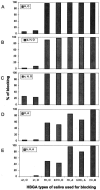
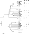
Noroviruses, an important cause of acute gastroenteritis, have been found to recognize human histo-blood group antigens (HBGAs) as receptors. Four strain-specific binding patterns to HBGAs have been described in our previous report. In this study, we have extended the binding patterns to seven based on 14 noroviruses examined. The oligosaccharide-based assays revealed additional epitopes that were not detected by the saliva-based assays. The seven patterns have been classified into two groups according to their interactions with three major epitopes (A/B, H, and Lewis) of human HBGAs: the A/B-binding group and the Lewis-binding group. Strains in the A/B binding group recognize the A and/or B and H antigens, but not the Lewis antigens, while strains in the Lewis-binding group react only to the Lewis and/or H antigens. This classification also resulted in a model of the norovirus/HBGA interaction. Phylogenetic analyses showed that strains with identical or closely related binding patterns tend to be clustered, but strains in both binding group can be found in both genogroups I and II. Our results suggest that noroviruses have a wide spectrum of host range and that human HBGAs play an important role in norovirus evolution. The high polymorphism of the human HBGA system, the involvement of multiple epitopes, and the typical protein/carbohydrate interaction between norovirus VLPs and HBGAs provide an explanation for the virus-ligand binding diversities.
Figures


Similar articles
-
Noroviruses bind to human ABO, Lewis, and secretor histo-blood group antigens: identification of 4 distinct strain-specific patterns.J Infect Dis. 2003 Jul 1;188(1):19-31. doi: 10.1086/375742. Epub 2003 Jun 12. J Infect Dis. 2003. PMID: 12825167
-
Genogroup IV and VI canine noroviruses interact with histo-blood group antigens.J Virol. 2014 Sep;88(18):10377-91. doi: 10.1128/JVI.01008-14. Epub 2014 Jul 9. J Virol. 2014. PMID: 25008923 Free PMC article.
-
Bovine Nebovirus Interacts with a Wide Spectrum of Histo-Blood Group Antigens.J Virol. 2018 Apr 13;92(9):e02160-17. doi: 10.1128/JVI.02160-17. Print 2018 May 1. J Virol. 2018. PMID: 29467317 Free PMC article.
-
Norovirus and its histo-blood group antigen receptors: an answer to a historical puzzle.Trends Microbiol. 2005 Jun;13(6):285-93. doi: 10.1016/j.tim.2005.04.004. Trends Microbiol. 2005. PMID: 15936661 Review.
-
Norovirus and histo-blood group antigens.Jpn J Infect Dis. 2011;64(2):95-103. Jpn J Infect Dis. 2011. PMID: 21519121 Review.
Cited by
-
Epochal evolution of GGII.4 norovirus capsid proteins from 1995 to 2006.J Virol. 2007 Sep;81(18):9932-41. doi: 10.1128/JVI.00674-07. Epub 2007 Jul 3. J Virol. 2007. PMID: 17609280 Free PMC article.
-
Human Milk Oligosaccharides: Potential Applications in COVID-19.Biomedicines. 2022 Feb 1;10(2):346. doi: 10.3390/biomedicines10020346. Biomedicines. 2022. PMID: 35203555 Free PMC article. Review.
-
Development of a Surrogate Neutralization Assay for Norovirus Vaccine Evaluation at the Cellular Level.Viruses. 2018 Jan 5;10(1):27. doi: 10.3390/v10010027. Viruses. 2018. PMID: 29304015 Free PMC article.
-
Vero Cells as a Mammalian Cell Substrate for Human Norovirus.Viruses. 2020 Apr 14;12(4):439. doi: 10.3390/v12040439. Viruses. 2020. PMID: 32295124 Free PMC article.
-
The First Norovirus Longitudinal Seroepidemiological Study From Sub-Saharan Africa Reveals High Seroprevalence of Diverse Genotypes Associated With Host Susceptibility Factors.J Infect Dis. 2018 Jul 24;218(5):716-725. doi: 10.1093/infdis/jiy219. J Infect Dis. 2018. PMID: 29912471 Free PMC article.
References
-
- Farkas, T., T. Berke, G. Reuter, G. Szucs, D. O. Matson, and X. Jiang. 2002. Molecular detection and sequence analysis of human caliciviruses from acute gastroenteritis outbreaks in Hungary. J. Med. Virol. 67:567-573. - PubMed
-
- Farkas, T., S. A. Thornton, N. Wilton, W. Zhong, M. Altaye, and X. Jiang. 2003. Homologous versus heterologous immune responses to Norwalk-like viruses among crew members after acute gastroenteritis outbreaks on 2 US Navy vessels. J. Infect. Dis. 187:187-193. - PubMed
-
- Green, K. Y., T. Ando, M. S. Balayan, T. Berke, I. N. Clarke, M. K. Estes, D. O. Matson, S. Nakata, J. D. Neill, M. J. Studdert, and H. J. Thiel. 2000. Taxonomy of the caliciviruses. J. Infect. Dis. 181(Suppl. 2):S322-S330. - PubMed
Publication types
MeSH terms
Substances
Grants and funding
LinkOut - more resources
Full Text Sources
Other Literature Sources
Medical

