. 2005 Jun;79(11):6631-43.
doi: 10.1128/JVI.79.11.6631-6643.2005.

Long-range RNA-RNA interactions circularize the dengue virus genome

Affiliations

Long-range RNA-RNA interactions circularize the dengue virus genome

Diego E Alvarez et al. J Virol. 2005 Jun.

Abstract

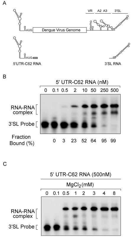
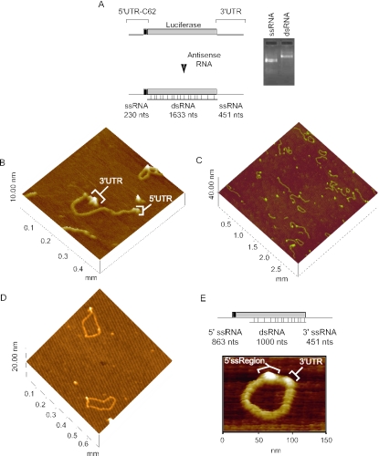
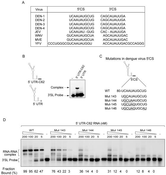
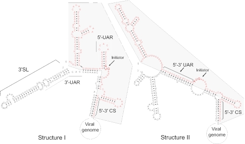
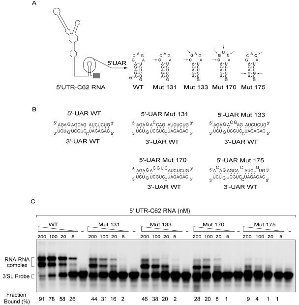
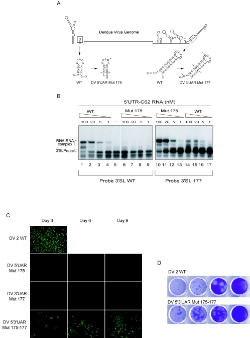
Secondary and tertiary RNA structures present in viral RNA genomes play essential regulatory roles during translation, RNA replication, and assembly of new viral particles. In the case of flaviviruses, RNA-RNA interactions between the 5' and 3' ends of the genome have been proposed to be required for RNA replication. We found that two RNA elements present at the ends of the dengue virus genome interact in vitro with high affinity. Visualization of individual molecules by atomic force microscopy revealed that physical interaction between these RNA elements results in cyclization of the viral RNA. Using RNA binding assays, we found that the putative cyclization sequences, known as 5' and 3' CS, present in all mosquito-borne flaviviruses, were necessary but not sufficient for RNA-RNA interaction. Additional sequences present at the 5' and 3' untranslated regions of the viral RNA were also required for RNA-RNA complex formation. We named these sequences 5' and 3' UAR (upstream AUG region). In order to investigate the functional role of 5'-3' UAR complementarity, these sequences were mutated either separately, to destroy base pairing, or simultaneously, to restore complementarity in the context of full-length dengue virus RNA. Nonviable viruses were recovered after transfection of dengue virus RNA carrying mutations either at the 5' or 3' UAR, while the RNA containing the compensatory mutations was able to replicate. Since sequence complementarity between the ends of the genome is required for dengue virus viability, we propose that cyclization of the RNA is a required conformation for viral replication.


Figures

FIG. 1.
FIG. 1.
Biochemical assays reveal RNA-RNA complex formation between the end sequences of dengue virus RNA. (A) Schematic representation of the dengue virus genome showing the predicted secondary structures at the 5′ and 3′ UTR. The four domains of the 3′ UTR, variable region (VR), A2, A3, and 3′ stem-loop (3′ SL), are indicated. The schematic representation of RNA molecules used for binding assays, 5′ UTR-C62 and 3′ SL, are also shown. (B) Mobility shift assays showing RNA-RNA associations. Uniformly labeled 3′ SL RNA, corresponding to the last 106 nucleotides of dengue virus type 2, was incubated with increasing concentrations of the 5′ UTR-C62 RNA corresponding to the first 160 nucleotides of the viral genome. The 5′ UTR-C62 RNA was used from 0 to 500 nM as indicated on the top of the gel. The locations of the 3′ SL probe (Probe) and the RNA-RNA complex are shown. Quantification of the fraction of probe bound for each concentration of the RNA is also indicated at the bottom of the gel. (C) RNA-RNA complex is formed only in the presence of Mg2+. The 3′ SL probe was incubated with an excess of the 5′ UTR-C62 RNA (500 nM), and complex formation was examined in the presence of increasing concentrations of Mg2+, from 0 to 8 mM.
FIG. 2.
FIG. 2.
Single-molecule analysis reveals cyclization of an RNA molecule carrying the 5′- and 3′-end sequences of dengue virus. (A) Schematic representation of a model RNA molecule of 2.3 kb showing the 5′ and 3′ dengue virus sequences flanking the luciferase coding sequence. Annealing of an antisense RNA of 1,633 nucleotides is shown. The resulting molecule bears single-stranded overhangs in the 5′ and 3′ ends of 230 and 451 nucleotides, respectively. On the right, purified single-stranded RNA (ssRNA) and double-stranded RNA were resolved on a 1% agarose gel and visualized by ethidium bromide staining. (B) Visualization of the model RNA molecules by AFM. A single RNA molecule is shown in a linear conformation. The double-stranded RNA region is flanked by single-stranded regions corresponding to the 5′ UTR-C62 and 3′ UTR of dengue virus. (C) An image of a representative field of RNA molecules deposited on mica obtained by tapping-mode AFM. Circular, linear, and head-to-tail dimers were observed. (D) Image of individual RNA molecules in circular conformation is shown. Contacts between the 5′ and 3′ single-stranded regions of the molecules can be observed. (E) Schematic representation of the same RNA molecule shown in A hybridized with an antisense RNA molecule of 1 kb. The double-stranded region of 1 kb is flanked by a 5′ single-stranded region of 863 nucleotides that contained the 160 nucleotides of the 5′ end of dengue virus and a 3′ single-stranded region that corresponds to the 3′ UTR of dengue virus. At the bottom, a representative image of an individual molecule with a double-stranded region of 1 kb is shown in circular conformation.
FIG. 3.
FIG. 3.
Visualization of genome-length dengue virus RNA by AFM. (A) Schematic representation of the dengue virus genome. An antisense RNA anneals to the molecule, resulting in a 3,302-bp double-strand species with single-stranded regions of 6,970 and 451 nucleotides at the 5′ and 3′ ends, respectively. (B) Visualization of representative images of individual genome-length RNA molecules obtained by tapping-mode AFM.
FIG. 4.
FIG. 4.
Interaction between the 5′ and 3′ CS is required for RNA-RNA complex formation. (A) The nucleotide sequence of the 5′ and 3′ CS is conserved among different mosquito-borne flaviviruses (DEN, dengue virus; JEV, Japanese encephalitis virus; WNV, West Nile virus; MVE, Murray Valley encephalitis virus; and YFV, yellow fever virus). (B) On the left, a schematic representation of the two RNA molecules used, 5′ UTR-C62 and the RNA with a deletion of the 5′ CS (5′ UTR), is shown. On the right, mobility shift assay of the 3′ SL probe in the presence of 5′ UTR-C62 or 5′ UTR RNAs is shown. (C) Nucleotide sequences of the wild type and mutated 5′ CS are indicated. Underlined letters indicate the specific substitutions in mutants 143, 144, 145, and 146. (D) RNA mobility shift analysis showing the effect of mutations within the 5′ CS in the 5′ UTR-C62 RNA on binding to the 3′ SL RNA. Uniformly labeled 3′ SL RNA was incubated with increasing concentrations of wild-type and mutated 5′ UTR-C62 RNAs as indicated at the top of the gel. The mobility of the 3′ SL probe and the RNA-RNA complex is indicated on the left. Quantification of the fraction of probe bound for each concentration of the WT and mutated RNAs is also indicated at the bottom of the gel.
FIG. 5.
FIG. 5.
Computer-generated secondary structures of the last 106 nucleotides (letters in black) and the first 143 nucleotides (letters in red) of dengue virus genomic RNA (65). The rest of the viral genome is represented schematically linking the 5′ and 3′ ends of the molecule. The predicted secondary structures I and II are those with the lowest ΔG (−90 and −91 kcal mol−1, respectively). The conserved sequences 5′-3′ CS and 5′-3′ UAR and the 3′ SL are shown. The initiator AUG codon is indicated with arrows.
FIG. 6.
FIG. 6.
Interaction between 5′ and 3′UAR sequences of dengue virus is required for RNA-RNA association. (A) Schematic representation of the predicted secondary structure of the first 160 nucleotides of dengue virus RNA. The stem-loop that contained the 16-nucleotide-long 5′ UAR is shown. The location of specific mutations (Mut) within the 5′ UAR are indicated with arrows, and the nucleotide changes are shown for mutants 131, 133, 170, and 175. (B) Base pairing between sequences corresponding to nucleotides 80 to 95 (5′ UAR) and 10642 to 10658 (3′ UAR) of the dengue virus type 2 genome are shown for the wild-type and mutated RNAs. (C) RNA mobility shift analysis showing the effect of mutations within the 5′ UAR in the 5′ UTR-C62 RNA on binding to the 3′ SL RNA. Uniformly labeled 3′ SL RNA was incubated with increasing concentrations of wild-type and mutated 5′ UTR-C62 RNAs as indicated at the top of the gel. The mobility of the 3′ SL probe and the RNA-RNA complex is indicated on the left. Quantifications of the fraction of probe bound for each concentration of the WT and mutated RNAs are also indicated at the bottom of the gel.
FIG. 7.
FIG. 7.
Specific mutations within the 5′ or 3′ UAR abolish viral replication, while reconstitution of 5′-3′ UAR complementarity restores the viral function. (A) Schematic representation of the dengue virus genome showing the predicted secondary structure of the RNA elements containing 5′ and 3′UAR (shown in boxes). Nucleotide sequences of wild-type and mutant (Mut) DV 5′UAR 175 and DV 3′UAR 177 are shown. Mutations are highlighted and marked by arrows. Mutations were designed to maintain the secondary structure of the stem-loop upstream of the initiator AUG at the 5′ end and the stem of the 3′ SL at the 3′ end of the genome. (B) RNA mobility shift assays showing that reconstitution of 5′-3′ UAR complementarity restores RNA-RNA complex formation. Two different 3′ SL probes, WT and 177, were incubated with decreasing concentrations of WT or mutant 175 unlabeled 5′ UTR-C62 RNAs as indicated in each case on the top of the gel. The mobility of the probe and the RNA-RNA complex is indicated on the left. (C) Expression of dengue virus proteins in cells transfected with wild-type and mutated viral RNAs was monitored by IFA using dengue virus type 2 murine hyperimmune ascitic fluid. BHK cells grown in 60-mm culture plates were transfected with 3 μg of in vitro-transcribed full-length dengue virus RNA. Cells were trypsinized on days 3, 6, and 9 after transfection and reseeded to a coverslip for IFA analysis. Transfection of RNA with mutations only at the 5′ or 3′ UAR (DV 5′UAR 175 and DV 3′UAR 177, respectively) did not show immunofluorescence, while the double mutant DV 5′3′UAR 175-177 showed positive IFA at day 3. Cells transfected with wild-type dengue virus RNA after day 4 are not shown because they were dead due to viral infection. (D) Comparison of plaque size formed by dengue virus wild type (DV2 WT) and double mutant that reconstitute 5′-3′ UAR complementarity (DV 5′3′UAR 175-177). Serial dilutions of DV2 WT and DV 5′3′UAR 175-177 (obtained from supernatants of cells 9 days after transfection) were used to infect a monolayer of BHK cells. The monolayer was stained 9 days after infection.

Similar articles

Cited by

References

    1. Andersen, E. S., S. A. Contera, B. Knudsen, C. K. Damgaard, F. Besenbacher, and J. Kjems. 2004. Role of the trans-activation response element in dimerization of HIV-1 RNA. J. Biol. Chem. 279:22243-22249. - PubMed
    1. Andreev, I. A., S. H. Kim, N. O. Kalinina, D. V. Rakitina, A. G. Fitzgerald, P. Palukaitis, and M. E. Taliansky. 2004. Molecular interactions between a plant virus movement protein and RNA: force spectroscopy investigation. J. Mol. Biol. 339:1041-1047. - PubMed
    1. Barends, S., H. H. Bink, S. H. van den Worm, C. W. Pleij, and B. Kraal. 2003. Entrapping ribosomes for viral translation: tRNA mimicry as a molecular Trojan horse. Cell 112:123-129. - PubMed
    1. Barton, D. J., B. J. O'Donnell, and J. B. Flanegan. 2001. 5′ cloverleaf in poliovirus RNA is a cis-acting replication element required for negative-strand synthesis. EMBO J. 20:1439-1448. - PMC - PubMed
    1. Blackwell, J. L., and M. A. Brinton. 1997. Translation elongation factor-1 alpha interacts with the 3′ stem-loop region of West Nile virus genomic RNA. J. Virol. 71:6433-6444. - PMC - PubMed

Publication types

MeSH terms

LinkOut - more resources