. 2005 Jun;79(11):6598-609.
doi: 10.1128/JVI.79.11.6598-6609.2005.

Vaccinia virus nonstructural protein encoded by the A11R gene is required for formation of the virion membrane

Affiliations

Vaccinia virus nonstructural protein encoded by the A11R gene is required for formation of the virion membrane

Wolfgang Resch et al. J Virol. 2005 Jun.

Abstract

The vaccinia virus A11R gene has orthologs in all known poxvirus genomes, and the A11 protein has been previously reported to interact with the putative DNA packaging protein A32 in a yeast two-hybrid screen. Using antisera raised against A11 peptides, we show that the A11 protein was (i) expressed at late times with an apparent mass of 40 kDa, (ii) not incorporated into virus particles, (iii) phosphorylated independently of the viral F10 kinase, (iv) coimmunoprecipitated with A32, and (v) localized to the viral factory. To determine the role of the A11 protein and test whether it is indeed involved in DNA packaging, we constructed a recombinant vaccinia virus with an inducible A11R gene. This recombinant was dependent on inducer for single-cycle growth and plaque formation. In the absence of inducer, viral late proteins were produced at normal levels, but proteolytic processing and other posttranslational modifications of some proteins were inhibited, suggesting a block in virus particle assembly. Consistent with this observation, electron microscopy of cells infected in the absence of inducer showed virus factories with abnormal electron-dense viroplasms and intermediate density regions associated with membranes and containing the D13 protein. However, no viral membrane crescents, immature virions, or mature virions were produced. The requirement for nonvirion protein A11 in order to make normal viral membranes was an unexpected and exciting finding, since neither the origin of these membranes nor their mechanism of formation in the cytoplasm of infected cells is understood.


Figures
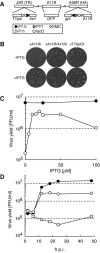
FIG. 1.
FIG. 1.
Construction and characterization of a recombinant VAC with an inducible A11R ORF (vA11Ri). (A) Genome structure of vA11Ri. The three loci at which vA11Ri differs from the WR strain are shown as boxes: J2R (thymidine kinase; TK), A11R and A56R (hemagglutinin; HA). Below the boxes are schematics of the modifications. Additional abbreviations: T7pol, bacteriophage T7 RNA polymerase gene; lacO, lac operator; P11, a VAC late promoter; P7.5, a VAC early/late promoter; lacI, E. coli lac repressor gene; gpt, E. coli guanine phosphoribosyltransferase gene; PT7, bacteriophage T7 promoter;EMC, encephalomyocarditis virus cap-independent translation enhancer element. (B) Plaque phenotype of vA11Ri. BS-C-1 cells were infected with vA11Ri, vA11Ri/A11R and vT7lacOI in the absence or presence of 25 μM IPTG. After 48 h, the cells were fixed and stained with crystal violet. (C) Dependency of vA11Ri replication on IPTG. BS-C-1 cells were infected with 10 PFU per cell of vT7lacOI (•) or with vA11Ri (○) in the presence of 0 to 100 μM IPTG, and the viral yield was determined after 24 h. (D) One-step growth curve of vA11Ri. BS-C-1 cells were infected with 10 PFU per cell of vT7lacOI (•) or vA11Ri in the absence (□) or presence (○) of 25 μM IPTG, and the virus yield was determined from 2 to 48 h postinfection (h p.i.).
FIG. 2.
FIG. 2.
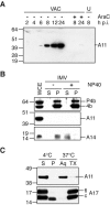
Synthesis of A11 and analysis of IMV. (A) A11 expression kinetics. BS-C-1 cells were infected with 10 PFU per cell of VAC and whole-cell extracts were prepared at the indicated times. Extracts from uninfected cells (U) and cells infected in the presence of cytosine arabinoside (AraC) were also prepared, and all extracts were analyzed by SDS-PAGE and Western blotting with a anti-A11 antiserum. The position and mass in kilodaltons of marker proteins are indicated on the left. Note that the apparent molecular mass of A11 is slightly larger than predicted. (B) Absence of A11 in purified IMV. Purified IMV was extracted with NP-40 (+) or mock treated (−) and separated into soluble (S) and pellet (P) fractions. Proteins in both fractions were separated by SDS-PAGE, followed by Western blotting with anti-P4b, anti-A11, or anti-A14 antisera. Whole-cell extract (WCE) containing similar amounts of P4b/4b and A14 was included in the analysis. The position and mass in kilodaltons of marker proteins are indicated on the left. (C) Phase separation of A11 in Triton X-114. BS-C-1 cells were infected with 3 PFU per cell of VAC. After 20 h, cells were harvested in cold lysis buffer containing 1% Triton X-114. The lysate was separated into soluble (S) and pellet (P) fractions, and the soluble fraction was separated into aqueous (Aq) and detergent (TX) phases. All samples were adjusted to equal volumes and subjected to SDS-PAGE, followed by Western blotting with anti-A11 or anti-A17 antisera. The position and mass in kilodaltons of marker proteins are indicated on the left. Lines on the right point to the full-length (fl) and cleaved (c) forms of the A17 protein.
FIG. 3.
FIG. 3.
Phosphorylation of A11. BS-C-1 cells were infected with 10 PFU per cell of the indicated recombinant VAC (rVAC) in the presence or absence of 50 μM IPTG. Infected cells were metabolically labeled either with a mixture of [35S]methionine and [35S]cysteine (35S) or with [32P]orthophosphate (32P) from 1 h until 20 h after infection. After labeling, cytoplasmic extracts were prepared, and proteins were captured with either anti-A11 (A11) or anti-V5 (F10V5) antibodies. The antibody-bound proteins were resolved by SDS-PAGE and visualized by autoradiography. The bands corresponding to A11 and F10V5 are indicated on the right. Note that the lower band seen in immunoprecipitations from vA11Ri-infected cells represents an internal translation initiation product. Position and mass of marker proteins are shown on the left in kilodaltons.
FIG. 4.
FIG. 4.
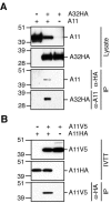
Coimmunoprecipitation of A11 with A32 and itself. (A) Coimmunoprecipitation of A11 and A32HA. BS-C-1 cells were infected with 10 PFU per cell of vA11Ri in the presence (A11+) or absence (A11−) of IPTG. Cells were then transfected with either pcDNA-A32HA (A32HA+) or empty vector (A32HA−). After 24 h, cytoplasmic extracts were prepared and proteins were precipitated with either anti-HA antibody or anti-A11 antiserum bound to agarose beads. Lysate and precipitate were analyzed by Western blotting. Position and mass in kilodaltons of marker proteins are indicated on the left. (B) Coimmunoprecipitation of differently tagged forms of A11. IVTT reactions were programmed with PCR products encoding A11HA and/or A11V5. After 90 min of protein synthesis, the IVTT reactions were subjected to immunoprecipitation with anti-HA antibody bound to agarose beads and reactions, and precipitates were analyzed by Western blotting with the anti-V5 antibody directly conjugated to horseradish peroxidase.
FIG. 5.
FIG. 5.
Localization of A11 by confocal microscopy. HeLa cells were infected with 5 PFU per cell of vA11Ri in the presence or absence of 25 μM IPTG. After 8 h cells were fixed, permeabilized, and stained with (i) anti-A11 antiserum, followed by rhodamine red-conjugated goat anti-rabbit antibody, and (ii) the DNA stain DAPI. Shown are single optical sections with scale bars recorded by confocal microscopy. Colors: blue, DAPI (nuclei and viral factories); red, A11.
FIG. 6.
FIG. 6.
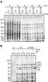
Synthesis and processing of viral proteins. (A) Pulse-labeling of synthesized proteins. BS-C-1 cells were infected with vT7lacOI or vA11Ri in the absence or presence of 25 μM IPTG at a multiplicity of 5 PFU per cell. Infected cells were labeled with [35S]methionine and [35S]cysteine for 30 min at 3 to 24 h postinfection (h p.i.), whole-cell lysates were prepared, and proteins were separated by SDS-PAGE, followed by autoradiographic visualization. Uninfected cells (U) were similarly labeled to differentiate viral from cellular proteins. Migration and mass in kilodaltons of marker proteins are indicated on the left. Note that several proteins indicated by filled circles on the right are synthesized at rates that differ between the three conditions. (B) Proteolytic processing of core proteins. BS-C-1 cells were infected with vT7lacOI in the absence or presence of 100 μg of rifampin(RIF)/ml or with vA11Ri in the absence or presence of 25 μM IPTG at a multiplicity of infection of 5 PFU per cell. Uninfected cells (U) were used as a control. After pulse-labeling at 9 h, the cells were either harvested directly (P) or shifted into chase media (C) containing excess unlabeled amino acids for 14 h before harvest. Proteins in whole-cell extracts of harvested cells were separated by SDS-PAGE and visualized by autoradiography. Major core protein precursors (P4a and P4b) and their mature processed products (4a and 4b) are indicated on the right. Position and mass in kilodaltons of marker proteins are shown on the left.
FIG. 7.
FIG. 7.
Effects of A11 repression on proteins involved in early morphogenesis. BS-C-1 cells were infected with 10 PFU per cell of vA11Ri in the presence of the indicated concentrations of IPTG. After 24 h, whole-cell extracts were prepared, fractionated by SDS-PAGE, and analyzed by Western blotting with anti-A11, anti-A17-N (detecting full-length [fl] and proteolytically cleaved [c] forms of A17), anti-pTyr (A17-PY), anti-F10, anti-A14-C (detecting glycosylated [g] and unglycosylated [ug] forms of A14) and anti-P4b/4b antisera. Loading of similar amounts of extract was verified by the detection of similar levels of P4b.
FIG. 8.
FIG. 8.
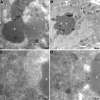
Electron microscopy of infected cells. BS-C-1 cells were infected with 3 PFU per cell of vA11Ri in the presence (A) or absence (B to D) of 25 μM IPTG. Cells were fixed and prepared for transmission electron microscopy at 20 h after infection. Electron micrographs are shown with their scale indicated by the bars. Abbreviations: c, crescent; IV, immature virion; nu, nucleoid within an IV; IMV, intracellular mature virion; IEV, intracellular enveloped virion; CEV, cell-associated enveloped virion; V, viroplasm; ✽, intermediate density area; N, nucleus.
FIG. 9.
FIG. 9.
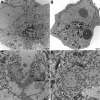
Localization of A17, A3 (P4b), D13, and PDI by immunoelectron microscopy. Cells were infected with 3 PFU per cell of vA11Ri in the absence of IPTG. After 24 h, cells were fixed, cryosectioned, and incubated with anti-D13 (A), anti-P4b/4b (B), anti-A17-N (C), or anti-PDI antisera (D), followed by an appropriate secondary antibody and colloidal gold coupled to protein A. Electron micrographs are shown with scale bars. Abbreviations: V, viroplasm; ✽, intermediate density area.

Similar articles

Cited by

References

    1. Alexander, W. A., B. Moss, and T. R. Fuerst. 1992. Regulated expression of foreign genes in vaccinia virus under the control of bacteriophage T7 RNA polymerase and the Escherichia coli lac repressor. J. Virol. 66:2934-2942. - PMC - PubMed
    1. Ansarah-Sobrinho, C., and B. Moss. 2004. Role of the I7 protein in proteolytic processing of vaccinia virus membrane and core components. J. Virol. 78:6335-6343. - PMC - PubMed
    1. Betakova, T., E. J. Wolffe, and B. Moss. 1999. Regulation of vaccinia virus morphogenesis: phosphorylation of the A14L and A17L membrane proteins and C-terminal truncation of the A17L protein are dependent on the F10L protein kinase. J. Virol. 73:3534-3543. - PMC - PubMed
    1. Byrd, C. M., T. C. Bolken, and D. E. Hruby. 2002. The vaccinia virus I7L gene product is the core protein proteinase. J. Virol. 76:8973-8976. - PMC - PubMed
    1. Cassetti, M. C., M. Merchlinsky, E. J. Wolffe, A. S. Weisberg, and B. Moss. 1998. DNA packaging mutant: repression of the vaccinia virus A32 gene results in noninfectious, DNA-deficient, spherical, enveloped particles. J. Virol. 72:5769-5780. - PMC - PubMed

MeSH terms

LinkOut - more resources