

Sources of fatty acids stored in liver and secreted via lipoproteins in patients with nonalcoholic fatty liver disease
- PMID: 15864352
- PMCID: PMC1087172
- DOI: 10.1172/JCI23621
Sources of fatty acids stored in liver and secreted via lipoproteins in patients with nonalcoholic fatty liver disease
Abstract
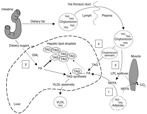
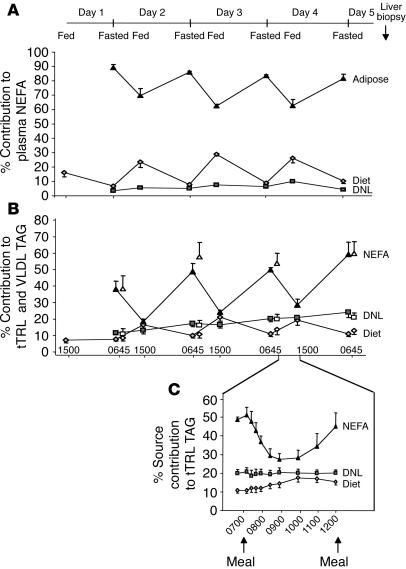
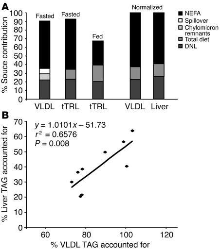
Nonalcoholic fatty liver disease (NAFLD) is characterized by the accumulation of excess liver triacylglycerol (TAG), inflammation, and liver damage. The goal of the present study was to directly quantify the biological sources of hepatic and plasma lipoprotein TAG in NAFLD. Patients (5 male and 4 female; 44 +/- 10 years of age) scheduled for a medically indicated liver biopsy were infused with and orally fed stable isotopes for 4 days to label and track serum nonesterified fatty acids (NEFAs), dietary fatty acids, and those derived from the de novo lipogenesis (DNL) pathway, present in liver tissue and lipoprotein TAG. Hepatic and lipoprotein TAG fatty acids were analyzed by gas chromatography/mass spectrometry. NAFLD patients were obese, with fasting hypertriglyceridemia and hyperinsulinemia. Of the TAG accounted for in liver, 59.0% +/- 9.9% of TAG arose from NEFAs; 26.1% +/- 6.7%, from DNL; and 14.9% +/- 7.0%, from the diet. The pattern of labeling in VLDL was similar to that in liver, and throughout the 4 days of labeling, the liver demonstrated reciprocal use of adipose and dietary fatty acids. DNL was elevated in the fasting state and demonstrated no diurnal variation. These quantitative metabolic data document that both elevated peripheral fatty acids and DNL contribute to the accumulation of hepatic and lipoprotein fat in NAFLD.
Figures
Comment in
-
Contribution of adipose tissue and de novo lipogenesis to nonalcoholic fatty liver disease.J Clin Invest. 2005 May;115(5):1139-42. doi: 10.1172/JCI24930. J Clin Invest. 2005. PMID: 15864343 Free PMC article.
Similar articles
-
Palmitoleic acid is elevated in fatty liver disease and reflects hepatic lipogenesis.Am J Clin Nutr. 2015 Jan;101(1):34-43. doi: 10.3945/ajcn.114.092262. Epub 2014 Nov 19. Am J Clin Nutr. 2015. PMID: 25527748 Free PMC article. Clinical Trial.
-
A meal high in saturated fat evokes postprandial dyslipemia, hyperinsulinemia, and altered lipoprotein expression in obese children with and without nonalcoholic fatty liver disease.JPEN J Parenter Enteral Nutr. 2013 Jul;37(4):517-28. doi: 10.1177/0148607112467820. Epub 2012 Dec 5. JPEN J Parenter Enteral Nutr. 2013. PMID: 23223552
-
Alterations in adipose tissue and hepatic lipid kinetics in obese men and women with nonalcoholic fatty liver disease.Gastroenterology. 2008 Feb;134(2):424-31. doi: 10.1053/j.gastro.2007.11.038. Epub 2007 Nov 28. Gastroenterology. 2008. PMID: 18242210 Free PMC article.
-
Clinical assessment of hepatic de novo lipogenesis in non-alcoholic fatty liver disease.Lipids Health Dis. 2016 Sep 17;15(1):159. doi: 10.1186/s12944-016-0321-5. Lipids Health Dis. 2016. PMID: 27640119 Free PMC article. Review.
-
Mechanisms of hepatic triglyceride accumulation in non-alcoholic fatty liver disease.J Gastroenterol. 2013 Apr;48(4):434-41. doi: 10.1007/s00535-013-0758-5. Epub 2013 Feb 9. J Gastroenterol. 2013. PMID: 23397118 Free PMC article. Review.
Cited by
-
Polyoxometalates Ameliorate Metabolic Dysfunction-Associated Steatotic Liver Disease by Activating the AMPK Signaling Pathway.Int J Nanomedicine. 2024 Oct 25;19:10839-10856. doi: 10.2147/IJN.S485084. eCollection 2024. Int J Nanomedicine. 2024. PMID: 39479173 Free PMC article.
-
β2-Adrenergic receptor agonist induced hepatic steatosis in mice: modeling nonalcoholic fatty liver disease in hyperadrenergic states.Am J Physiol Endocrinol Metab. 2021 Jul 1;321(1):E90-E104. doi: 10.1152/ajpendo.00651.2020. Epub 2021 May 24. Am J Physiol Endocrinol Metab. 2021. PMID: 34029162 Free PMC article.
-
Historical narrative from fatty liver in the nineteenth century to contemporary NAFLD - Reconciling the present with the past.JHEP Rep. 2021 Feb 26;3(3):100261. doi: 10.1016/j.jhepr.2021.100261. eCollection 2021 Jun. JHEP Rep. 2021. PMID: 34036255 Free PMC article. Review.
-
Hepatic Steatosis as a Marker of Metabolic Dysfunction.Nutrients. 2015 Jun 19;7(6):4995-5019. doi: 10.3390/nu7064995. Nutrients. 2015. PMID: 26102213 Free PMC article. Review.
-
A novel ChREBP isoform in adipose tissue regulates systemic glucose metabolism.Nature. 2012 Apr 19;484(7394):333-8. doi: 10.1038/nature10986. Nature. 2012. PMID: 22466288 Free PMC article.
References
-
- Ludwig J, Viggiano TR, McGill DB, Oh BJ. Nonalcoholic steatohepatitis: Mayo Clinic experiences with a hitherto unnamed disease. Mayo Clinic. Proc. 1980;55:434–438. - PubMed
-
- Clark JM, Brancati FL, Diehl AM. Nonalcoholic fatty liver disease. Gastroenterology. 2002;122:1649–1657. - PubMed
-
- Lee RG. Nonalcoholic steatohepatitis: a study of 49 patients. Hum. Path. 1989;20:594–598. - PubMed
-
- Bacon BR, Farahvash MJ, Janney CG, Neuschwander-Tetri BA. Nonalcoholic steatohepatitis: an expanded clinical entity. Gastroenterology. 1994;107:1103–1109. - PubMed
-
- James O, Day C. Non-alcoholic steatohepatitis: another disease of affluence. Lancet. 1999;353:1634–1636. - PubMed
Publication types
MeSH terms
Substances
Grants and funding
LinkOut - more resources
Full Text Sources
Other Literature Sources
