Comparative Study
. 2005 Apr 27;25(17):4420-34.
doi: 10.1523/JNEUROSCI.0382-05.2005.

A novel RNA-binding protein in neuronal RNA granules: regulatory machinery for local translation

Affiliations
Comparative Study

A novel RNA-binding protein in neuronal RNA granules: regulatory machinery for local translation

Nobuyuki Shiina et al. J Neurosci. .

Abstract

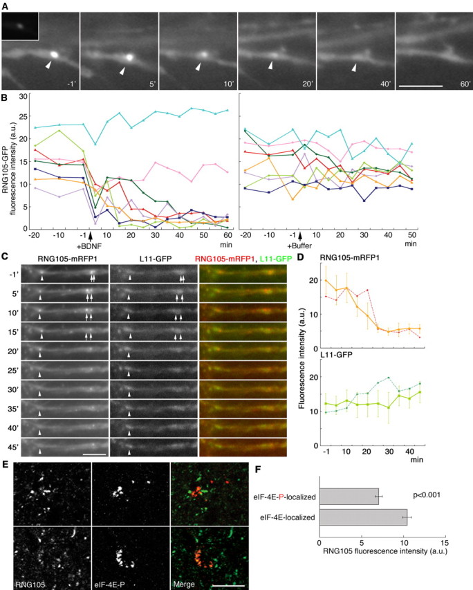
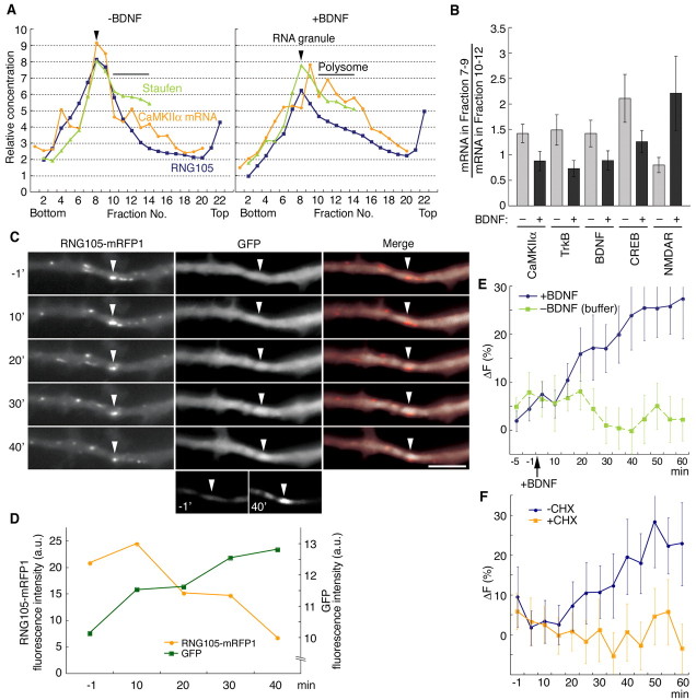
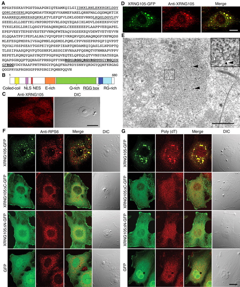
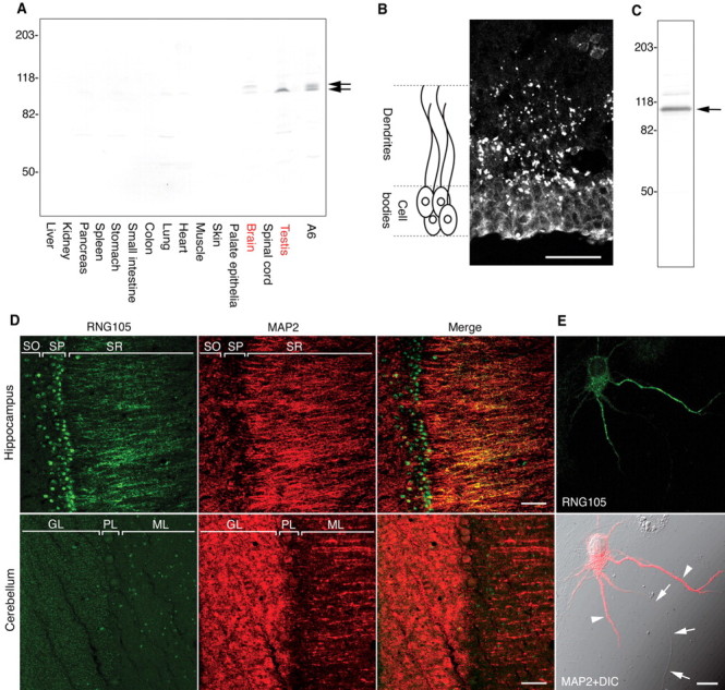
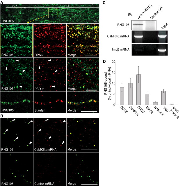
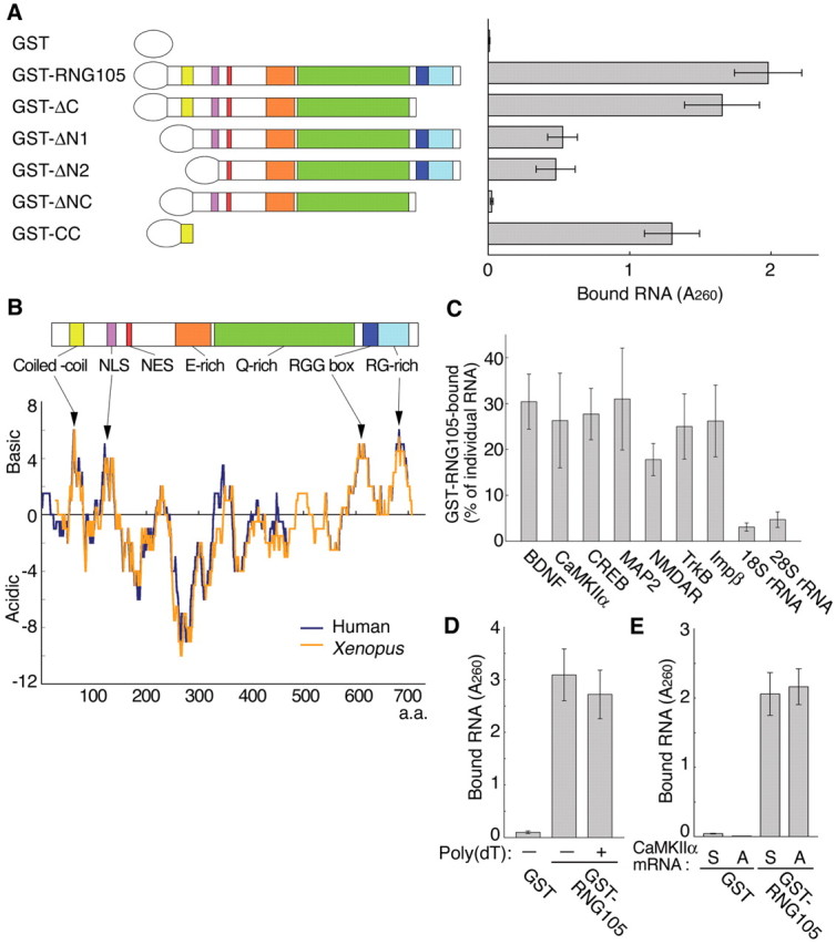
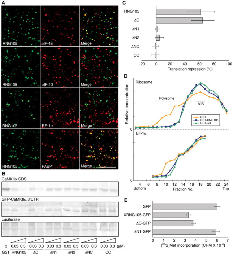
Local translation in neuronal dendrites is an important basis for long-term synaptic plasticity, and RNA granules in the dendrites are involved in the local translation. Here, we identify RNG105 (RNA granule protein 105), a novel RNA-binding protein, as a component of the RNA granules in dendrites of hippocampal neurons. The RNG105-localizing RNA granules contain mRNAs, the translational products of which play key roles in synaptic plasticity. RNG105 has an ability to repress translation both in vitro and in vivo, consistent with the finding that the RNA granule is translationally arrested in the basal conditions. Dissociation of RNG105 from the RNA granules is induced by BDNF, a growth factor responsible for synaptic plasticity. The RNG105 dissociation is coincident with the induction of local translation near the granules. These findings suggest that RNG105 is a translational repressor in the RNA granules and provide insight into the link between RNG105 dynamics and local translational regulation.


Figures

Figure 6.
Figure 6.
Dissociation of RNG105 from the RNA granules by BDNF stimulation. A, Time-lapse imaging of RNG105-GFP in the dendrites of cultured hippocampal neurons. The time after BDNF treatment is indicated. Arrowheads denote an RNG105-localizing RNA granule, the localization of which was stationary during the imaging period. Enhanced images are shown, and the inset is the original image of the same magnification. Scale bar, 5 μm. B, Left, Profile of RNG105-GFP fluorescence intensity in the granule during the experiment in A. Results obtained from eight granules are shown. RNG105-GFP fluorescence intensity was reduced in the granules, except for those near the cell bodies (pale blue and pink). Right, Control experiments with buffer (PBS) treatment. C, Time-lapse imaging of RNG105-mRFP1 and ribosomal L11-GFP in the dendrites. The time after BDNF treatment is indicated. Arrowheads and arrows denote RNG105-localizing RNA granules, the locations of which were unchanged during the experiment. At the arrowhead and the left arrow, RNG105-mRFP1 signals were significantly reduced after 25 and 15 min, respectively. In contrast, L11-GFP signals at the positions were rather constant during the experiment. Scale bar, 5 μm. D, Fluorescence intensities of RNG105-mRFP1 and L11-GFP in the granule denoted by the arrowheads in C were plotted (dashed lines). Results obtained from eight granules are shown as solid lines (means ± SE). E, F, Rat hippocampal slices were immunostained with anti-RNG105 antibody (E, left) and costained with antibody to phosphorylated eIF-4E (eIF-4E-P; E, middle). Although the slices were not treated with BDNF, some areas were stained with anti-eIF-4E-P antibody, which may be because these areas had been activated when the animals were alive. Dendritic areas are shown. RNG105 fluorescence in the granules in which eIF-4E-P was localized was relatively faint compared with that in other RNA granules. F, Fluorescence intensity of RNG105 in the eIF-4E-P-localizing granules was measured and compared with that in the eIF-4E-localizing granules. a.u., Arbitrary units. Scale bar, 10 μm. Error bars represent SEM.
Figure 7.
Figure 7.
BDNF induces mRNA shift from the RNA granules to polysomes and local translation near the granules. A, Extracts from control or BDNF-stimulated rat hippocampal slices were fractionated by sucrose density gradient centrifugation and then RNG105, staufen, and CaMKIIα mRNA we reanalyzed semiquantitatively (means from 3 independent experiments). B, The indicated mRNAs were analyzed as in A. The ratios of the mRNA in the RNA granule fractions to that in the lighter fractions (polysomes) are shown. C, Rat cultured hippocampal neurons were transfected with both GFP reporter fused to the CaMKIIα 3′UTR and RNG105-mRFP1. Time-lapse images of GFP and RNG105-mRFP1 proteins after BDNF stimulation are shown. Arrowheads denote the position of a granule with a stationary localization during the imaging period. RNG105-mRFP1 in the granule was reduced after BDNF stimulation, which coincided with the increase in GFP signal near the granule. To image clearly the increase in GFP signal near the granules, enhanced images of the GFP signal -1 and 40 min after the stimulation are shown at the bottom. Other granules were motile and changed their positions during the experiment. Scale bar, 5 μm. D, Profiles of RNG105-mRFP1 and GFP fluorescence intensity in the granule indicated by arrowheads in C. a.u., Arbitrary units. E, Profile of GFP fluorescence changes during the experiment in C. The changes in GFP fluorescence intensities near the RNG105-localizing RNA granules between the indicated time points and t = -10 min are plotted. Data are means ± SE from eight granules of two independent experiments. F, The same experiment as in E, except for the addition of 10 μg/ml cycloheximide (CHX) 10 min before the BDNF stimulation. Error bars represent SEM.
Figure 1.
Figure 1.
RNG105 is a novel protein with an RNA-binding motif and promotes the organization of cytoplasmic granules containing ribosomes and mRNAs. A, Predicted amino acid sequence of XRNG105. Solid line, Coiled-coil domain; dotted line, NLS; double line, nuclear export signal (NES); wavy line and bold letters, RGG box, an RNA-binding motif. B, Schematic diagram of XRNG105. E-rich, Glutamic acid-rich region; Q-rich, glutamine-rich region; RG-rich, arginine- and glycine-rich region. Other regions are described in A. C, Immunostaining of A6 cells with anti-XRNG105 antibody. Right, Differential interference contrast (DIC) image. Scale bar, 10 μm. D, A6 cells expressing XRNG105-GFP were stained with anti-XRNG105 antibody. Scale bar, 10 μm. Merge, Merged images. E, Immunoelectron microscopy of A6 cells expressing XRNG105-GFP stained with anti-XRNG105 antibody. Spots of immunogold (black arrowheads) are seen in a ribosome-rich area (∼1 μm in diameter) in the cytoplasm. White arrowheads denote ribosomes (inset). Scale bar, 500 nm. F, G, A6 cells expressing XRNG105-GFP, XRNG105ΔC-GFP, XRNG105ΔN-GFP, and GFP were stained with anti-RPS6 antibody (F) or stained by in situ hybridization with poly(dT) for mRNA detection (G). The right panels are DIC images. Scale bar, 10 μm.
Figure 2.
Figure 2.
RNG105 is highly expressed in dendrites of hippocampal neurons. A, Western blotting of extracts (30 μg each) from Xenopus tissues and A6 cells with anti-XRNG105 antibody. Arrows denote XRNG105. In brain and A6 cells, XRNG105 was detected as doublets, although the reasons for this are not known. B, Immunostaining of slices from the lateral pallium of Xenopus forebrain with anti-XRNG105 antibody. A schematic drawing is shown on the left. Scale bar, 50 μm. C, Rat brain extracts were immunoblotted with an antibody directed against mammalian RNG105. Arrow, RNG105. D, Slices from rat hippocampus (top) and cerebellum (bottom) were immunostained with anti-RNG105 antibody (green) and costained with anti-MAP2 antibody, which locates dendrites (red). RNG105 was concentrated in the dendritic region of the stratum radiatum (SR) and in the nuclei of pyramidal neurons [stratum pyramidale (SP)] in the hippocampus. In contrast, RNG105 was not detectable in the dendrites of Purkinje cells in the molecular layer (ML) of the cerebellum. Merge, Merged images; SO, stratum oriens; GL, granule cell layer; PL, Purkinje cell layer. Scale bars, 50 μm. E, Rat cultured hippocampal neurons were immunostained with anti-RNG105 antibody (top) and costained with anti-MAP2 antibody (bottom). Bottom, Composite with a differential interference contrast (DIC) image. Arrowheads indicate dendrites; arrows indicate an axon. Scale bar, 10 μm.
Figure 3.
Figure 3.
RNG105-localizing granules exhibit features common to RNA granules in neuronal dendrites. A, Rat hippocampal slices (top and middle) or cultured hippocampal neurons (bottom) were immunostained with anti-RNG105 antibody (green) and costained with anti-RPS6, anti-PSD-95, or anti-staufen antibodies (red). Dendritic areas are magnified and shown. Arrowheads denote colocalization. Merge, Merged images; SP, stratum pyramidale; SR, stratum radiatum. Scale bars, 10 μm. B, Rat hippocampal slices were immunostained with anti-RNG105 antibody (green) and costained by in situ hybridization with probes for CaMKIIα mRNA or control probes (CaMKIIα sense RNAs) (red). Dendritic areas are shown. Arrowheads denote colocalization. Scale bars, 10 μm. C, Rat hippocampal extracts were subjected to immunoprecipitation (IP) with anti-RNG105 antibody or control IgG. Precipitants were analyzed for RNG105 by Western blotting and for CaMKIIα mRNA and control importin β (Impβ) mRNA semiquantitatively by RT-PCR. D, Indicated mRNAs precipitated with RNG105 were analyzed by semiquantitative RT-PCR as in C. The ratio of the RNG105-bound mRNA compared with the input amount of the mRNA is shown. Error bars represent SEM.
Figure 4.
Figure 4.
RNG105 binds directly to mRNAs in vitro. A, Recombinant GST-RNG105 deletion mutants were incubated with RNAs isolated from rat hippocampus. RNAs bound to the recombinant proteins were extracted and quantified. ΔC, Deletion of C terminus (from RGG box to C-end); ΔN1, deletion of N terminus (from N-end to coiled-coil region); ΔN2, deletion of N terminus (from N-end to NLS); ΔNC, deletion of N and C termini (from N-end to coiled-coil region and from RGG box to C-end); CC, coiled-coil region. B, Acidity-basicity analysis of human and Xenopus RNG105. Scores of +1, +0.5, and -1 were given to Lys and Arg, His, and Asp and Glu, respectively. The sum of the scores of 21 aa (residues X -10 to X +10) is plotted against a residue number X. Arrows denote clusters of basic residues. NES, Nuclear export signal. C, Indicated RNAs bound to GST-RNG105 in A were analyzed by semiquantitative RT-PCR. The ratio of the GST-RNG105-bound RNA compared with the input amount of the RNA is shown. Impβ, Importin β. D, Recombinant proteins were incubated with the hippocampal RNAs in the presence or absence of poly(dT), and RNAs bound to the recombinant proteins were quantified. E, Recombinant proteins were incubated with sense (S) or antisense (A) CaMKIIα mRNA, which had been transcribed in vitro. Bound RNA was quantified. Error bars represent SEM.
Figure 5.
Figure 5.
The RNA granule is translationally arrested, and RNG105 negatively regulates translation. A, Rat hippocampal slices were immunostained with anti-RNG105 antibody (green) and costained with anti-eIF-4E, anti-eIF-4G, anti-EF-1α, or anti-PABP antibody (red). Dendritic areas are shown. Merge, Merged image. Scale bar, 10 μm. B, RNG105 inhibits translation in vitro. The CaMKIIα CDS, GFP-CaMKIIα 3′UTR, and luciferase mRNAs were translated in the presence of indicated recombinant proteins in rabbit reticulocyte lysates and detected by SDS-PAGE. CC, Coiled coil. C, The bands in B were quantified, and translational repression activity of each recombinant protein (0.3 μm) was calculated. Data are means ± SE of translational repression of the three mRNAs by the recombinant proteins. D, The lysates incubated with the recombinant proteins (0.3 μm) in B were fractionated by sucrose density gradient centrifugation. Elution profiles of ribosomes and EF-1α are shown. E, RNG105 inhibits translation in vivo. A6 cells expressing XRNG105-GFP, ΔC-GFP, ΔN1-GFP, or control GFP were labeled with [35S]methionine for 10 min and then proteins were isolated from the cells by TCA precipitation. Incorporation of [35S]methionine ([35S]met) into newly synthesized proteins is shown. CPM, Counts per minute. Error bars represent SEM.

Similar articles

Cited by

References

    1. Aakalu G, Smith WB, Nguyen N, Jiang C, Schuman EM (2001) Dynamic visualization of local protein synthesis in hippocampal neurons. Neuron 30: 489-502. - PubMed
    1. Angenstein F, Evans AM, Settlage RE, Moran ST, Ling S-C, Klintsova AY, Shabanowitz J, Hunt DF, Greenough WT (2002) A receptor for activated C kinase is part of messenger ribonucleoprotein complexes associated with polyA-mRNAs in neurons. J Neurosci 22: 8827-8837. - PMC - PubMed
    1. Burd CG, Dreyfuss G (1994) Conserved structures and diversity of functions of RNA-binding proteins. Science 265: 615-621. - PubMed
    1. Burgin KE, Waxham MN, Rickling S, Westgate SA, Mobley WC, Kelly PT (1990) In situ hybridization histochemistry of Ca2+/calmodulin-dependent protein kinase in developing rat brain. J Neurosci 10: 1788-1798. - PMC - PubMed
    1. Cahuzac B, Berthonneau E, Birlirakis N, Guittet E, Mirande M (2000) A recurrent RNA-binding domain is appended to eukaryotic aminoacyl-tRNA synthetases. EMBO J 19: 445-452. - PMC - PubMed

Publication types

MeSH terms

Substances