. 2005 Apr 25;579(11):2387-96.
doi: 10.1016/j.febslet.2005.03.039.

Sumoylation of the nucleocapsid protein of severe acute respiratory syndrome coronavirus

Affiliations

Sumoylation of the nucleocapsid protein of severe acute respiratory syndrome coronavirus

Frank Qisheng Li et al. FEBS Lett. .

Abstract

Severe acute respiratory syndrome coronavirus (SARS-CoV) encodes a highly basic nucleocapsid (N) protein of 422 amino acids. Similar to other coronavirus N proteins, SARS-CoV N protein is predicted to be phosphorylated and may contain nuclear localization signals, serine/arginine-rich motif, RNA binding domain and regions responsible for self-association and homo-oligomerization. In this study, we demonstrate that the protein is posttranslationally modified by covalent attachment to the small ubiquitin-like modifier. The major sumoylation site was mapped to the (62)lysine residue of the N protein. Further expression and characterization of wild type N protein and K62A mutant reveal that sumoylation of the N protein drastically promotes its homo-oligomerization, and plays certain roles in the N protein-mediated interference of host cell division. This is the first report showing that a coronavirus N protein undergoes posttranslational modification by sumoylation, and the functional implication of this modification in the formation of coronavirus ribouncleoprotein complex, virion assembly and virus-host interactions.


Figures

Figure 1
Figure 1
(A) Schematic representation of the SARS‐CoV N protein, showing the SUMO‐1 conjugation site (K62), three putative nucleolar localization signals, the S/R‐rich motif, the RNA‐binding domain and self‐association domain. (B) Analysis of the expression of N protein in bacterial and mammalian cells. Plasmids pGEX‐5X‐1 and pGEX‐N were transformed into E. coli BL21, and expressed by induction with IPTG. The GST (lane 1) and GST‐N (lane 2) proteins were affinity‐purified and separated on an SDS–10% polyacrylamide gel. The protein was visualized by staining the gel with Coomassie blue. Total cell lysates prepared from HeLa cells transfected with pKT0‐Flag empty plasmid (lane 3) and Flag‐tagged N protein (lane 4) were separated by SDS–PAGE and analyzed by Western blotting with anti‐Flag antibody. Numbers on the left and right indicate molecular masses in kilodaltons.
Figure 2
Figure 2
Modification of SARS‐CoV N protein by SUMO‐1. (A) Analysis of sumoylation of N protein by Western blotting. Cell lysates prepared from HeLa cells overexpressing either the Flag‐tagged N protein alone (lanes 1 and 2) or together with SUMO‐1 (lanes 3, 4, 5, and 6) were prepared either in the presence (lanes 2, 3 and 5) or absence (lanes 1 and 4) of the isopeptidase inhibitors IAA and NEM or by direct lysis in preheated SDS loading buffer (lane 6). The polypeptides were separated by SDS–PAGE under either reducing (lanes 1–3) or non‐reducing (lanes 4–6) conditions and analyzed by Western blotting with anti‐Flag antibody. The three major isoforms of N protein are indicated by brackets and the major SUMO‐1 modified form of N protein is indicated by asterisks. Numbers on the left indicate molecular masses in kilodaltons. (B) Analysis of sumoylation of N protein by immunoprecipitation and Western blotting. Total cell lysates were prepared, in the presence of the isopeptidase inhibitors IAA and NEM, from HeLa cells expressing SUMO‐1 (lanes 1 and 4), Flag‐N (lanes 2 and 5), and Flag‐N + SUMO‐1 (lanes 3 and 6), and immunoprecipitated with either anti‐Flag (lanes 1, 2 and 3) or anti‐SUMO‐1 antibody (lanes 4, 5, and 6). The immunoprecipitated proteins were separated by SDS–PAGE under nonreducing conditions and analyzed by Western blotting with anti‐Flag antibody. The major SUMO‐1 modified forms of N protein are indicated by asterisks, and the immunoglobulin is indicated by Ig.
Figure 3
Figure 3
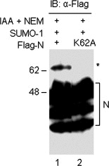
Mapping of the major sumoylation site on SARS‐CoV N protein. Cell lysates from HeLa cells overexpressing wild type (lane 1) and K62A mutant N protein (lane 2) together with SUMO‐1 were prepared in the presence of IAA and NEM. The polypeptides were separated by SDS–PAGE under nonreducing conditions and analyzed by Western blotting with anti‐Flag antibody. The three major isoforms of N protein are indicated by brackets and the major SUMO‐1 modified form of N protein is indicated by asterisks. Numbers on the left indicate molecular masses in kilodaltons.
Figure 4
Figure 4
Analysis of the homo‐oligomerization of SARS‐CoV N protein. (A) HeLa cells overexpressing wild type N protein only (lane 1) or together with SUMO‐1 (lane 2) were lysed in the absence (lane 1) or presence of IAA and NEM (lane 2). The polypeptides were separated by SDS–PAGE under reducing conditions and analyzed by Western blotting with anti‐Flag antibody. The three major isoforms of N protein are indicated by brackets and the major SUMO‐1 modified form of N protein is indicated by asterisks. The dimers are also indicated. Numbers on the left indicate molecular masses in kilodaltons. (B) HeLa cells overexpressing wild type N protein only (lanes 1 and 2), wild type + SUMO‐1 (lanes 3 and 4) or the K62A mutant N protein + SUMO‐1 (lane 5) were lysed in the presence of IAA and NEM (lanes 1, 3 and 5) or with preheated SDS loading buffer (lanes 2 and 4). The polypeptides were separated by SDS–PAGE under nonreducing conditions and analyzed by Western blotting with anti‐Flag antibody. The three major isoforms of N protein are indicated by brackets and the major SUMO‐1 modified form of N protein is indicated by asterisks. The dimers and tetramers are also indicated. Numbers on the left indicate molecular masses in kilodaltons.
Figure 5
Figure 5
Further analysis of the homo‐oligomerization of SARS‐C‐V N protein. (A) HeLa cells overexpressing the empty pKT0‐Flag (lane 1), wild type N protein (lane 2), or the K62A mutant N protein (lane 3) were lysed in the presence of IAA and NEM. The lysates were immunoprecipitated with anti‐Flag antibody. The precipitated polypeptides were separated by SDS–PAGE under nonreducing conditions and analyzed by Western blotting with anti‐Flag antibody. The three major isoforms of N protein, the N protein dimer, and the immunoglobulin are indicated. Numbers on the left indicate molecular masses in kilodaltons. (B) HeLa cells overexpressing the Flag‐tagged wild type N protein alone (lane 1), the c‐Myc‐tagged wild type N protein alone (lane 2), the Flag‐tagged wild type and c‐Myc‐tagged wild type N protein (lane 3), and the Flag‐tagged K62A mutant and c‐Myc‐tagged wild type N protein (lane 4) were lysed in the presence of IAA and NEM. Polypeptides were immunoprecipitated with anti‐Myc antibody, separated by SDS–PAGE under nonreducing conditions, and analyzed by Western blotting with anti‐Flag antibody. The N protein dimer and the immunoglobulin are indicated. A band migrating at approximately 50 kDa, which may represent the antibody heavy chain, is also indicated. (C) HeLa cells overexpressing the Flag‐tagged wild type N protein alone (lanes 1 and 5), the c‐Myc‐tagged wild type N protein alone (lanes 2 and 6), the Flag‐tagged wild type and c‐Myc‐tagged wild type N protein (lanes 3 and 7), and the Flag‐tagged K62A mutant and c‐Myc‐tagged wild type N protein (lanes 4 and 8) were lysed in the presence of IAA and NEM. Polypeptides were separated by SDS–PAGE under nonreducing conditions, and analyzed by Western blotting with either anti‐Flag antibody (lanes 1–4), or anti‐Myc antibody (lanes 5–8). The three major isoforms of N protein and the dimer are indicated.
Figure 6
Figure 6
Subcellular localization of SARS‐CoV N protein. HeLa cells expressing EGFP (A), N‐EGFP (B) and K62A‐EGFP fusion protein (C) were detected directly under the fluorescence microscope at 36 h posttransfection. Indirect immunofluorescent staining of HeLa cells transfected with empty plasmid (D), wild type N protein (E) and K62A mutant (F), was carried out at 18 h posttransfection with rabbit anti‐SARS N antisera and FITC‐labeled goat anti‐rabbit antibodies. The multinucleated cells are indicated by arrows.
Figure 7
Figure 7
Nucleolar localization of SARS‐CoV N protein. Indirect immunofluorescent staining of HeLa cells expressing the Flag‐tagged N protein was carried out at 18 h posttransfection with mouse anti‐Flag (A) and rabbit anti‐fibrillarin (B) antisera. The N protein was then detected by TRITC‐conjugated anti‐mouse secondary antibodies and fibrillarin was detected by FITC‐labeled anti‐rabbit antibodies. (C) represents the merged images. All images were taken using a Zeiss LSM510 META laser scanning confocal microscope.
Figure 8
Figure 8
Effects of sumoylation of SARS‐CoV N protein on its interference of host cell division. Percentages of multinucleated cells among HeLa cells expressing EGFP, N‐EGFP, N‐EGFP + SUMO‐1, and K62A‐EGFP + SUMO‐1 were calculated by counting the multinucleated cells among 300 green cells under the fluorescence microscope. The percentages and S.D. are results of three repeated experiments.

Similar articles

Cited by

References

    1. Baric R.S., Nelson G.W., Fleming J.O., Deans R.J., Keck J.G., Casteel N., Stohlman S.A., Interactions between coronavirus nucleocapsid protein and viral RNAs: implications for viral transcription. J. Virol., 62, (1988), 4280– 4287. - PMC - PubMed
    1. Chang L.K., Lee Y.H., Cheng T.S., Hong Y.R., Lu P.J., Wang J.J., Wang W.H., Kuo C.W., Li S.S., Liu S.T., Posttranslational modification of Rta of Epstein–Barr virus by SUMO-1. J. Biol. Chem., 279, (2004), 38803– 38812. - PubMed
    1. Chang R.Y., Brian D.A., cis requirement for N-specific protein sequence in bovine coronavirus defective interfering RNA replication. J. Virol., 70, (1996), 2210– 2217. - PMC - PubMed
    1. Chen H., Wurm T., Britton P., Brooks G., Hiscox J.A., Interaction of the coronavirus nucleocapsid with nucleolar antigens and the host cell. J. Virol., 76, (2002), 5233– 5250. - PMC - PubMed
    1. Davies H.A., Dourmashkin R.R., MacNaughton R., Ribonucleoprotein of avian infectious bronchitis virus. J. Gen. Virol., 53, (1981), 67– 74. - PubMed

Publication types

MeSH terms

Substances