

Voxel-based analysis of MRI detects abnormal visual cortex in children and adults with amblyopia
- PMID: 15846772
- PMCID: PMC6871714
- DOI: 10.1002/hbm.20109
Voxel-based analysis of MRI detects abnormal visual cortex in children and adults with amblyopia
Abstract
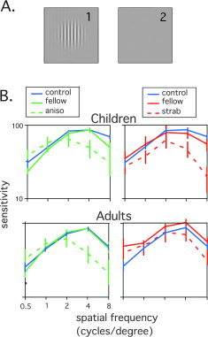
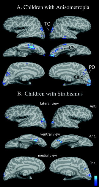
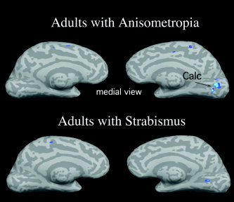
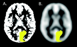
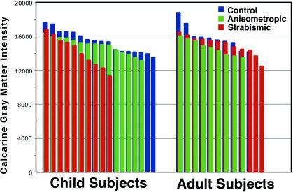
Amblyopia, sometimes called "lazy eye," is a relatively common developmental visual disorder well characterized behaviorally; however, the neural substrates associated with amblyopia in humans remain unclear. We hypothesized that abnormalities in the cerebral cortex of subjects with amblyopia exist, possibly as a result of experience-dependent neuronal plasticity. Anatomic magnetic resonance imaging (MRI) and psychophysical vision testing was carried out on 74 subjects divided into two age ranges, 7-12 years and 18-35 years, and three diagnoses, strabismic amblyopia, anisometropic amblyopia, and normal vision. We report a behavioral impairment in contrast sensitivity for subjects with amblyopia, consistent with previous reports. When the high-resolution MRI brain images were analyzed quantitatively with optimized voxel-based morphometry, results indicated that adults and children with amblyopia have decreased gray matter volume in visual cortical regions, including the calcarine sulcus, known to contain primary visual cortex. This finding was confirmed with a separate region-of-interest analysis. For the children with amblyopia, additional gray matter reductions in parietal-occipital areas and ventral temporal cortex were detected, consistent with recent reports that amblyopia can result in spatial location and object processing deficits. These data are the first to provide possible neuroanatomic bases for the loss of binocularity and visual sensitivity in children and adults with amblyopia.
Figures
Similar articles
-
Combination of blood oxygen level-dependent functional magnetic resonance imaging and visual evoked potential recordings for abnormal visual cortex in two types of amblyopia.Mol Vis. 2012;18:909-19. Epub 2012 Apr 11. Mol Vis. 2012. PMID: 22539870 Free PMC article.
-
Neuronal correlates of amblyopia in the visual cortex of macaque monkeys with experimental strabismus and anisometropia.J Neurosci. 1998 Aug 15;18(16):6411-24. doi: 10.1523/JNEUROSCI.18-16-06411.1998. J Neurosci. 1998. PMID: 9698332 Free PMC article.
-
Binocularity and spatial frequency dependence of calcarine activation in two types of amblyopia.Neurosci Res. 2001 Jun;40(2):147-53. doi: 10.1016/s0168-0102(01)00220-6. Neurosci Res. 2001. PMID: 11377753
-
The Role of Binocularity in Anisometropic Amblyopia.J Binocul Vis Ocul Motil. 2019 Oct-Dec;69(4):141-152. doi: 10.1080/2576117X.2019.1656034. Epub 2019 Sep 5. J Binocul Vis Ocul Motil. 2019. PMID: 31486743 Review.
-
Amblyopia and the binocular approach to its therapy.Vision Res. 2015 Sep;114:4-16. doi: 10.1016/j.visres.2015.02.009. Epub 2015 Apr 20. Vision Res. 2015. PMID: 25906685 Review.
Cited by
-
Visual imagery and functional connectivity in blindness: a single-case study.Brain Struct Funct. 2016 May;221(4):2367-74. doi: 10.1007/s00429-015-1010-2. Epub 2015 Feb 18. Brain Struct Funct. 2016. PMID: 25690326 Free PMC article.
-
Retinotopic maps and foveal suppression in the visual cortex of amblyopic adults.J Physiol. 2007 Aug 15;583(Pt 1):159-73. doi: 10.1113/jphysiol.2007.136242. Epub 2007 Jul 12. J Physiol. 2007. PMID: 17627994 Free PMC article.
-
Visuomotor Behaviour in Amblyopia: Deficits and Compensatory Adaptations.Neural Plast. 2019 Jun 9;2019:6817839. doi: 10.1155/2019/6817839. eCollection 2019. Neural Plast. 2019. PMID: 31281344 Free PMC article. Review.
-
Changes in cortical grey matter density associated with long-standing retinal visual field defects.Brain. 2009 Jul;132(Pt 7):1898-906. doi: 10.1093/brain/awp119. Epub 2009 May 25. Brain. 2009. PMID: 19467992 Free PMC article.
-
Altered spontaneous activity in anisometropic amblyopia subjects: revealed by resting-state FMRI.PLoS One. 2012;7(8):e43373. doi: 10.1371/journal.pone.0043373. Epub 2012 Aug 24. PLoS One. 2012. PMID: 22937041 Free PMC article.
References
-
- Ashburner J, Friston KJ (2000): Voxel‐based morphometry: the methods. Neuroimage 11: 805–821. - PubMed
-
- Ashburner J, Friston KJ (2001): Why voxel‐based morphometry should be used. Neuroimage 14: 1238–1243. - PubMed
-
- Asper L, Crewther D, Crewther SG (2000): Strabismic amblyopia. Part 2. Neural processing. Clin Exp Optom 83: 200–211. - PubMed
-
- Baddini‐Caramelli C, Hatanaka M, Polati M, Umino AT, Susanna R Jr (2001): Thickness of the retinal nerve fiber layer in amblyopic and normal eyes: a scanning laser polarimetry study. J AAPOS 5: 82–84. - PubMed
Publication types
MeSH terms
Grants and funding
LinkOut - more resources
Full Text Sources
Medical
