. 2005 May;25(9):3506-18.
doi: 10.1128/MCB.25.9.3506-3518.2005.

SUMO-1 modification of PIASy, an E3 ligase, is necessary for PIASy-dependent activation of Tcf-4

Affiliations

SUMO-1 modification of PIASy, an E3 ligase, is necessary for PIASy-dependent activation of Tcf-4

Motomasa Ihara et al. Mol Cell Biol. 2005 May.

Abstract

We have previously shown that modification of Tcf-4, a transcription factor in the Wnt pathway, with SUMO by PIASy, a SUMO E3 ligase, enhances its transcriptional activity. Since PIASy itself was also modified with SUMO-1, we studied the role of sumoylation of PIASy in the regulation of Tcf-4. Lys(35) was found to be a sumoylation site of PIASy. PIASy(K35R), in which Lys(35) was mutated to Arg, did not enhance sumoylation of Tcf-4, although this PIASy mutant did not lose the ligase activity of sumoylation for other proteins. Wild-type PIASy and PIASy(K35R) showed a distinct distribution in the nucleus, although both were colocalized with Tcf-4. Promyelocytic leukemia protein, which is involved in transcriptional regulation, was associated with PIASy(K35R) more frequently than wild-type PIASy in the nucleus. PIASy(K35R) could not stimulate the transcriptional activity of Tcf-4 under the conditions in which wild-type PIASy enhanced it. Conjugation of SUMO-1 to the amino terminus of PIASy(K35R) neither enhanced sumoylation of Tcf-4 nor stimulated the transcriptional activity of Tcf-4. These results suggest that sumoylation of Lys(35) in PIASy determines the nuclear localization of PIASy and that it is necessary for PIASy-dependent sumoylation and transcriptional activation of Tcf-4.


Figures
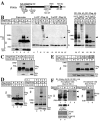
FIG. 1.
FIG. 1.
Modification of PIASy with SUMO. (A) Schematic representation of PIASy used in the present study. (B) Modification of PIASy with SUMO-1. 293 cells expressing the indicated proteins were incubated with 10% TCA to precipitate the proteins. The resulting precipitates were dissolved in Laemmli sample buffer and probed with the anti-Flag and anti-HA antibodies (lanes 1 to 5). Lysates (200 μg of protein) prepared by extraction with RIPA buffer were immunoprecipitated with the anti-Flag antibody, and the immunoprecipitates were probed with the anti-HA antibody (lanes 6 to 10). The immunoprecipitates were further treated with SDS and immunoprecipitated with the anti-Flag antibody again. The second immunoprecipitates were also probed with the anti-HA antibody (lanes 11 to 15), and some of the supernatant (sup) and precipitates (ppt) were probed with the anti-HA and anti-Flag antibodies (lanes 16 to 21). The reason why immunoglobulin (lanes 18 and 21) and Flag-PIASy (lane 21) were detected in the supernatant of the second immunoprecipitation is that the immunoprecipitation efficiency by the anti-Flag antibody isca. 70%. Arrowheads indicate Flag-PIASy modified with HA-SUMO-1. Ab, antibody; IP, immunoprecipitation; IB, immunoblotting; WT, wild type; ΔGG, SUMO-1-(1-95). (C) 293 cells expressing the indicated proteins were incubated with TCA to precipitate the proteins, and the precipitates were probed with the anti-Flag and anti-HA antibodies. CA, a PIASy RING mutant in which Cys342 and Cys347 are changed to Ala. (D) Effects of SUMO-1 and SUMO-3 on sumoylation of PIASy and Tcf-4. Precipitates were prepared from 293 cells expressing the indicated proteins by adding TCA and probed with the anti-Flag and anti-HA antibodies (lanes 1 to 3) or the anti-Tcf-4 and anti-HA antibodies (lanes 4 to 6). (E) Identification of sumoylation site of PIASy. Precipitates were prepared from 293 cells expressing the indicated proteins by adding TCA and probed with the anti-Flag and anti-HA antibodies. K35R, a PIASy mutant in which Lys35 is changed to Arg; K128R, a PIASy mutant in which Lys128 is changed to Arg. K35R/K128R, a PIASy mutant in which both Lys35 and Lys128 are changed to Arg. (F) Modification of endogenous PIASy with endogenous SUMO-1. HeLa S3 cells were transfected with (lanes 2, 4, and 6) or without (lanes 1, 3, and 5) a dsRNAi oligonucleotide for Axam. The nuclear extracts (300 μg of protein) were incubated with His6-SUMO-1 immobilized on nickel-agarose. After His6-SUMO-1 was precipitated by centrifugation, the precipitates were probed with the anti-PIASy (lanes 1 and 2) or anti-SUMO-1 (lanes 3 and 4) antibody. Arrowheads indicate PIASy modified with SUMO-1, and the arrow indicates PIASy. Bottom panel shows a longer exposure of the same immunoblot. The lysates (20 μg of protein) were directly probed with the anti-Axam and anti-GSK-3β antibodies (lanes 5 and 6). To compare the molecular masses of endogenous PIASy and Flag-PIASy, the nuclear extracts expressing Flag-PIASy (10 μg of protein) (lane 7) and the nuclear extracts without expression of Flag-PIASy (50 μg of protein) (lane 8) were incubated with His6-SUMO-1 immobilized on nickel-agarose. After His6-SUMO-1 was precipitated by centrifugation, the precipitates were probed with the anti-PIASy antibody. The results shown are representative of three independent experiments.
FIG. 2.
FIG. 2.
Sumoylation of Tcf-4 by PIASyK35R. (A) Ability of PIASyK35R to enhance sumoylation. 293 cells expressing the indicated proteins were incubated with 10% TCA, and the precipitates were probed with the anti-HA and anti-Flag antibodies. IB, immunoblotting; Ab, antibody; WT, wild type; CA, PIASy RING mutant; ΔGG, SUMO-1-(1-95). (B) Sumoylation of Tcf-4 in vitro. GST-Tcf-4 was incubated with GST-Aos1/His6-Uba2, GST-SUMO-1(GG), His6-Ubc9, and MBP-PIASy, or MBP-PIASyK35R. After the incubation, the mixtures were probed with the anti-Tcf-4 antibody. Arrowheads indicate GST-Tcf-4 modified with GST-SUMO-1, and the arrow indicates GST-Tcf-4. (C) Sumoylation of Tcf-4 in intact cells. Precipitates were prepared from 293 cells expressing the indicated proteins by adding TCA and probed with the anti-Tcf-4 and anti-Flag antibodies. The arrow indicates Tcf-4. The results shown are representative of three independent experiments. (D) Interaction of Tcf-4 with PIASy. The lysates (20 μg of protein) of COS cells expressing the indicated proteins were probed with the anti-HA and anti-Flag antibodies (lanes 1 to 4). The same lysates (200 μg of protein) were immunoprecipitated with the anti-Flag antibody, and the immunoprecipitates were probed with the anti-HA and anti-Flag antibodies (lanes 5 to 7).
FIG. 3.
FIG. 3.
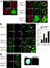
Localization of PIASy in the nucleus. (A) Sumoylation-dependent localization of PIASy. (a) Control COS cells were stained with the anti-PIASy antibody. (b to m) COS cells expressing Flag-PIASy WT alone (b), Flag-PIASyK35R alone (c), HA-SUMO-1 WT alone (d), Flag-PIASy WT and HA-SUMO-1 WT (e to g), Flag-PIASyK35R and HA-SUMO-1 WT (h to j), or Flag-PIASy WT and HA-SUMO-1(ΔGG) (k to m) were stained with the anti-Flag antibody to detect PIASy (b, c, e, h, and k) and the anti-HA antibody to detect SUMO-1 (d, f, i, and l). The merged images are shown in subpanels g, j, and m. Bars, 5 μm. (B) Localization of PIASy and PML in the nucleus. GFP-PIASy WT alone (a to d), GFP-PIASyK35R alone (e to h), GFP-PIASy WT and HA-SUMO-1 WT (i to l), or GFP-PIASyK35R and HA-SUMO-1 WT (m to p) were expressed in COS cells. The cells were viewed directly with a confocal scanning microscope to detect GFP-PIASy (a, e, i, and m) and stained with the anti-PML antibody to detect endogenous PML (b, f, j, and n). The merged images are shown in subpanels c, g, k, and o. The areas indicated by arrowheads are further enlarged (d, h, l, and p). Bars, 5 μm. (C) Percentages of PML associated with PIASy or PIASyK35R. The numbers of PML showing the colocalization or adjacent localization with PIASy or PIASyK35R were counted in the cells used in panel B. The black bars indicate the percentages of PML colocalized with or located next to PIASy or PIASyK35R. The white bars indicate the percentages of PML, which is present apart from PIASy or PIASyK35R. More than 100 cells were examined in three independent experiments. (D) Localization of PIASy in the nuclear matrix. After COS cells expressing Flag-PIASy WT and HA-SUMO-1 WT (a and b) or Flag-PIASyK35R and HA-SUMO-1 WT (c and d) were treated with DNase I, the cells were stained with the anti-Flag (a and c) and anti-HA (b and d) antibodies. The nuclei incubated with (f) or without (e) DNase I were stained blue with DAPI. (E) Biochemical subcellular fractionation. 293 cells expressing Flag-PIASy WT and HA-SUMO-1 WT (lanes 1 to 4) or Flag-PIASyK35R and HA-SUMO-1 WT (lanes 5 to 8) were fractionated into total homogenate (lanes 1 and 5), 3,300 × g supernatant fraction (3300 g sup) (lanes 2 and 6), nuclear soluble fraction (N. soluble) (lanes 3 and 7), and nuclear insoluble fraction (N. insoluble) (lanes 4 and 8). Proteins from the fractions were probed with the anti-Flag, anti-α-tubulin, and anti-histone H1 antibodies. The results shown are representative of four independent experiments. WT, wild type.
FIG. 3.
FIG. 3.
Localization of PIASy in the nucleus. (A) Sumoylation-dependent localization of PIASy. (a) Control COS cells were stained with the anti-PIASy antibody. (b to m) COS cells expressing Flag-PIASy WT alone (b), Flag-PIASyK35R alone (c), HA-SUMO-1 WT alone (d), Flag-PIASy WT and HA-SUMO-1 WT (e to g), Flag-PIASyK35R and HA-SUMO-1 WT (h to j), or Flag-PIASy WT and HA-SUMO-1(ΔGG) (k to m) were stained with the anti-Flag antibody to detect PIASy (b, c, e, h, and k) and the anti-HA antibody to detect SUMO-1 (d, f, i, and l). The merged images are shown in subpanels g, j, and m. Bars, 5 μm. (B) Localization of PIASy and PML in the nucleus. GFP-PIASy WT alone (a to d), GFP-PIASyK35R alone (e to h), GFP-PIASy WT and HA-SUMO-1 WT (i to l), or GFP-PIASyK35R and HA-SUMO-1 WT (m to p) were expressed in COS cells. The cells were viewed directly with a confocal scanning microscope to detect GFP-PIASy (a, e, i, and m) and stained with the anti-PML antibody to detect endogenous PML (b, f, j, and n). The merged images are shown in subpanels c, g, k, and o. The areas indicated by arrowheads are further enlarged (d, h, l, and p). Bars, 5 μm. (C) Percentages of PML associated with PIASy or PIASyK35R. The numbers of PML showing the colocalization or adjacent localization with PIASy or PIASyK35R were counted in the cells used in panel B. The black bars indicate the percentages of PML colocalized with or located next to PIASy or PIASyK35R. The white bars indicate the percentages of PML, which is present apart from PIASy or PIASyK35R. More than 100 cells were examined in three independent experiments. (D) Localization of PIASy in the nuclear matrix. After COS cells expressing Flag-PIASy WT and HA-SUMO-1 WT (a and b) or Flag-PIASyK35R and HA-SUMO-1 WT (c and d) were treated with DNase I, the cells were stained with the anti-Flag (a and c) and anti-HA (b and d) antibodies. The nuclei incubated with (f) or without (e) DNase I were stained blue with DAPI. (E) Biochemical subcellular fractionation. 293 cells expressing Flag-PIASy WT and HA-SUMO-1 WT (lanes 1 to 4) or Flag-PIASyK35R and HA-SUMO-1 WT (lanes 5 to 8) were fractionated into total homogenate (lanes 1 and 5), 3,300 × g supernatant fraction (3300 g sup) (lanes 2 and 6), nuclear soluble fraction (N. soluble) (lanes 3 and 7), and nuclear insoluble fraction (N. insoluble) (lanes 4 and 8). Proteins from the fractions were probed with the anti-Flag, anti-α-tubulin, and anti-histone H1 antibodies. The results shown are representative of four independent experiments. WT, wild type.
FIG. 4.
FIG. 4.
Colocalization of PIASy and Tcf-4 in the nucleus. (A) Representative patterns of Tcf-4 distribution. COS cells expressing GFP-Tcf-4 and HA-SUMO-1 WT (a to c), GFP-Tcf-4, Flag-PIASy WT, and HA-SUMO-1 WT (d to g), or GFP-Tcf-4, Flag-PIASyK35R, and HA-SUMO-1 WT (h to k) were stained with the anti-HA antibody to detect SUMO-1 (b, f, and j), and the anti-Flag antibody to detect PIASy (e and i). Some cells were viewed directly with a confocal scanning microscope to detect GFP-Tcf-4 (a, d, and h). The merged images are shown in panels c, g, and k. Bars, 5 μm. (B) Percentages of the cells showing the different distribution of Tcf-4. The numbers of the cells showing different distribution of GFP-Tcf-4 were counted among the cells used in Fig. 4A. The black, hatched, and white bars indicate the diffuse, small punctate, and large granule patterns, respectively. More than 100 cells were examined in four independent experiments. WT, wild type.
FIG. 5.
FIG. 5.
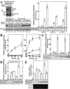
Activation of Tcf-4 by PIASy and SUMO-1. (A) dsRNAi for PIASy. The nuclear extracts (50 μg of protein) of HeLa S3 cells transfected with a dsRNAi oligonucleotide (lane 2) or an ss oligonucleotide (lane 1) for PIASy were precipitated with His6-SUMO-1, and the precipitates were probed with the anti-PIASy antibody. The same nuclear extracts (5 μg of protein) were probed with the anti-histone H1 antibody. Lysates from HeLa S3 cells transfected with pCMV5-Flag/PIASy (lanes 4 and 5), pEF-BOS-HA/PIAS3 (lanes 7 and 8), or pCGN/PIASxα (lanes 10 and 11), and a dsRNAi oligonucleotide (lanes 4, 7, and 10) or an ss oligonucleotide (lanes 5, 8, and 11) for PIASy were probed with the anti-Flag or anti-HA and anti-GSK-3β antibodies. Lysates from HeLa S3 cells transfected with pCMV5-Flag/PIASy (lane 3), pEF-BOS-HA/PIAS3 (lane 6), or pCGN/PIASxα (lane 9) alone were used as a control. After HeLa S3 cells had been transfected with or without the indicated RNAi oligonucleotide for PIASy, the cells were further transfected with pEF-BOS/hTcf-4E (0.1 μg), TOP-fos-Luc (0.5 μg), pCGN/SUMO-1 (0.5 μg) and pUC/EF-1α/β-cateninSA (0.5 μg) (right panel). The luciferase activity was measured and expressed as the fold increase compared to the level observed in cells transfected with TOP-fos-Luc and pEF-BOS/hTcf-4E. All of the transfection experiments were performed at least five times, and the results shown are means ± the standard error. ds, double-stranded oligonucleotide; ss, single-stranded sense oligonucleotide. (B) SUMO-1-dependent activation of Tcf-4. The indicated amounts of pCMV5-Flag/PIASy, pEF-BOS/hTcf-4E (0.1 μg), and TOP-fos-Luc (0.5 μg) were transfected into 293 cells with pCGN/SUMO-1 (•), pCGN/SUMO-3 (▪), or pCGN/SUMO-1(ΔGG) (□) (0.5 μg) or without these plasmids (○) (left panel). The indicated amounts of pCMV5-Flag/PIASy, pEF-BOS/hTcf-4E (0.1 μg), and TOP-fos-Luc (0.5 μg) were transfected into 293 cells with pCGN/SUMO-1 (0.5 μg) (•), pUC/EF-1α/β-cateninSA (0.1 μg) (□), or pCGN/SUMO-1 and pUC/EF-1α/β-cateninSA (▪) or without these plasmids (○) (right panel). (C) Effects of sumoylation of PIASy on Tcf-4. pCGN/SUMO-1 (0.5 μg), pUC/EF-1α/β-cateninSA (0.1 μg), pEF-BOS/hTcf-4E (0.1 μg), and TOP-fos-Luc or FOP-fos-Luc (0.5 μg) were transfected into 293 cells with pCMV5-Flag/PIASy, pCMV5-Flag/PIASyK35R, pCMV5-Flag/PIASyK128R, or pCMV5-Flag/PIASyK35R/K128R (0.1 μg). (D) Effects of PIASy on the Tcf activity for natural Tcf-responsive promoters. Cyclin D1 (−163)-Luc (0.5 μg), pCGN/SUMO-1 (0.1 μg), pUC/EF-1α/β-cateninSA (0.5 μg), and pEF-BOS/hTcf-4E (0.1 μg) were transfected into 293 cells with pCMV5-Flag/PIASy or pCMV5-Flag/PIASyK35R (0.5 μg). The luciferase activity was measured and is expressed as the fold increase compared to the level observed in the cells transfected with cyclin D1 (−163)-Luc, pCGN/SUMO-1, and pEF-BOS/hTcf-4E. (E) RT-PCR analysis of expression of endogenous cyclin D1 mRNA. Total RNA from 293 cells transfected with pCGN/SUMO-1 (0.1 μg), pUC/EF-1α/β-cateninSA (0.5 μg), and pCMV5-Flag/PIASy or pCMV5-Flag/PIASyK35R (0.5 μg) was subjected to semiquantitative RT-PCR at 24 h after transfection. The result for cyclin D1 mRNA levels was normalized by that for GAPDH and is expressed as the fold increase compared to that of cells transfected with control vectors alone (top panel). The results shown are means ± the standard errors of the mean from four independent experiments. Middle panel, cyclin D1; bottom panel, GAPDH. WT, wild type.
FIG. 6.
FIG. 6.
Effect of SUMO-1 conjugation to N terminus of PIASy. (A) SUMO-1 conjugated PIASy. SUMO-1 (amino acids 1 to 96) was directly fused to the N terminus of wild-type PIASy or PIASyK35R. (B) Effect of SUMO-1-PIASyK35R on sumoylation of Tcf-4 in intact cells. Precipitates from 293 cells expressing the indicated proteins were prepared by incubating with TCA and they were probed with the anti-Tcf-4 antibody. S-35R, SUMO-1-PIASyK35R; S-WT, SUMO-1-PIASy WT. (C) Subnuclear localization of SUMO-1-PIASyK35R. COS cells expressing Flag-SUMO-1-PIASyK35R (a) and GFP-Tcf-4 and Flag-SUMO-1-PIASyK35R (b to d) were stained with the anti-Flag antibody to detect SUMO-1-PIASyK35R (a and c). (b) GFP-Tcf-4 was directly viewed. The merged image is shown in subpanel d. Bars, 5 μm. (D) Effect of SUMO-1-PIASyK35R on Tcf transcriptional activity. pCGN/SUMO-1 (0.5 μg), pUC/EF-1α/β-cateninSA (0.1 μg), pEF-BOS/hTcf-4E (0.1 μg), and TOP-fos-Luc (0.5 μg) were transfected into 293 cells with pCMV5-Flag, pCMV5-Flag/SUMO-1-PIASy WT, or pCMV5-Flag/SUMO-1-PIASyK35R (0.1 μg). The luciferase activity was measured and expressed as the fold increase compared to the level observed in cells transfected with TOP-fos-Luc and pEF-BOS/hTcf-4E. All of the transfection experiments were performed five times, and the results shown are means ± the standard errors of the mean. WT, wild type.

Similar articles

Cited by

References

    1. Abmayr, S. M., and J. L. Workman. 1989. Preparation of nuclear and cytoplasmic extracts from mammalian cells, p. 12.1.1-12.1.9. In F. M. Ausubel, R. Brent, R. E. Kingston, D. D. Moore, J. G. Seidman, J. A. Smith, and K. Struhl (ed.), Current protocols in molecular biology, vol. 2. Wiley, New York, N.Y. - PubMed
    1. Azuma, Y., A. Arnaoutov, and M. Dasso. 2003. SUMO-2/3 regulates topoisomerase II in mitosis. J. Cell Biol. 163:477-487. - PMC - PubMed
    1. Bienz, M., and H. Clevers. 2000. Linking colorectal cancer to Wnt signaling. Cell 103:311-320. - PubMed
    1. Dobreva, G., J. Dambacher, and R. Grosschedl. 2003. SUMO modification of a novel MAR-binding protein, SATB2, modulates immunoglobulin μ gene expression. Genes Dev. 17:3048-3061. - PMC - PubMed
    1. Fey, E. G., K. M. Wan, and S. Penman. 1984. Epithelial cytoskeletal framework and nuclear matrix-intermediate filament scaffold: three-dimensional organization and protein composition. J. Cell Biol. 98:1973-1984. - PMC - PubMed

Publication types

MeSH terms