Aged BALB/c mice as a model for increased severity of severe acute respiratory syndrome in elderly humans
- PMID: 15827197
- PMCID: PMC1082763
- DOI: 10.1128/JVI.79.9.5833-5838.2005
Aged BALB/c mice as a model for increased severity of severe acute respiratory syndrome in elderly humans
Abstract
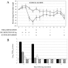
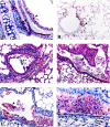
Advanced age has repeatedly been identified as an independent correlate of adverse outcome and a predictor of mortality in cases of severe acute respiratory syndrome (SARS). SARS-associated mortality may exceed 50% for persons aged 60 years or older. Heightened susceptibility of the elderly to severe SARS and the ability of SARS coronavirus to replicate in mice led us to examine whether aged mice might be susceptible to disease. We report here that viral replication in aged mice was associated with clinical illness and pneumonia, demonstrating an age-related susceptibility to SARS disease in animals that parallels the human experience.
Figures


Similar articles
-
A mouse-adapted SARS-coronavirus causes disease and mortality in BALB/c mice.PLoS Pathog. 2007 Jan;3(1):e5. doi: 10.1371/journal.ppat.0030005. PLoS Pathog. 2007. PMID: 17222058 Free PMC article. Clinical Trial.
-
Molecular determinants of severe acute respiratory syndrome coronavirus pathogenesis and virulence in young and aged mouse models of human disease.J Virol. 2012 Jan;86(2):884-97. doi: 10.1128/JVI.05957-11. Epub 2011 Nov 9. J Virol. 2012. PMID: 22072787 Free PMC article.
-
Early upregulation of acute respiratory distress syndrome-associated cytokines promotes lethal disease in an aged-mouse model of severe acute respiratory syndrome coronavirus infection.J Virol. 2009 Jul;83(14):7062-74. doi: 10.1128/JVI.00127-09. Epub 2009 May 6. J Virol. 2009. PMID: 19420084 Free PMC article.
-
Severe acute respiratory syndrome (SARS): development of diagnostics and antivirals.Ann N Y Acad Sci. 2006 May;1067(1):500-5. doi: 10.1196/annals.1354.072. Ann N Y Acad Sci. 2006. PMID: 16804033 Free PMC article. Review.
-
Severe acute respiratory syndrome (SARS): an old virus jumping into a new host or a new creation?J Biosci. 2003 Jun;28(4):359-60. doi: 10.1007/BF02705108. J Biosci. 2003. PMID: 12799480 Free PMC article. Review. No abstract available.
Cited by
-
Influence of Th1 versus Th2 immune bias on viral, pathological, and immunological dynamics in SARS-CoV-2 variant-infected human ACE2 knock-in mice.EBioMedicine. 2024 Oct;108:105361. doi: 10.1016/j.ebiom.2024.105361. Epub 2024 Sep 30. EBioMedicine. 2024. PMID: 39353281 Free PMC article.
-
Immune mRNA Expression and Fecal Microbiome Composition Change Induced by Djulis (Chenopodium formosanum Koidz.) Supplementation in Aged Mice: A Pilot Study.Medicina (Kaunas). 2024 Sep 20;60(9):1545. doi: 10.3390/medicina60091545. Medicina (Kaunas). 2024. PMID: 39336586 Free PMC article.
-
The immunobiology of SARS-CoV-2 infection and vaccine responses: potential influences of cross-reactive memory responses and aging on efficacy and off-target effects.Front Immunol. 2024 Feb 26;15:1345499. doi: 10.3389/fimmu.2024.1345499. eCollection 2024. Front Immunol. 2024. PMID: 38469293 Free PMC article. Review.
-
SARS-CoV-2 Aerosol and Intranasal Exposure Models in Ferrets.Viruses. 2023 Nov 29;15(12):2341. doi: 10.3390/v15122341. Viruses. 2023. PMID: 38140582 Free PMC article.
-
Characterization of a mouse-adapted strain of bat severe acute respiratory syndrome-related coronavirus.J Virol. 2023 Sep 28;97(9):e0079023. doi: 10.1128/jvi.00790-23. Epub 2023 Aug 21. J Virol. 2023. PMID: 37607058 Free PMC article.
References
-
- Booth, C. M., L. M. Matukas, G. A. Tomlinson, A. R. Rachlis, D. B. Rose, H. A. Dwosh, S. L. Walmsley, T. Mazzulli, M. Avendano, P. Derkach, I. E. Ephtimios, I. Kitai, B. D. Mederski, S. B. Shadowitz, W. L. Gold, L. A. Hawryluck, E. Rea, J. S. Chenkin, D. W. Cescon, S. M. Poutanen, and A. S. Detsky. 2003. Clinical features and short-term outcomes of 144 patients with SARS in the greater Toronto area. JAMA 289:2801-2809. (Erratum, 290:334, 2003.) - PubMed
-
- Bruunsgaard, H., M. Pedersen, and B. K. Pedersen. 2001. Aging and proinflammatory cytokines. Curr. Opin. Hematol. 8:131-136. - PubMed
-
- Bukreyev, A., E. W. Lamirande, U. J. Buchholz, L. N. Vogel, W. R. Elkins, M. St. Claire, B. R. Murphy, K. Subbarao, and P. L. Collins. 2004. Mucosal immunisation of African green monkeys (Cercopithecus aethiops) with an attenuated parainfluenza virus expressing the SARS coronavirus spike protein for the prevention of SARS. Lancet 363:2122-2127. - PMC - PubMed
Publication types
MeSH terms
LinkOut - more resources
Full Text Sources
Other Literature Sources
Molecular Biology Databases
Miscellaneous

