Comparative Study
. 2005 Apr 6;25(14):3539-50.
doi: 10.1523/JNEUROSCI.0081-05.2005.

Transgenic mouse model of tau pathology in astrocytes leading to nervous system degeneration

Affiliations
Comparative Study

Transgenic mouse model of tau pathology in astrocytes leading to nervous system degeneration

Mark S Forman et al. J Neurosci. .

Abstract

Filamentous tau inclusions in neurons and glia are neuropathological hallmarks of sporadic and familial tauopathies. Because tau gene mutations are pathogenic for the autosomal dominant tauopathy "frontotemporal dementia and parkinsonism linked to chromosome 17," tau abnormalities are implicated directly in the onset and/or progression of disease. Although filamentous tau aggregates are acknowledged to play roles in degenerative mechanisms resulting in neuron loss, the contributions of glial tau pathology to neurodegeneration remain essentially unexplored. To begin to elucidate the role of glial pathology in tauopathies, we generated a transgenic (Tg) mouse model of astrocytic tau pathology by expressing the human tau protein driven by the glial fibrillary acidic protein (GFAP) promoter. Whereas endogenous tau was not detected in astrocytes of control mice, in GFAP/tau Tg mice there was robust astrocytic tau expression that was associated with a redistribution of the GFAP network. Subsequently, there was an age-dependent accumulation of tau pathology in astrocytes that was Gallyas and variably thioflavine S positive as observed in many tauopathies. The tau pathology in these Tg mice was abnormally phosphorylated, ubiquitinated, and filamentous, and the emergence of this pathology coincided with accumulation of insoluble tau protein. Furthermore, in regions with robust astrocytic tau pathology, there was mild blood- brain barrier disruption, induction of low-molecular-weight heat shock proteins, and focal neuron degeneration. Thus, these Tg mice recapitulate key features of astrocytic pathology observed in human tauopathies and demonstrate functional consequences of this pathology including neuron degeneration in the absence of neuronal tau inclusions.


Figures

Figure 1.
Figure 1.
Expression of human tau in GFAP/tau Tg mice. A-C, Western blot analysis of HS-TBS-soluble proteins (10 μg per lane) extracted from cortex, brainstem (BS), and spinal cord (SC) samples of 2-month-old GFAP/tau Tg and control mice [non-Tg (NTg)]. Immunoblots were detected with antibodies that detect human tau only (T14; A), murine tau only (T49; B), or both human and murine tau (T46; C). D, Western blot analysis of native (-) and dephosphorylated (+) HS-TBS-soluble proteins extracted from brainstem and probed with T46 that recognizes both mouse and human tau. Arrows in C and D indicate migration of T34 human tau that is specifically expressed in GFAP/tau Tg mice. Molecular weight standards are indicated to the left of each panel. WT, Wild type; DeP, dephosphorylated.
Figure 2.
Figure 2.
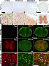
Astrocyte-specific regional tau expression in GFAP/tau Tg mice. A-H, Immunohistochemistry was performed with the human tau-specific MAb OT12 on 6-month-old non-Tg (A-D) and GFAP/tau Tg line 4 (E-H) mice from the CNS regions as indicated. Consistent with the biochemical analysis (Fig. 1), the highest levels of expression were observed consistently in the gray matter of the spinal cord. The inset in E shows morphology of cortical astrocytes at high magnification. Scale bars: (in A) A-C, E-G, 200 μm; (in D) D, H, 400 μm; E, inset, 25 μm. I-Q, Spinal cord sections from 6-month-old non-Tg (I-K) and GFAP/tau Tg, line 4 (L-Q) mice were double-labeled with OT12 (green; I, L, O) and GFAP (red; J, M, P). Merged images are depicted in K, N, and Q. Prominent GFAP staining is observed in astrocytes within the gray matter of Tg mice that is not detected in non-Tg animals. Arrowheads in O indicate the junction of the gray and white matter in the anterior horn of the spinal cord. Scale bars: (in K) I-N, 400 μm; (in Q) O-Q, 100 μm.
Figure 3.
Figure 3.
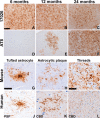
Accumulation of tau pathology in aged GFAP/tau Tg mice that resembles astrocytic pathology in human tauopathies. A-F, Immunohistochemical analysis was performed on sections of medulla from 6-month-old (A, D), 12-month-old (B, E), and 24-month-old (C, F) GFAP/tau Tg mice, line 4, as indicated with antibodies to recombinant tau (17026; A-C) or tau phosphorylated at Ser202 and Thr205 (AT8; D-F). There is an age-dependent accumulation of phosphorylated tau epitopes detected in GFAP/tau Tg mice. G-L, High-power photomicrographs of astrocytic pathology of Tg mice (G-I) and human tauopathies (J-L). The astrocytic pathology in the Tg mice resembles the tufted astrocytes (G, J), astrocytic plaques (H, K), and thread pathology (I, L) observed in tauopathies such as PSP and CBD. Scale bars: (in A) A-F, 200 μm; (in G) G, H, J, K, 40 μm; (in I) I, L, 80 μm.
Figure 4.
Figure 4.
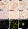
Characterization of tau pathology in GFAP/tau Tg mice. A-E, Immunohistochemical analysis was performed on sections of pons (A, B) and thalamus (C-E) from 22-month-old GFAP/tau Tg mice, line 4 with a panel of antibodies to distinct phosphorylated tau epitopes (A-D) and ubiquitin, as indicated. Ubiquitin (E) is detected only in a subset of tau pathologies. F-I, Gallyas silver impregnation (F) and thioflavine S (ThioS) (G-I) staining of astrocytic tau pathology in brainstem of 22-month-old GFAP/tau Tg mice. There is robust Gallyas-positive tau pathology in both astrocytic processes and cell soma. The thioflavine S stain detects only a subset of the pathology observed with antibodies to tau phosphoepitopes or Gallyas stains. Scale bars: (in A) A-G, 80 μm; (in I) H, I, 25 μm.
Figure 5.
Figure 5.
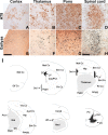
Distribution of tau pathology in GFAP/tau Tg mice. A-H, Immunohistochemical analysis with AT8 (A-D) and Gallyas silver impregnation stain (E-H) of CNS regions indicated from 24-month-old GFAP/tau Tg mouse. Robust pathology was observed consistently in the spinal cord, brainstem, and thalamus, whereas cortical pathology was highly variable. Scale bars: (in E) A-C, E-G, 100 μm; (in H) D, H, 200 μm. I, Diagrammatic summary of astrocytic tau pathology shown as coronal sections of the mouse neuroaxis. The density of dots corresponds to frequency and quantity of observed pathology in aged Tg mice. Amyg, Amygdala; Aq, aqueduct; Aud Cx, auditory cortex; Cau/Put, caudate-putamen; CC, corpus callosum; Cereb, cerebellum; DH, dorsal horn; Ent Cx, entorhinal cortex; Hip, hippocampus; Hypo, hypothalamus; LV, lateral ventricle; Mam, mamillary body; Med, medulla; Mid, midbrain; Mot Cx, motor cortex; Olf Cx, olfactory cortex; PC, posterior column; Pir Cx, piriform cortex; Sen Cx, sensory cortex; SN, substantia nigra; Vis Cx, visual cortex; VH, ventral horn; third, third ventricle; fourth, fourth ventricle.
Figure 8.
Figure 8.
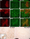
Phenotypic changes associated with tau pathology in astrocytes. A-I, Two-color immunofluorescence staining of spinal cord from 24-month-old GFAP/tau Tg mice. OT12, which is specific for exon 2 of human tau, colocalizes with GFAP (G-I). In contrast, in regions with AT8-positive tau pathology in astrocytes, there is a relative reduction of stainable GFAP (A-F). J, K, Immunohistochemistry for hsp27, from the thalamus of 24-month-old GFAP/tau Tg and non-Tg (NTg) mice. Tau pathology (J, inset) is associated with increased staining of hsp27. Scale bars: (in A) A-C, G-I, 400 μm; (in D) D-F, 100 μm; (in K) J, K, 200 mm.
Figure 6.
Figure 6.
Age-dependent accumulation of insoluble, aggregated, and phosphorylated tau protein in GFAP/tau Tg mice. Cortex (Cx), brainstem (BS), and spinal cord (SC) from pairs of non-Tg (NTg) and GFAP/tau Tg mice at 6, 12, 18, and 24 months of age were sequentially extracted as described in Materials and Methods. A, Immunoblot analysis of 10 μg of soluble protein extracted from the brainstem of GFAP/tau Tg mice at the age indicated and detected with the human-specific anti-tau MAb T14. There is only mild and variable change in tau protein expression with increasing age. B, Immunoblot analysis of insoluble tau corresponding to 25 mg of starting wet tissue weight extracted from GFAP/tau Tg mice at the age indicated and detected with the human tau-specific MAb T14. C, Immunoblot analysis of insoluble tau corresponding to 25 mg of starting wet tissue weight extracted from the spinal cord of non-Tg and GFAP/tau Tg mice at the age indicated and detected with polyclonal antibody 17026. Insoluble and heavily aggregated tau is detected in brainstem and spinal cord by 18 months of age. Insoluble tau is not detected in non-Tg mice. Arrowheads indicate nonspecific bands that are not recognized by other tau antibodies. D, Immunoblot analysis of insoluble tau extracted from cortex, brainstem, and spinal cord of 24-month-old GFAP/tau Tg mice and detected with a panel of tau-specific MAb as indicated. In contrast to B and C above, samples corresponding to only 5 mg starting wet tissue weight were loaded on the gels to reduce the smearing associated with heavily aggregated, insoluble tau protein, which facilitated the visualization of the predominant tau protein band at ∼66 kDa. Insoluble tau is phosphorylated at multiple sites (PH1, AT8, and PHF6) and is composed of human tau only (compare human tau-specific antibody T14 to murine tau-specific antibody T49). The position of molecular weight markers are depicted on the left. WT, Wild type.
Figure 7.
Figure 7.
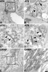
Immuno-EM demonstrates filamentous tau accumulation in astrocytes of aged GFAP/tau Tg mice. A, Immuno-EM of filamentous inclusion in spinal cord gray matter of a 22-month-old GFAP/tau Tg mouse labeled with AT8. B, High-power photomicrograph of the boxed area in A showing tau-positive filaments. C, D, High-power photomicrographs of the tau-positive filaments depicted by the arrowhead (C) and the arrow (D) in B. Arrowheads in C andD indicate filaments that are immunolabeled with the AT8. E, Immuno-EM of GFAP-positive filaments in astrocytes of spinal cord from same mouse depicted in A-D. F, High-power photomicrograph of the boxed area in E showing coarsely bundled GFAP-positive intermediate filaments. Scale bar: (in F) A, 1.25 mm; B, E, 500 nm; C, D, F, 100 nm.
Figure 9.
Figure 9.
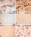
Astrocytic tau pathology is associated with mild BBB disruption. Histochemical analysis of 24-month-old GFAP/tau Tg (A, B, D-F) and non-Tg (NTg; C). A, B, There is prominent tau pathology in astrocytic foot processing surrounding blood vessels detected by both immunostaining with AT8 (A) and Gallyas silver stains (B). C-F, The perivascular astrocytic tau pathology is associated with amild disruption of the BBB. Immunohistochemistry performed on the indicated brain regions with prominent AT8-positive tau pathology showed focal somatic and perivascular staining with a polyclonal rabbit anti-mouse IgG antibody (D-F). No staining was detectable with age-matched control non-Tg mice (C) or in brain regions of Tg mice without tau pathology (data not shown). Scale bars: (in A) A, B, F, 40 μm; (in C) C-E, 100 μm.
Figure 10.
Figure 10.
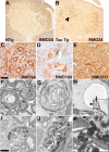
Neuronal degeneration associated with tau pathology in GFAP/tau Tg mice. A-E, Immunohistochemical analysis of spinal cord from 24-month-old GFAP/tau Tg (B-E) and non-Tg (NTg; A) mice with a panel of antibodies to phosphorylated neurofilament epitopes. In regions with robust astrocytic tau pathology, there is abnormal expression of phosphorylated neurofilament epitopes (C-E). The arrowhead in B indicates region examined at higher magnification in C-E. Scale bars: (in C) A, B, 160 μm; C-E, 40 μm. F-K, Transmission EM demonstrating axonal degeneration in 24-month-old GFAP/tau Tg mouse. F, There is prominent axonal degeneration (a) with loss of compact myelin and occasional axonal inclusions (i). G, H, High-power photomicrographs of axonal degeneration with myelin breakdown (G) and axonal inclusion (H). Inset shows edge of axonal inclusion with axonal inclusion on right and comparatively normal axon on left with both neurofilaments (arrowhead) and microtubules (arrow). These inclusions did not stain with antibodies to the tau protein. I, Axonal degeneration with myelin breakdown in association with putative astrocyte nucleus (n). J, K, High-power photomicrographs of degenerative changes shown in I. Inset in K shows fragment of degenerating myelin sheath. Scale bars (in I) I, 1.5 μm; F, 1 μm; J, 600 nm; G, H, 250 nm; K, H, inset, 150 nm; K, inset, 75 nm.

Similar articles

Cited by

References

    1. Biernat J, Gustke N, Drewes G, Mandelkow EM, Mandelkow E (1993) Phosphorylation of Ser262 strongly reduces binding of tau to microtubules: distinction between PHF-like immunoreactivity and microtubule binding. Neuron 11: 153-163. - PubMed
    1. Bramblett GT, Goedert M, Jakes R, Merrick SE, Trojanowski JQ, Lee VM-Y (1993) Abnormal tau phosphorylation at Ser396 in Alzheimer's disease recapitulates development and contributes to reduced microtubule binding. Neuron 10: 1089-1099. - PubMed
    1. Brenner M, Kisseberth WC, Su Y, Besnard F, Messing A (1994) GFAP promoter directs astrocyte-specific expression in transgenic mice. J Neurosci 14: 1030-1037. - PMC - PubMed
    1. Brenner M, Johnson AB, Boespflug-Tanguy O, Rodriguez D, Goldman JE, Messing A (2001) Mutations in GFAP, encoding glial fibrillary acidic protein, are associated with Alexander disease. Nat Genet 27: 117-120. - PubMed
    1. Claudio L (1996) Ultrastructural features of the blood-brain barrier in biopsy tissue from Alzheimer's disease patients. Acta Neuropathol (Berl) 91: 6-14. - PubMed

Publication types

MeSH terms

LinkOut - more resources