

Increase in number of circulating disseminated epithelial cells after surgery for non-small cell lung cancer monitored by MAINTRAC(R) is a predictor for relapse: A preliminary report
- PMID: 15801980
- PMCID: PMC1087511
- DOI: 10.1186/1477-7819-3-18
Increase in number of circulating disseminated epithelial cells after surgery for non-small cell lung cancer monitored by MAINTRAC(R) is a predictor for relapse: A preliminary report
Abstract
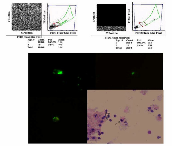
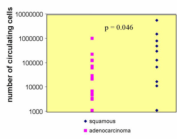
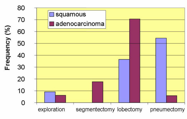
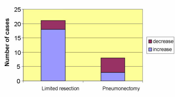
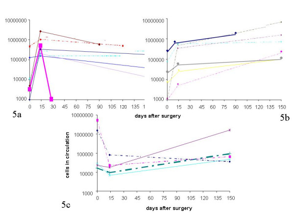
BACKGROUND: Lung cancer still remains one of the most commonly occurring solid tumors and even in stage Ia, surgery fails in 30% of patients who develop distant metastases. It is hypothesized that these must have developed from occult circulating tumor cells present at the time of surgery, or before. The aim of the present study was to detect such cells in the peripheral blood and to monitor these cells following surgery. METHODS: 30 patients treated for lung cancer with surgery were monitored for circulating epithelial cells (CEC) by taking peripheral blood samples before, 2 weeks and 5 months after surgery and/or radiotherapy (RT) chemotherapy (CT) or combined RT/CT using magnetic bead enrichment and laser scanning cytometry (MAINTRAC(R)) for quantification of these cells. RESULTS: In 86% of the patients CEC were detected before surgery and in 100% at 2 weeks and 5 months after surgery. In the control group, which consisted of 100 normal donors without cancer, 97 % were negative for CEC. A significantly higher number of CEC was found preoperatively in patients with squamous cell carcinoma than in those with adenocarcinoma. In correlation to the extent of parenchymal manipulation 2 weeks after surgery, an increase in numbers of CEC was observed with limited resections (18/21) whereas pneumonectomy led to a decrease (5/8) of CEC, 2 weeks after surgery. The third analysis done 5 months after surgery identified 3 groups of patients. In the group of 5 patients who received neo- or adjuvant chemo/radiotherapy there was evidence that monitoring of CEC can evaluate the effects of therapy. Another group of 7 patients who underwent surgery only showed a decrease of CEC and no signs of relapse. A third group of 11 patients who had surgery only, showed an increase of CEC (4 with an initial decrease after surgery and 7 with continuous increase). In the group with a continuous increase during the following 24 months, 2 early relapses in patients with stage Ia adenocarcinoma were observed. The increase of CEC preceded clinical detection by six months. CONCLUSION: We consider, therefore, that patients with adenocarcinoma and a continuous increase of CEC after complete resection for lung cancer are at an increased risk of early relapse.
Figures
Similar articles
-
[Pelvic lymphadenectomy as an alternative to adjuvant radiotherapy in early stage endometrial cancer at high risk of recurrent lymphatic metastases (stage I)].Minerva Ginecol. 2009 Feb;61(1):1-12. Minerva Ginecol. 2009. PMID: 19204656 Italian.
-
Increased Circulating Epithelial Tumor Cells (CETC/CTC) over the Course of Adjuvant Radiotherapy Is a Predictor of Less Favorable Outcome in Patients with Early-Stage Breast Cancer.Curr Oncol. 2022 Dec 24;30(1):261-273. doi: 10.3390/curroncol30010021. Curr Oncol. 2022. PMID: 36661670 Free PMC article. Clinical Trial.
-
Patterns of disease failure after trimodality therapy of nonsmall cell lung carcinoma pathologic stage IIIA (N2). Analysis of Cancer and Leukemia Group B Protocol 8935.Cancer. 1996 Jun 1;77(11):2393-9. doi: 10.1002/(SICI)1097-0142(19960601)77:11<2393::AID-CNCR31>3.0.CO;2-Q. Cancer. 1996. PMID: 8635112 Clinical Trial.
-
Should aggressive surgery ever be part of the management of small cell lung cancer?Thorac Surg Clin. 2004 May;14(2):271-81. doi: 10.1016/S1547-4127(04)00004-0. Thorac Surg Clin. 2004. PMID: 15382303 Review.
-
The present status of surgery for lung cancer.Acta Chir Belg. 1996 Nov-Dec;96(6):245-51. Acta Chir Belg. 1996. PMID: 9008764 Review.
Cited by
-
Circulating tumor cells as emerging tumor biomarkers in lung cancer.J Thorac Dis. 2012 Oct;4(5):438-9. doi: 10.3978/j.issn.2072-1439.2012.08.23. J Thorac Dis. 2012. PMID: 23050098 Free PMC article. No abstract available.
-
Fourier ptychographic microscopy for filtration-based circulating tumor cell enumeration and analysis.J Biomed Opt. 2014 Jun;19(6):066007. doi: 10.1117/1.JBO.19.6.066007. J Biomed Opt. 2014. PMID: 24949708 Free PMC article.
-
Propofol inhibits growth and invasion of pancreatic cancer cells through regulation of the miR-21/Slug signaling pathway.Am J Transl Res. 2016 Oct 15;8(10):4120-4133. eCollection 2016. Am J Transl Res. 2016. PMID: 27829997 Free PMC article.
-
Circulating Tumor Cells Identify Early Recurrence in Patients with Non-Small Cell Lung Cancer Undergoing Radical Resection.PLoS One. 2016 Feb 25;11(2):e0148659. doi: 10.1371/journal.pone.0148659. eCollection 2016. PLoS One. 2016. PMID: 26913536 Free PMC article.
-
Circulating tumor cells (CTCs) in lung cancer: current status and future perspectives.Lung Cancer (Auckl). 2010 Jul 3;1:77-84. doi: 10.2147/lctt.s6828. eCollection 2010. Lung Cancer (Auckl). 2010. PMID: 28210108 Free PMC article. Review.
References
-
- Takita H, Pitoniak RF. Induction therapy for locoregional lung cancer using paclitaxel combination. A preliminary report. J Exp Clin Cancer Res. 2000;19:291–293. - PubMed
LinkOut - more resources
Full Text Sources
Other Literature Sources
