

Homeobox protein Hop functions in the adult cardiac conduction system
- PMID: 15790958
- PMCID: PMC1615918
- DOI: 10.1161/01.RES.0000163108.47258.f3
Homeobox protein Hop functions in the adult cardiac conduction system
Abstract
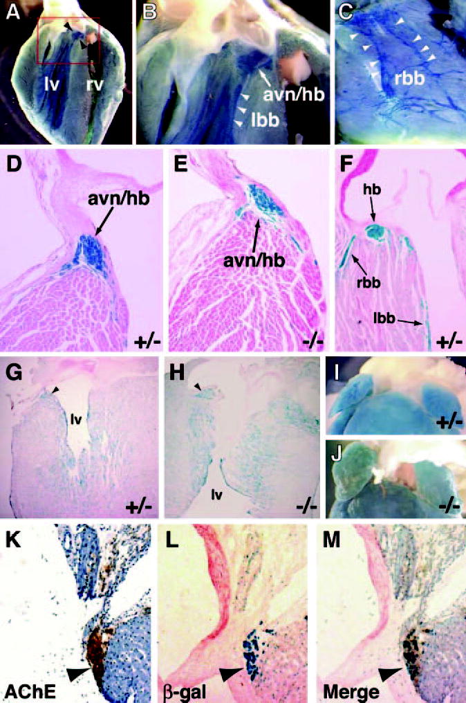
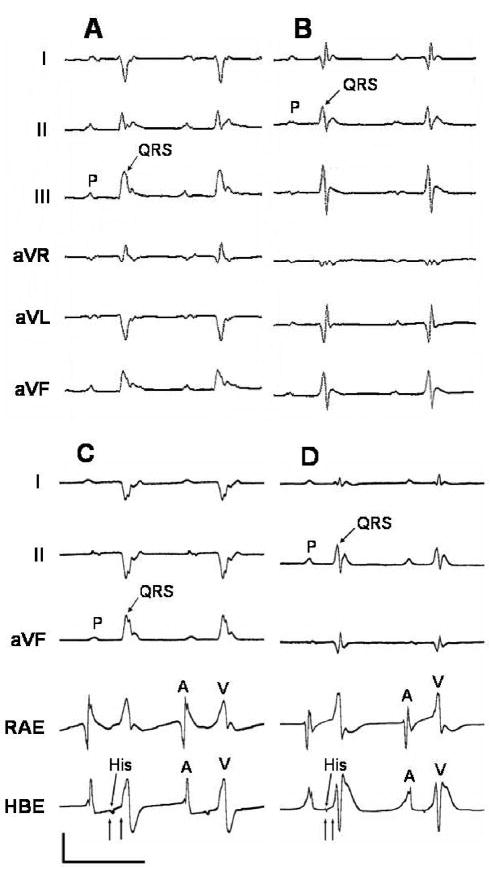
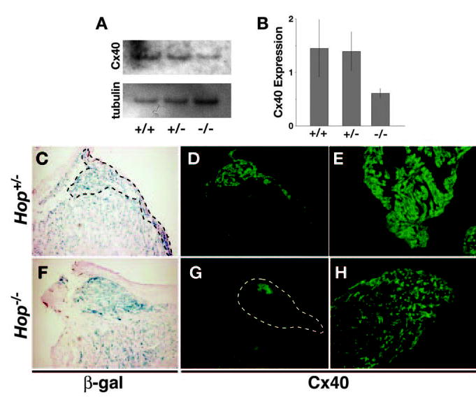
Hop is an unusual homeobox gene expressed in the embryonic and adult heart. Hop acts downstream of Nkx2-5 during development, and Nkx2-5 mutations are associated with cardiac conduction system (CCS) defects. Inactivation of Hop in the mouse is lethal in half of the expected null embryos. Here, we show that Hop is expressed strongly in the adult CCS. Hop-/- adult mice display conduction defects below the atrioventricular node (AVN) as determined by invasive electrophysiological testing. These defects are associated with decreased expression of connexin40. Our results suggest that Hop functions in the adult CCS and demonstrate conservation of molecular hierarchies between embryonic myocardium and the specialized conduction tissue of the mature heart.
Figures
Comment in
-
Understanding conduction system development: a hop, skip and jump away?Circ Res. 2005 Apr 29;96(8):809-11. doi: 10.1161/01.RES.0000165653.83279.20. Circ Res. 2005. PMID: 15860760 No abstract available.
Similar articles
-
Function follows form: cardiac conduction system defects in Nkx2-5 mutation.Anat Rec A Discov Mol Cell Evol Biol. 2004 Oct;280(2):966-72. doi: 10.1002/ar.a.20102. Anat Rec A Discov Mol Cell Evol Biol. 2004. PMID: 15368343 Review.
-
Nkx2-5 mutation causes anatomic hypoplasia of the cardiac conduction system.J Clin Invest. 2004 Apr;113(8):1130-7. doi: 10.1172/JCI19846. J Clin Invest. 2004. PMID: 15085192 Free PMC article.
-
Targeted mutation reveals essential functions of the homeodomain transcription factor Shox2 in sinoatrial and pacemaking development.Circulation. 2007 Apr 10;115(14):1830-8. doi: 10.1161/CIRCULATIONAHA.106.637819. Epub 2007 Mar 19. Circulation. 2007. PMID: 17372176
-
Understanding conduction system development: a hop, skip and jump away?Circ Res. 2005 Apr 29;96(8):809-11. doi: 10.1161/01.RES.0000165653.83279.20. Circ Res. 2005. PMID: 15860760 No abstract available.
-
Role of homeodomain-only protein in the cardiac conduction system.Trends Cardiovasc Med. 2006 Aug;16(6):193-8. doi: 10.1016/j.tcm.2006.03.009. Trends Cardiovasc Med. 2006. PMID: 16839862 Free PMC article. Review.
Cited by
-
Characterization of sinoatrial node in four conduction system marker mice.J Mol Cell Cardiol. 2007 May;42(5):946-53. doi: 10.1016/j.yjmcc.2007.02.008. Epub 2007 Feb 22. J Mol Cell Cardiol. 2007. PMID: 17459410 Free PMC article.
-
GATA-dependent regulatory switches establish atrioventricular canal specificity during heart development.Nat Commun. 2014 Apr 28;5:3680. doi: 10.1038/ncomms4680. Nat Commun. 2014. PMID: 24770533 Free PMC article.
-
Cardiac melanocytes influence atrial reactive oxygen species involved with electrical and structural remodeling in mice.Physiol Rep. 2015 Sep;3(9):e12559. doi: 10.14814/phy2.12559. Physiol Rep. 2015. PMID: 26400986 Free PMC article.
-
GATA-Binding Factor 6 Contributes to Atrioventricular Node Development and Function.Circ Cardiovasc Genet. 2015 Apr;8(2):284-93. doi: 10.1161/CIRCGENETICS.113.000587. Epub 2015 Jan 22. Circ Cardiovasc Genet. 2015. PMID: 25613430 Free PMC article.
-
Myocardial Notch signaling reprograms cardiomyocytes to a conduction-like phenotype.Circulation. 2012 Aug 28;126(9):1058-66. doi: 10.1161/CIRCULATIONAHA.112.103390. Epub 2012 Jul 26. Circulation. 2012. PMID: 22837163 Free PMC article.
References
-
- Cheng G, Litchenberg WH, Cole GJ, Mikawa T, Thompson RP, Gourdie RG. Development of the cardiac conduction system involves recruitment within a multipotent cardiomyogenic lineage. Development. 1999;126:5041–5049. - PubMed
-
- Pashmforoush M, Lu JT, Chen H, Amand TS, Kondo R, Pradervand S, Evans SM, Clark B, Feramisco JR, Giles WR, Ho SY, Benson DW, Siberbach M, Shou W, Chien KR. Nkx2–5 pathways and congenital heart disease: loss of ventricular myocyte lineage specification leads to progressive cardiomyopathy and complete heart block. Cell. 2004;117:378–386. - PubMed
Publication types
MeSH terms
Substances
Grants and funding
LinkOut - more resources
Full Text Sources
Molecular Biology Databases
Research Materials
