. 2005 Mar 21;201(6):937-47.
doi: 10.1084/jem.20041901.

Asthmatic bronchial epithelial cells have a deficient innate immune response to infection with rhinovirus

Affiliations

Asthmatic bronchial epithelial cells have a deficient innate immune response to infection with rhinovirus

Peter A B Wark et al. J Exp Med. .

Abstract

Rhinoviruses are the major trigger of acute asthma exacerbations and asthmatic subjects are more susceptible to these infections. To investigate the underlying mechanisms of this increased susceptibility, we examined virus replication and innate responses to rhinovirus (RV)-16 infection of primary bronchial epithelial cells from asthmatic and healthy control subjects. Viral RNA expression and late virus release into supernatant was increased 50- and 7-fold, respectively in asthmatic cells compared with healthy controls. Virus infection induced late cell lysis in asthmatic cells but not in normal cells. Examination of the early cellular response to infection revealed impairment of virus induced caspase 3/7 activity and of apoptotic responses in the asthmatic cultures. Inhibition of apoptosis in normal cultures resulted in enhanced viral yield, comparable to that seen in infected asthmatic cultures. Examination of early innate immune responses revealed profound impairment of virus-induced interferon-beta mRNA expression in asthmatic cultures and they produced >2.5 times less interferon-beta protein. In infected asthmatic cells, exogenous interferon-beta induced apoptosis and reduced virus replication, demonstrating a causal link between deficient interferon-beta, impaired apoptosis and increased virus replication. These data suggest a novel use for type I interferons in the treatment or prevention of virus-induced asthma exacerbations.


Figures

Figure 1.
Figure 1.
ICAM-1 expression of normal and asthmatic BECs before and after RV infection. (A and B) ICAM-1 expression was measured by flow cytometry immediately before RV-16 infection (A) or 24 h after infection (B). Data are expressed as mean fluorescence intensity (MFI). Before infection, asthmatic cells had a tendency to a lower median MFI 31 IQR (12, 80) compared with healthy control cells 67 (34, 83) but this was not significant (P = 0.4; A). ICAM-1 was significantly induced in both asthmatic and healthy control cells by 24 h (B), asthmatic cells had an MFI of 54.6 (27.6, 145.2) healthy control cells 110.4 (65, 195.3; P = 0.3). In these and all later box whisker plots, the line inside the box represents the median, upper box border represents 75th quartile, lower 25th quartile, whiskers are 5th and 95th centiles, dots represent outliers. (C and D) IL-6 and RANTES production was measured 48 h after infection in the supernatant of cells by ELISA. In C there was virus-specific release of IL-6 from RV infected in asthmatic cells with a median fold increase from baseline of 14 (IQR 5.2, 19.8) and in healthy control cells of 4 (4, 22.4). This was significantly greater than in cells treated with medium alone or UV-inactivated RV (P < 0.01), but there was no difference between the groups. In D there was virus-specific release of RANTES from RV infected in asthmatic cells with a median fold increase from baseline of 66 (IQR 33.2, 125.8) and in healthy control cells of 82.3 (25.2, 215.9). This was significantly greater than in cells treated with medium alone or UV-inactivated RV (P < 0.01), but there was no difference between the groups. *, Significantly different from cells treated with medium alone and UV-inactivated RV-16, P < 0.05. Shaded bars, asthma; open bars, healthy controls.
Figure 2.
Figure 2.
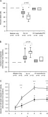
RV-16 replication and release from normal and asthmatic BECs. (A) RV-16 vRNA production was measured by qPCR after 8 h of infection. Median (IQR) production (×106) from asthmatic cells was significantly increased at 2.1 (0.16, 9.7) compared with 0.04 (0.009, 0.06) from healthy controls (P = 0.007). (B and C) Late cell lysis as a consequence of RV-16 infection was determined by analysis of LDH activity in culture supernatants; values represent the fold induction from baseline. Data points represent the mean and the SD. In asthmatic cells LDH activity progressively increased over time and was significantly increased from baseline at both 24 h (P = 0.01) and 48 h (P = 0.003) whereas in healthy control cells there was no significant increase even at 48 h (P = 0.2; B). By 48 h, the mean LDH activity from asthmatic cells was significantly greater than in normal cells, at 3.4 (0.1)-fold increased over baseline compared with only a 1.34 (0.1)-fold increase in the healthy control cells (P = 0.0001; C). Induction of LDH activity was shown to be virus replication dependent in that no significant change in LDH activity was seen in cells treated with medium alone or UV-inactivated RV (C). (D) RV-16 release into the supernatant of infected cells was determined by calculating the TCID50 × 104/ml by titration assay in Ohio HeLa cells. The supernatant from 14 ICSs requiring asthmatics and 10 healthy controls were examined on individual titration plates. Data points represent the mean and the standard deviation. By 48 h significantly more RV was detected from asthmatic cells with a mean TCID50 × 104/ml of 3.99 (0.8), compared with 0.54 (0.12) in healthy control cells (P = 0.001). *, Significantly different from cells treated with UV inactivated RV or medium alone. **, Results from asthmatic cells and healthy controls significantly different (P < 0.01). +, Significantly increased above baseline (P < 0.001). Shaded bars, asthma; open bars, healthy controls.
Figure 3.
Figure 3.
Differences in cell viability and apoptotic response after RV-16 infection in asthmatic and normal BECs. (A) Viable (AxV/7AAD) cell number was determined 8 h after infection and expressed as percent viability compared with cells treated with medium alone. Infection with RV-16 led to a significant reduction in median (IQR) cell viability in both asthmatic and control cells compared with both medium alone (P = 0.03 and 0.02, respectively) and UV-inactivated RV-16 (P = 0.02 and 0.001, respectively). In asthmatic cells there was significantly better viability, median 80% (74, 86), compared with healthy controls 63% (51, 69; P = 0.02). (B) Apoptotic (AxV+/7AAD) cells were also analyzed 8 h after RV-16 infection. Data are expressed as median (IQR) fold change in apoptosis from baseline. There was a significant and virus-specific increase in cell apoptosis in response to infection in cells from both groups, however, this response was significantly impaired in asthmatic cells with a fold increase of only 1.4 (1.3, 1.7), compared with 2.2 (2.1, 2.3) in healthy controls (P = 0.02). (C) The time course for activation of caspase 3/7 by RV-16 was determined in cells from 10 ICSs requiring asthmatics and 10 healthy controls, all conditions were done in quadruplicate, values represent the fold induction from baseline. There was significant mean (SD) induction of active caspase 3/7 in response to infection in normal cells at 4, 8, and 12 h, induction reached a plateau at 8 h. In asthmatic cells the induction was later, being significantly increased above baseline only at 8 and 12 h and was of significantly reduced magnitude at each time point compared with normal cells. At 8 h there was a significantly impaired induction of active caspase 3/7 in asthmatic cells (mean [SD] = 1.47 [0.13]) compared with healthy controls (mean [SD] =2.16 [0.34]; P = 0.004). *, Significantly different from cells treated with UV-inactivated RV-16 and medium only (P < 0.01). **, Asthmatic cells significantly different from healthy controls (P < 0.05). +, Significantly different from baseline (P < 0.05). Shaded bars, asthma; open bars, healthy controls.
Figure 4.
Figure 4.
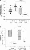
Inhibition of caspase activity inhibits apoptosis and increases RV-16 replication. (A) The effect of inhibition of caspase-3 using the inhibitor, ZVD-fmk, was measured by flow cytometry. Cells were treated with RV-16 alone or with ZVD-fmk, before and after infection with RV-16. Results are expressed as the fold change in apoptotic cells compared with cells treated with medium alone. In asthmatic cells were there was a median (IQR) induction of apoptosis above baseline of 1.4 (1.3, 1.7) with RV-16 alone; pretreatment of cells with the ZVD-fmk, had little effect on apoptosis (median (IQR) = 1.12 (1.01, 1.8); (P = 0.4). However, in healthy controls cells, RV-16 infection resulted in a median (IQR) fold induction of apoptosis above baseline of 2.2 (2.1, 2.3) and this was abolished by pretreatment with ZVD-fmk (median [IQR] 0.82 [0.76, 0.86; P = 0.03]). (B) The effect of caspase-3 inhibition on RV-16 production was measured by HeLa titration assay on the BEC supernatant removed after 48 h of infection. There was no difference seen in the TCID50 × 104/ml in the supernatant removed from asthmatic cells infected with RV-16 (median [IQR] = 3.56 [3.50–3.62]) compared with infected cells treated with ZVD-fmk (median [IQR] = 3.5 [3.45–3.62]; P = 0.94). However, for healthy control BECs, the TCID50 × 104/ml increased greater than fourfold, from a median (IQR) value of 0.6 (0.4, 0.63) with infection alone to 2.78 (0.63, 6.32; P = 0.01) in the presence of RV-16 and ZVD-fmk. Shaded bars, asthma; open bars, healthy controls.
Figure 5.
Figure 5.
Impaired IFN-β production in asthma and its role in restoring apoptotic and antiviral response in asthmatic cells. (A) Induction of IFN-β mRNA was measured by qPCR after 8 h of RV-16 infection. There was no significant induction of IFN-β mRNA from asthmatic cells 8 h after infection with RV-16, median (IQR) fold induction from baseline control of 0.3 (0.3, 0.8), which was not significantly different from cells treated with medium alone or UV inactivated RV-16. In contrast there was a 3.6 (3.4, 3.6)-fold increase in IFN-β mRNA expression in cells from healthy controls (P = 0.004). (B) Release of IFN-β into culture supernatants 48 h after infection was measured by ELISA. For asthmatic BECs, median (IQR) IFN-β levels were significantly reduced at 721 (464, 1,290) pg/ml, compared with 1,854 pg/ml (758, 3,766; P = 0.03) in healthy controls. In both groups there was a significant increase above cells treated with medium alone and UV-inactivated RV-16 (unpublished data). (C) The ability of IFN-β to restore induction of apoptosis in RV-16–infected asthmatic cells was measured by FACS analysis as described in the legend to Fig. 3. Asthmatic cells were either pretreated with IFN-β (100 IU) for 24 h or simultaneously exposed to RV-16 and IFN-β. To mimic the presence of viral RNA, cells were also exposed to poly(I)/poly(C) a synthetic double-stranded RNA oligonucleotide, instead of RV-16. Results are expressed as the median (IQR) fold increase in apoptotic cells from baseline at 8 h after infection. There was no significant increase in apoptosis in cells exposed to either IFN-β 1.2 (1.1, 1.8; P = 0.3), RV-16 1.7 (1.3, 1.9; P = 0.2) or poly(I)/poly(C) 1.9 (1.7, 3.5; P = 0.08) alone. In cells simultaneously treated with RV-16 and IFN-β there was a tendency to increased apoptosis 3.8 (1.7, 5.0; P = 0.11) whereas in those pretreated with IFN-β for 24 h and then infected there was a significant induction of apoptosis 5.6 (3.9, 5.7; P = 0.02, compared with virus alone). In cells exposed to poly(I)/poly(C) alone there was a similar trend toward an increase in apoptosis 1.9 (1.7, 3.5; P = 0.08) which was significantly enhanced by simultaneous treatment with IFN-β 5.1 (3.9, 5.6; P = 0.01) and further enhanced by 24 h pretreatment with IFN-β 9.3 (6.6, 9.3; P = 0.001), compared with poly(I)/poly(C) alone. (D) The effect of IFN-β on virus release from asthmatic cells was measured by HeLa titration assay of asthmatic BEC culture supernatants removed 48 h after infection. Cells were either pretreated with IFN-β (100 IU) for 12 h and then exposed to RV-16 or were treated with IFN-β immediately after infection. There was a significant reduction in virus release in cells treated with IFN-β after infection median TCID50 × 104/ml 2.78 (2, 3.56; P = 0.04) and a further reduction in cells pretreated with IFN-β 1.12 (0.28, 1.34) compared with cells infected with RV-16 alone 3.56 (3.5–3.62; P = 0.012). *, Significantly different from medium alone. Shaded bars, asthma; open bars, healthy controls.
Figure 6.
Figure 6.
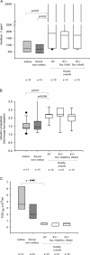
The effect of corticosteroids on interferon-β release apoptotic responses and virus replication. The results from asthmatic cells (those requiring ICSs) and healthy controls were compared with asthmatic subjects who had never used corticosteroids (ICSs naive). Cells from healthy controls and ICSs naive asthmatics were treated with RV-16 and pretreated for 24 h with dexamethasone (Dex) at 10 and 100 nM and then infected with RV-16. only the results from healthy control cells are displayed in the figures. (A) Release of IFN-β into culture supernatants 48 h after infection was measured by ELISA. As noted in asthmatic BECs, median (IQR) IFN-β levels were significantly reduced at 721 (464, 1,290) pg/ml, compared with 1,854 pg/ml (758, 3,766; P = 0.01) in healthy controls. This was also seen in ICSs naive asthmatic cells 666 (387, 1,039) compared with healthy controls (P = 0.02). The addition of Dex did significantly alter these levels. In healthy control cells; 10 nM 1717 (720, 1,990) compared with infected control cells (P = 0.55) or 100 nM 990 (721, 2,656). This was also seen in ICSs-naive asthma; 10 nM 772 (581, 1,220) compared with ICSs-naive asthma cells infected alone (P = 0.7), though at 100 nM; 449 (389, 609) there appeared to be a reduction, this did not reach statistical significance (P = 0.2). (B) Apoptotic (AxV+/7AAD) cells were also analyzed 8 h after RV-16 infection. Data are expressed as median (IQR) fold change in apoptosis from baseline. Healthy controls cells had significantly more virus-specific apoptosis 2.19 (1.99, 2.2), compared with asthmatic cells (ICSs requiring) 1.39 (1.35, 1.67; P = 0.01) and ICSs naive asthmatic cells 1.36 (1.2, 1.5; P = 0.004). The addition of Dex to healthy control cells had no significant impact on apoptosis compared with cells not pretreated, at either 10 nM 2.16 (1.86, 2.6; P = 0.96) or 100 nM 1.87 (1.65, 2.14; P = 0.09). This was also seen in ICSs-naive asthma cells at 10nM 1.26 (1.2, 1.8) compared with ICSs naive cells infected alone (P = 0.97) and 100 nM 1.16 (1.1, 1.4; P = 0.08). (C) RV-16 release into the supernatant of infected cells was determined by calculating the TCID50 × 104/ml by titration assay in Ohio HeLa cells. By 48 h significantly more RV was detected from asthmatic cells with a mean TCID50 × 104/ml of 3.99 (0.8), compared with 0.54 (0.12) in healthy control cells (P = 0.002). There was a trend to higher titer in ICSs naive asthmatics 2.2 (1.2), but this was not significantly different from either ICSs requiring asthmatics or healthy controls. The addition of dexamethasone 10 nM in control cells 0.46 (0.3) and ICSs-naive asthma cells 2.1 (1.3) or 100 nM in control cells 0.42 (0.3) and ICSs-naive asthma 2.1 (1.3) had no significant effect. Shaded bars, asthma (ICSs treated); hatched bars, asthma (ICSs naive); open bars, healthy controls.

Similar articles

Cited by

References

    1. Johnston, S.L., P.K. Pattemore, G. Sanderson, S. Smith, F. Lampe, L. Josephs, P. Symington, S. O'Toole, S.H. Myint, D.A.J. Tyrell, and S.T. Holgate. 1995. Community study of the role of viral infections in exacerbations of asthma in 9–11-year old children. BMJ. 310:1225–1228. - PMC - PubMed
    1. Nicholson, K.G., J. Kent, and D.C. Ireland. 1993. Respiratory viruses and exacerbations of asthma in adults. BMJ. 307:982–986. - PMC - PubMed
    1. Johnston, S.L., P.K. Pattemore, G. Sanderson, S. Smith, M.J. Campbell, L.K. Josephs, A. Cunningham, B.S. Robinson, S.H. Myint, M.E. Ward, et al. 1996. The relationship between upper respiratory infections and hospital admissions for asthma: a time-trend analysis. Am. J. Respir. Crit. Care Med. 154:654–660. - PubMed
    1. Reddel, H., S. Ware, G. Marks, C. Salome, C. Jenkins, and A. Woolcock. 1999. Differences between asthma exacerbations and poor asthma control. Lancet. 353:364–369. - PubMed
    1. Doull, I.J.M., F.C. Lampe, S. Smith, J. Schreiber, N.J. Freezer, and S.T. Holgate. 1997. Effect of inhaled corticosteroids on episodes of wheezing associated with viral infection in school age children: randomised double blind placebo controlled trial. BMJ. 315:858–862. - PMC - PubMed

Publication types

MeSH terms