. 2005 May;16(5):2191-206.
doi: 10.1091/mbc.e04-09-0844. Epub 2005 Feb 23.

A Dictyostelium homologue of WASP is required for polarized F-actin assembly during chemotaxis

Affiliations

A Dictyostelium homologue of WASP is required for polarized F-actin assembly during chemotaxis

Scott A Myers et al. Mol Biol Cell. 2005 May.

Abstract

The actin cytoskeleton controls the overall structure of cells and is highly polarized in chemotaxing cells, with F-actin assembled predominantly in the anterior leading edge and to a lesser degree in the cell's posterior. Wiskott-Aldrich syndrome protein (WASP) has emerged as a central player in controlling actin polymerization. We have investigated WASP function and its regulation in chemotaxing Dictyostelium cells and demonstrated the specific and essential role of WASP in organizing polarized F-actin assembly in chemotaxing cells. Cells expressing very low levels of WASP show reduced F-actin levels and significant defects in polarized F-actin assembly, resulting in an inability to establish axial polarity during chemotaxis. GFP-WASP preferentially localizes at the leading edge and uropod of chemotaxing cells and the B domain of WASP is required for the localization of WASP. We demonstrated that the B domain binds to PI(4,5)P2 and PI(3,4,5)P3 with similar affinities. The interaction between the B domain and PI(3,4,5)P3 plays an important role for the localization of WASP to the leading edge in chemotaxing cells. Our results suggest that the spatial and temporal control of WASP localization and activation is essential for the regulation of directional motility.


Figures

Figure 1.
Figure 1.
Deduced sequence of Dictyostelium WASP. (A) Sequence comparison of Dictyostelium WASP with WASPs from other species. Deduced amino acid sequence of WASP from the 1200-base pair DNA clone is aligned with human (Hs), mouse (Mm), Caenorhabditis elegans (Ce), and Drosophila (Dm) WASP. (B) Schematic diagram of WASP domain structure. WH1, WASP homology I domain; B, Basic domain; GBD, Cdc42/Rac binding domain; V, Verprolin homology domain; C, central domain; A, acidic domain. (C) Northern blot showing the developmental time course of WASP expression. Total RNA of 16 μg/sample was resolved on a 1.0% denaturing agarose gel, blotted, and probed as described previously (Datta and Firtel, 1987). The 0-h time point is for vegetative cells. (D) Northern blot showing reduction of WASP transcript in WASPhypo cells and the lack of WASP transcript in WASPTK cells.
Figure 2.
Figure 2.
F-actin organization of cells expressing low levels of WASP. (A) F-actin organization revealed by phalloidin staining in WASPhypo and WASPTK cells under cAMP gradient. Stainings of two cells with representative phenotypes are shown. Arrows indicate the direction of cAMP gradient. Right panel shows ratio of intensity of F-actin staining at the foremost part (front) or rear end (back) of cells divided by the intensity of the center of cells. Intensity value was acquired by linescan of images in the direction of the gradient (n = 10). (B) In vivo actin polymerization assay measuring F-actin assembled in response to the chemoattractant (cAMP) stimulation. Note that F-actin polymerization upon cAMP stimulation is defective in WASPTK cells. (C) F-actin organization revealed with ABP-GFP in live cells. ABP-GFP fusion protein binds specifically and dynamically to the same F-actin structures that phalloidin recognizes in fixed cells. (D) Distribution of free barbed end in cells. Rhodamine-labeled actin (0.4 μM) was incorporated into permeabilized cells to visualize free barbed-ends for actin polymerization in wild-type cells or WASPTK cells. Linescans of a cell from point A to B were shown in bottom panels. Incorporation of Rhodamine-labeled G-actin was quantified by measuring the fluorescence intensity and shown in right panel (n = 6). (E) Axial polarity of wild-type and WASPTK cells. GFP fusion of N-terminal half of PAKa (PAKa-N-GFP) shows specific localization to uropod of polarized cells and was used as a reporter to examine cellular polarity. Aggregation competent wild-type cells are well polarized and showed biased localization of PAKa-N-GFP at the uropod, but WASPTK cells show almost equal distribution of PAKa-N-GFP, indicating lack of axial polarity. Polarity index was calculated by the ratio of PAKa-N-GFP intensity at the leading edge to GFP intensity at the uropod and shown in the right panel (n = 6). The polarity index of wild-type cells is significantly greater than that of WASPTK cells.
Figure 3.
Figure 3.
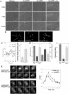
Abnormal chemotactic movement of cells expressing low levels of WASP. (A) DIC images of cells migrating toward a cAMP gradient. Wild-type cells are well polarized and retract uropod actively. WASPhypo cells have defects in protruding lamellipod and retracting rear cell body, resulting in narrow and long uropod. WASPTK cells do not polarize and actively migrate. WASPTK cells expressing tTA, a chimeric tetracycline-controlled transcriptional activator protein, recovers normal chemotactic movements. Images of cell at 0, 7, and 14 min are shown. (B) Chemotaxis of cells were analyzed by Metamorph software (Universal Imaging) and traces of cells chemotaxing toward cAMP source are shown. Asterisk represents the position of micropipette. (C) Cell body length of WASPhypo cells migrating toward cAMP gradient. Because of lack of active retraction at the uropod, the length of cell body extends until the uropod retracts by mechanical force generated by pseudopod extension. As an example, variations of cell body length of a wild-type cell and a WASPhypo mutant cell are shown in the left. Differences in longest and shortest cell body lengths of eight cells during chemotaxis were analyzed and are shown in the right. (D) Chemotaxis speed of wild-type, WASPTK, and WASPTK cells expressing tTA (n = 6). The chemotactic speed of WASPTK cells is significantly decreased, but recovered by the expression of tTA leading WASP expression. DIC images of migrating cells were taken in 6-s intervals for 15 min and analyzed with Metamorph software. Angular deviation shows deviations of the angle of the path taken by cells from frame to frame. Chemotaxis indices were calculated as described in Futrelle et al. (1982). If the cell moves directly toward the gradient source it is 1, if directly away it is –1. If movement is indifferent to gradient (random movement), it is 0. (E) Translocation of Akt/PH-GFP upon cAMP stimulation. Wild-type and WASPTK cells expressing PH domain of Akt/PKB (Akt/PH-GFP) were stimulated by the addition of a saturating dose of cAMP (10 μM). On cAMP stimulation, a dramatic translocation of Akt/PH-GFP from the cytosol to the plasma membrane is observed. Numbers in the lower right corner are seconds before and after stimulation. Right panel shows the translocation kinetics of Akt/PH-GFP obtained from time-lapse recordings. The fluorescence intensity of membrane-localized GFP fusion protein was quantitated as E(t) using the linescan module of Metamorph software (Universal Imaging). Et/Eo is plotted as a measure of the amount of membrane-associated protein relative to the starting conditions.
Figure 4.
Figure 4.
Motility defects of cells expressing low level of WASP in multicellular development. (A) Development of cells expressing a low level of WASP. Cells grown in axenic medium were washed and plated on nonnutrient agar plate. Photographs were taken at various developmental stages thereafter. Development of WASPhypo cells was delayed in mound stage, resulting in delayed formation of slugs and fruiting bodies. WASPTK cells showed a severe defect in chemotactic aggregation, resulting in few fruits formed. Ectopic expression of GFP-WASP rescued defects of WASPTK cells. (B) Motility defect of WASPhypo cells. Wild-type, WASPhypo, WASPhypo cells expressing GFP were mixed in a ratio of 8:1:1. GFP-labeled WASPhypo cells were uniformly distributed in the loose-aggregate at 8 h after the onset of development, but sorted out to the periphery of the tight mound (12 h), presumably due to the motility defect. (C) Motility defects of WASPTK cells. GFP-labeled WASPTK cells were mixed with unlabeled wild-type cells, and they showed significant defects in chemotactic aggregation. Most of GFP-labeled WASPTK cells were neither present in the mound nor polarized.
Figure 5.
Figure 5.
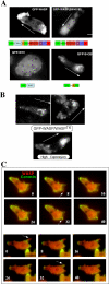
(A) Localization of GFP-WASP in Dictyostelium cells (WASPhypo) migrating toward a cAMP gradient. For examining GFP-WASP, cells were pulsed for 4.5 h at 6-min intervals. Fluorescence images were taken from live aggregation-competent cells migrating toward a chemoattractant gradient. GFP-WASP localizes to the leading edge and the uropod, which is recapitulated by GFP-B-GBD. GFP-WASP variant lacking WH1-B domains do not show prominent localization because GFP-WH1 shows uniform distribution. Arrows indicate the direction of gradient. Bar, 5 μm. (B) Localization of GFP-WASP in WASPTK cells. GFP-WASP still shows biased distribution at the leading edge and uropod, but appears to be associated with vesicular structures. Higher expression of GFP-WASP showed more diffuse distribution. (C) Localization of YFP-WASP to the leading edge. CFP-Coronin and YFP-WASP were coexpressed in wasphypo cells, and their localizations in chemotaxing cells were examined in time-lapse recording of alternate exposures (1 s) for CFP and YFP. Images were acquired and analyzed with multidimensional acquisition utility of Metamorph (Universal Imaging) software. Coronin localizes to sites of dynamic actin assembly and functions as a reporter for F-actin distribution in live cells.
Figure 6.
Figure 6.
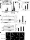
(A) The phospholipid binding properties of the basic domain of WASP. The indicated phospholipids were spotted onto a nitrocellulose membrane (PIP strip) that was then incubated with GST-PH/PLCδ1 and GST-B-GBD. The membranes were washed and the GST fusion proteins bound were detected by Western blot using a GST antibody. Inset shows a PIP strip and bar graph is an average of four PIP strips. Note that GST-PH/PLCδ1 specifically bound to PI(4,5)P2, but GST-B-GBD showed higher affinity to PI(3,4,5)P3 and PI(3,4)P2, which are products of PI3 kinase. Unexpectedly higher binding to PI(4)P and PI(5)P in the graph was due to having one blot showing unusually high binding to PI(4)P and PI(5)P. (B) PIP beads pull-down assay. GST-B-GBD fusion protein was incubated with agarose beads cross-linked to phosphoinositides and bound GST-fusion protein was pulled down and then probed by a Western blot. GST-B-GBD showed binding to PI(3,4,5)P3-beads with slightly higher affinity than to PI(4,5)P2-beads, whereas control beads showed minimal binding. Quantification of three experiments is shown. (C) Liposome cosedimentation assay with purified GST-YFP-B-GBD or GST-WASP protein and liposomes composed of 95% PC and 5% of either PI, PI(4)P, PI(4,5)P2, or PI(3,4,5)P3. GST-YFP-B-GBD protein and liposomes were mixed and subjected to centrifugation on an Optiprep gradient (5–30% Optiprep). Collected fractions were run on SDS-PAGE gel and probed with Western blot with anti-GST antibody. Ovalbumin was added in the binding mixture to block nonspecific binding and it also serves as an internal control for lower concentration of Optiprep fraction. Specific binding efficiency was quantified by summing of GST-YFP-B-GBD band intensity in the higher fractions than fractions where ovalbumin was present. (D) Disruption of polarized localization of GFP-WASP by the PI3K inhibitor LY294002. Dictyostelium chemotaxing cells expressing the Coronin-GFP or GFP-WASP were treated with 15 μM LY294002 and the change in the subcellular localization of Coronin-GFP and GFP-WASP was followed by time-lapse digital video microscopy. Arrows indicate the direction of movements and numbers shown are time (seconds) after LY treatment.
Figure 7.
Figure 7.
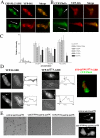
(A) Colocalization of YFP-B-GBD and CFP-PLCδ1PH in chemotaxing cells. CFP-PLCδ1PH binds to PI(4,5)P2 with very high specificity. CFP-PLCδ1PH showed punctate vesicle labeling, which appears to overlap with the YFP-B-GBD signal. Note that some YFP-B-GBD signal was found at the leading edge without colocalization with CFP-PLCδ1PH. (B) Colocalization of YFP-B-GBD and CFP-PhdA in chemotaxing cells. CFP-PhdA selectively binds to PI(3,4,5)P3 and accumulated at the leading edge of migrating cells. Wild-type cells migrating toward a cAMP source show a distinct colocalization of PhdA–GFP and YFP-B-GBD at the leading edge. (C) PIP strip assay of B domain mutants. Lys residues of the B domain were mutated to Ala and the binding efficiency of the GST fusion protein of these mutants to phosphoinositides were assessed by PIP strip assay. Lys156A and Lys157A appear to specifically block the binding to phosphoinositides. (D) Localization of YFP-B-GBD and YFP-B157A-GBD in chemotaxing cells. Phase contrast and fluorescence micrographs of cells expressing YFP-B-GBD and YFP-B157A-GBD are shown. Note that the YFP-B-GBD is localized at the leading edge, but the localization of YFP-B157A-GBD to the leading edge is significantly reduced. Fluorescence intensities of YFP were measured along a thin line through the central portion of the cell. Arrows indicate the position of the leading edge. Some of YFP-B-GBD signal was from intracellular vesicles, which appears to be blurry because optical focus was at the leading edge membrane, which cells tend to lift up. YFP-B156,157A-GBD showed essentially the same distribution as YFP-B157A-GBD. (E) Rescue of motility defects of WASPTK cells by expressing WASP B domain mutants. Wild-type WASP, K156AWASP, or K156,157AWASP were expressed in WASPTKcells and their development were examined. Cells expressing K156AWASP showed partial rescue of development, whereas K156,157AWASP did not rescue motility defects of WASPTK cells. Compared with polarized localization of GFP-WASP, YFP-K156AWASP did not show any biased localization.
Figure 8.
Figure 8.
F-actin organization of cells expressing WASP mutants. (A) Deletion mutations expected to abrogate the function of specific domains of WASP were created and F-actin organization of cells expressing these mutants were examined by TRITC-phalloidin staining. Cells under a cAMP gradient were fixed and stained with TRITC-phalloidin. Arrows indicate the direction of gradient. (B) Chemotaxis of cells expressing WASPΔProV and GFP-WASPΔV. DIC images of cells migrating toward a cAMP gradient at 0 and 15 min are shown. Average speed and angular deviation of these mutants are shown. (C) Developmental phenotypes of cells expressing WASP truncation mutants. Cells grown in axenic medium were washed and plated on nonnutrient agar plate. Photographs were taken at various developmental stages thereafter.

Similar articles

Cited by

References

    1. Aldrich, R. A., Steinberg, A. G., and Campbell, D. C. (1954). Pedigree demonstrating a sex linked recessive condition characterized by draining ears, eczematoid dermatitis, and bloody diarrhea. Pediatrics 13, 133–139. - PubMed
    1. Altman, L. C., Snyderman, R., and Blaese, R. M. (1974). Abnormalities of chemotactic lymphokine synthesis and mononuclear leukocyte chemotaxis in Wiskott-Aldrich syndrome. J. Clin. Invest. 54, 486–493. - PMC - PubMed
    1. Asano, S., Mishima, M., and Nishida, E. (2001). Coronin forms a stable dimer through its C-terminal coiled coil region: an implicated role in its localization to cell periphery. Genes Cells 6, 225–235. - PubMed
    1. Aubry, L., and Firtel, R. (1999). Integration of signaling networks that regulate Dictyostelium differentiation. Annu. Rev. Cell Dev. Biol. 15, 469–4517. - PubMed
    1. Badolato, R., Sozzani, S., Malacarne, F., Bresciani, S., Fiorini, M., Borsatti, A., Albertini, A., Mantovani, A., Ugazio, A. G., and Notarangelo, L. D. (1998). Monocytes from Wiskott-Aldrich patients display reduced chemotaxis and lack of cell polarization in response to monocyte chemoattractant protein-1 and formyl-methionyl-leucyl-phenylalanine. J. Immunol. 161, 1026–1033. - PubMed

Publication types

MeSH terms

LinkOut - more resources