Maturation of papillomavirus capsids
- PMID: 15709003
- PMCID: PMC548454
- DOI: 10.1128/JVI.79.5.2839-2846.2005
Maturation of papillomavirus capsids
Abstract
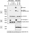
The papillomavirus capsid is a nonenveloped icosahedral shell formed by the viral major structural protein, L1. It is known that disulfide bonds between neighboring L1 molecules help to stabilize the capsid. However, the kinetics of inter-L1 disulfide bond formation during particle morphogenesis have not previously been examined. We have recently described a system for producing high-titer papillomavirus-based gene transfer vectors (also known as pseudoviruses) in mammalian cells. Here we show that papillomavirus capsids produced using this system undergo a maturation process in which the formation of inter-L1 disulfide bonds drives condensation and stabilization of the capsid. Fully mature capsids exhibit improved regularity and resistance to proteolytic digestion. Although capsid maturation for other virus types has been reported to occur in seconds or minutes, papillomavirus capsid maturation requires overnight incubation. Maturation of the capsids of human papillomavirus types 16 and 18 proceeds through an ordered accumulation of dimeric and trimeric L1 species, whereas the capsid of bovine papillomavirus type 1 matures into more extensively cross-linked forms. The presence of encapsidated DNA or the minor capsid protein, L2, did not have major effects on the kinetics or extent of capsid maturation. Immature capsids and capsids formed from L1 mutants with impaired disulfide bond formation are infectious but physically fragile. Consequently, capsid maturation is essential for efficient purification of papillomavirus-based gene transfer vectors. Despite their obvious morphological differences, mature and immature capsids are similarly neutralizable by various L1- and L2-specific antibodies.
Figures


Similar articles
-
Assembly of the herpes simplex virus capsid: characterization of intermediates observed during cell-free capsid formation.J Mol Biol. 1996 Nov 1;263(3):432-46. doi: 10.1006/jmbi.1996.0587. J Mol Biol. 1996. PMID: 8918599
-
Carboxyl terminus of bovine papillomavirus type-1 L1 protein is not required for capsid formation.Virology. 1996 Sep 1;223(1):238-44. doi: 10.1006/viro.1996.0473. Virology. 1996. PMID: 8806558
-
Infectious human papillomavirus type 18 pseudovirions.J Mol Biol. 1998 Oct 30;283(3):529-36. doi: 10.1006/jmbi.1998.2113. J Mol Biol. 1998. PMID: 9784363
-
Assembly, stability and dynamics of virus capsids.Arch Biochem Biophys. 2013 Mar;531(1-2):65-79. doi: 10.1016/j.abb.2012.10.015. Epub 2012 Nov 8. Arch Biochem Biophys. 2013. PMID: 23142681 Review.
-
[Research advances on the role of human papillomavirus structural proteins in viral infection].Bing Du Xue Bao. 2008 Jan;24(1):79-82. Bing Du Xue Bao. 2008. PMID: 18320829 Review. Chinese. No abstract available.
Cited by
-
Critical determinants of human α-defensin 5 activity against non-enveloped viruses.J Biol Chem. 2012 Jul 13;287(29):24554-62. doi: 10.1074/jbc.M112.354068. Epub 2012 May 25. J Biol Chem. 2012. PMID: 22637473 Free PMC article.
-
A comparative study of two different assay kits for the detection of secreted alkaline phosphatase in HPV antibody neutralization assays.Hum Vaccin Immunother. 2015;11(2):337-46. doi: 10.4161/21645515.2014.990851. Hum Vaccin Immunother. 2015. PMID: 25695397 Free PMC article.
-
Characterization of neutralizing epitopes within the major capsid protein of human papillomavirus type 33.Virol J. 2006 Oct 2;3:83. doi: 10.1186/1743-422X-3-83. Virol J. 2006. PMID: 17014700 Free PMC article.
-
The Known and Potential Intersections of Rab-GTPases in Human Papillomavirus Infections.Front Cell Dev Biol. 2019 Aug 14;7:139. doi: 10.3389/fcell.2019.00139. eCollection 2019. Front Cell Dev Biol. 2019. PMID: 31475144 Free PMC article. Review.
-
Pseudotyped Virus for Papillomavirus.Adv Exp Med Biol. 2023;1407:85-103. doi: 10.1007/978-981-99-0113-5_5. Adv Exp Med Biol. 2023. PMID: 36920693
References
-
- Bukrinskaya, A. G. 2004. HIV-1 assembly and maturation. Arch. Virol. 149:1067-1082. - PubMed
-
- Chen, X. S., R. L. Garcea, I. Goldberg, G. Casini, and S. C. Harrison. 2000. Structure of small virus-like particles assembled from the L1 protein of human papillomavirus 16. Mol. Cell 5:557-567. - PubMed
-
- Christensen, N. D., J. Dillner, C. Eklund, J. J. Carter, G. C. Wipf, C. A. Reed, N. M. Cladel, and D. A. Galloway. 1996. Surface conformational and linear epitopes on HPV-16 and HPV-18 L1 virus-like particles as defined by monoclonal antibodies. Virology 223:174-184. - PubMed
MeSH terms
Substances
LinkOut - more resources
Full Text Sources
Other Literature Sources
Research Materials

